Esta referencia está disponible actualmente en inglés. La versión en español está en preparación.
IGF-1 Receptor: How the Insulin-Family Receptor Binds Its Ligand
Por Benn Bluestein-Veyra, M.Sc. en Química Orgánica · Actualizado el 9 de noviembre de 2026
ExaVeyra Sciences es un proveedor B2B con sede en Miami que suministra exosomas para uso estético tópico a clínicas con licencia, y trabaja con una farmacia de preparados compuestos 503A que prepara formulaciones para prescriptores con licencia según prescripciones individuales.
| Pregunta | Respuesta |
|---|---|
| Qué cubre esta página | How the IGF-1 receptor is built as a disulfide-linked alpha2beta2 tetramer, how one IGF-1 binds it, and how it signals through IRS-1 and Shc. |
| Quién puede hacer pedidos | Clínicas y profesionales con licencia profesional activa y NPI verificados por ExaVeyra. Los pedidos de exosomas requieren además un reconocimiento firmado de uso profesional. |
| Qué se envía y cómo | El material de exosomas para uso estético tópico, caracterizado por lote, se envía en cadena de frío por exprés nocturno. La entrega puede tardar 1 a 3 días hábiles. Se emite un certificado de análisis para cada lote y se entrega a solicitud. |
The IGF-1 receptor (IGF-1R) binds its ligand asymmetrically: a single IGF-1 molecule sits between the L1 domain of one receptor half and the alpha-CT helix of the other, and that binding separates domains that otherwise hold the receptor in an inactive shape.[1][2] The receptor is a covalent dimer from the start, an alpha2beta2 tetramer held together by disulfide bonds, which is why its activation works differently from the single-chain receptor tyrosine kinases described on the PDGFR-beta and EGFR pages.[3]
This page covers how IGF-1R is built, how its structures show IGF-1, IGF-2 and insulin binding, how it pairs with the insulin receptor, what its kinase phosphorylates, how it is internalized and turned over, how binding proteins control its ligand, and what it does in normal growth.
IGF-1R is a preformed, disulfide-linked alpha2beta2 receptor
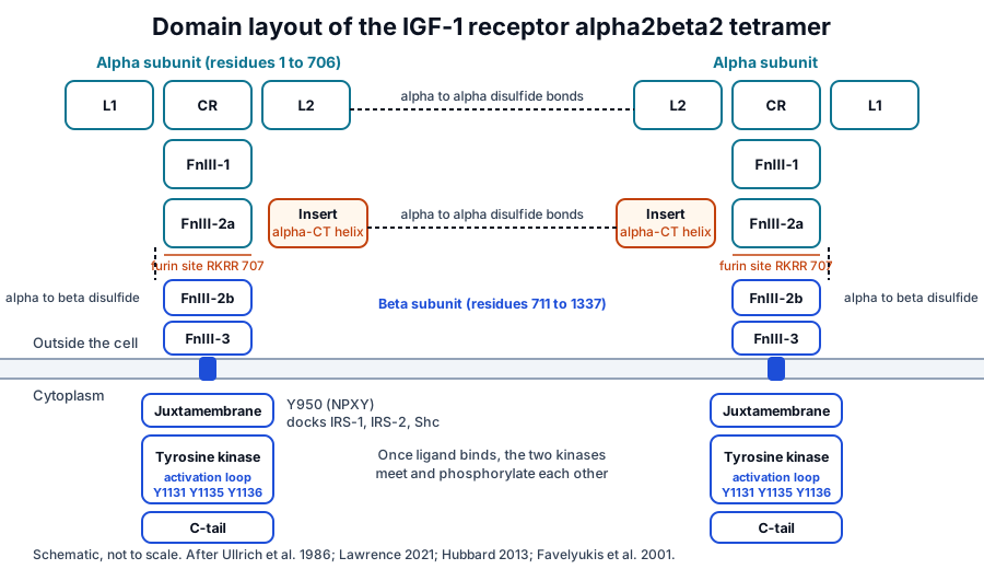
The human IGF1R gene on chromosome 15q26.3 encodes a precursor of 1,367 amino acids.[4] Ullrich and colleagues deduced that sequence from cloned cDNA in 1986: a 30-residue signal peptide is removed, and cleavage of an Arg-Lys-Arg-Arg sequence at position 707 splits the chain into an alpha subunit and a beta subunit, which run at about 135 and 90 kDa once glycosylated.[5]
Two alpha subunits and two beta subunits assemble into one receptor and are joined by disulfide bonds, so IGF-1R, like the insulin receptor, is a covalently linked tetramer before any ligand arrives.[3] Each alpha subunit carries the ligand-binding domains, and each beta subunit carries the membrane-spanning helix and the tyrosine kinase.[6]
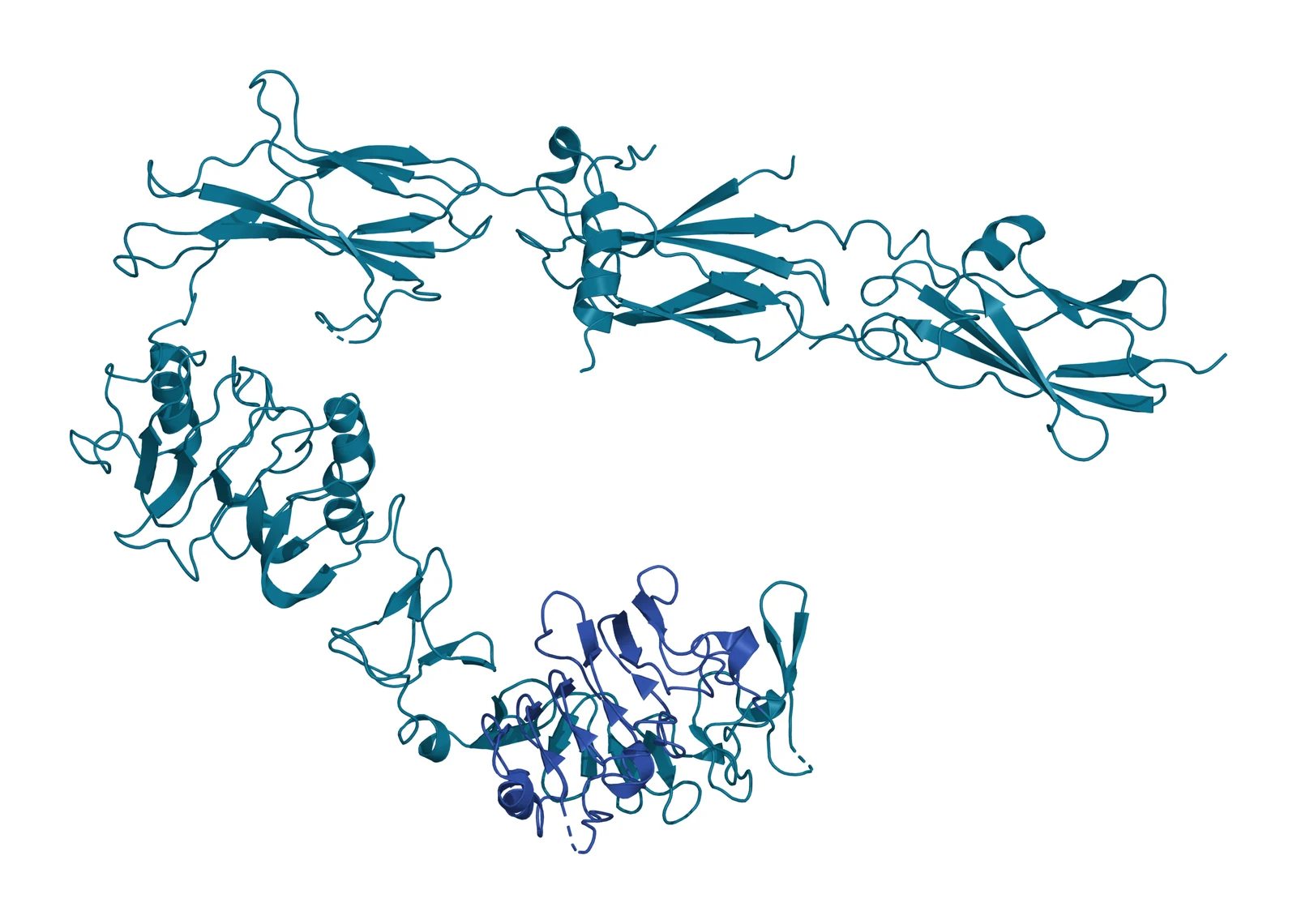
The extracellular half of each protomer is a chain of six domains: two leucine-rich-repeat domains, L1 and L2, flanking a cysteine-rich region, followed by three fibronectin type III domains, FnIII-1, FnIII-2 and FnIII-3.[7] The second fibronectin domain is interrupted by an insert that holds the alpha-CT segment, a short helix that completes the ligand-binding site, and the furin cleavage site that separates the two subunits (see Fig. 1 and Fig. 2).[1][7]
The L1-CR-L2 fragment outlines the ligand-binding pocket
Garrett and colleagues solved the first three domains of IGF-1R, residues 1 to 462, at 2.6 Å resolution in 1998. Each L domain is a single-stranded right-handed beta-helix, and the cysteine-rich region is built from eight disulfide-bonded modules, seven of which form a rod.[8]
The three domains surround a central space large enough to hold a ligand, and many of the residues that decide binding and ligand preference map to that site, even though the fragment on its own does not bind IGF-1.[8] A later structure of a hybrid receptor was consistent with the determinants for IGF binding residing in the IGF-1R cysteine-rich region.[9]
One IGF-1 binds between L1 of one protomer and the alpha-CT helix of the other
Xu and colleagues reported crystal structures of the IGF-1R ectodomain in its apo form, at 3.0 Å, and with IGF-1 bound after soaking, at 3.3 Å.[1] The ligand occupies the primary binding site, formed by L1 together with the alpha-CT helix contributed by the partner protomer, and binding separates receptor domains by induced fit (Fig. 3).[1]
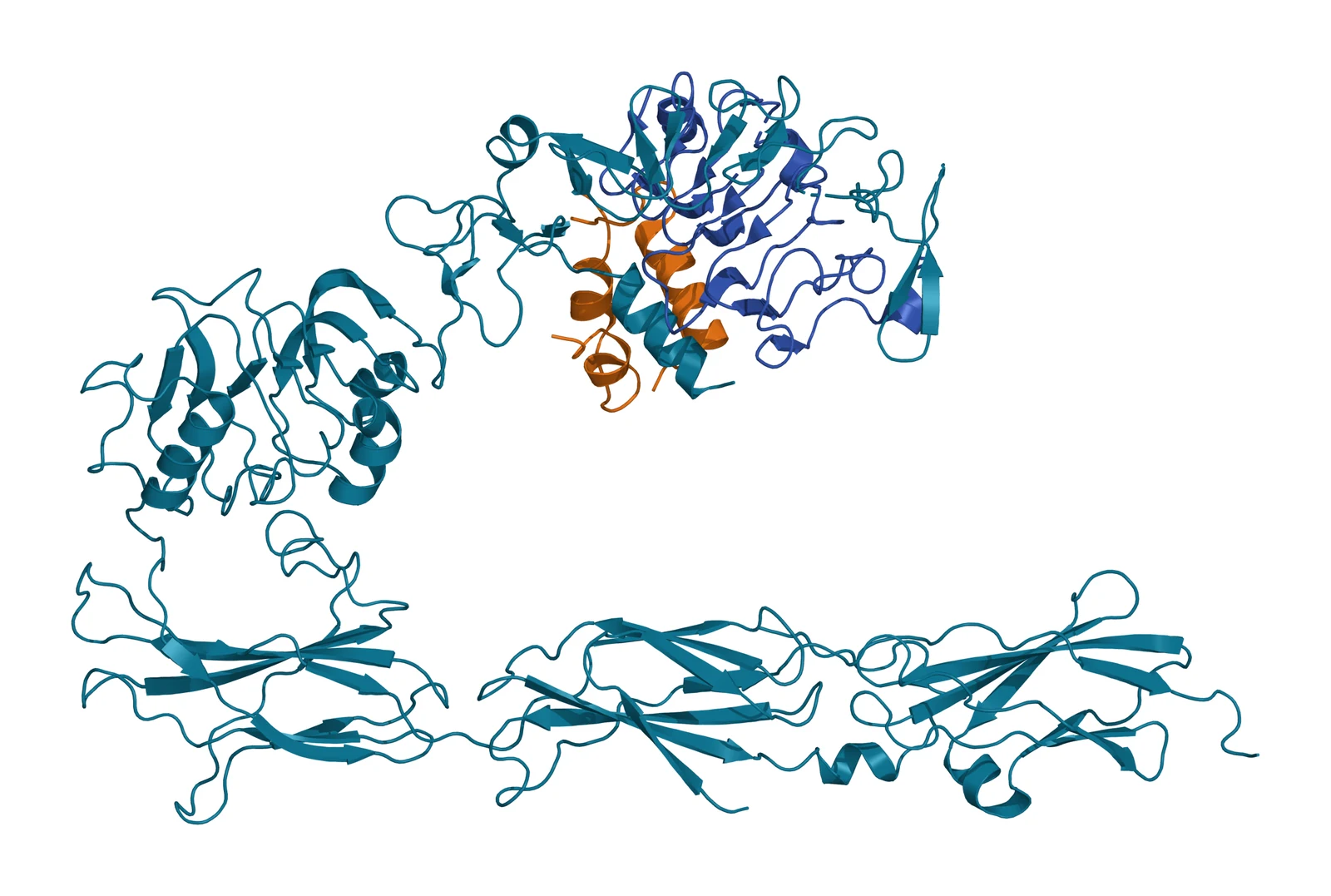
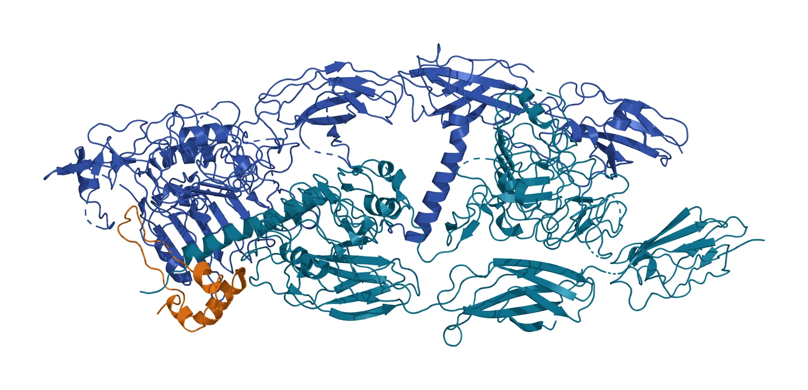
Fig. 3 | IGF-1 bound to the IGF-1R ectodomain and Full-length IGF-1R dimer with one IGF-1, cryo-EM.
Li and colleagues then solved the full-length receptor with IGF-1 by cryo-electron microscopy. Only one IGF-1 binds the Γ-shaped, asymmetric dimer, in a site made of the L1 and cysteine-rich domains of one protomer and the alpha-CT and FnIII-1 domains of the other (Fig. 4).[2] The liganded alpha-CT forms a rigid beam with the unliganded alpha-CT, which blocks the movement the second alpha-CT would need to bind another ligand, a structural source of the negative cooperativity long seen in binding studies.[2]
The same study found a contact between L1 and FnIII-2 that brings the membrane-proximal domains of the two protomers together, and receptor activation was reduced when that contact was disrupted.[2] Zhang and colleagues confirmed in the full-length receptor that a single insulin or a single IGF-1 molecule is enough to bind and activate it.[10]
Why does binding switch the kinase on? Kavran and colleagues showed that the ectodomain maintains an autoinhibited state in which the two transmembrane helices are held apart. Ligand binding releases that constraint, the helices associate, and the intracellular regions phosphorylate each other; once phosphorylated, the kinase is fully active even without the ligand or the extracellular region.[11]
IGF-2 and insulin use the same site with different contacts
IGF-2 binds IGF-1R with an affinity within an order of magnitude of IGF-1. Xu and colleagues solved IGF-2 bound to a stabilized ectodomain at up to 3.2 Å, in two conformations that differ in how far apart the two membrane entry points sit, and found that the C domain of IGF-2 contacts the receptor differently from the C domain of IGF-1.[12]
Alvino and colleagues showed earlier, by mutating Glu12 of IGF-2, that this residue bridges the two receptor halves and is needed for full activation.[13] Insulin also binds IGF-1R, at much lower affinity,[6] and the full-length receptor with insulin bound has been imaged at 4.7 Å.[10]
Hybrid receptors with the insulin receptor are common in tissues
Because IGF-1R and the insulin receptor are both made from alpha-beta half-receptors, a half of each can pair into a hybrid. Bailyes and colleagues measured how much of each receptor exists this way: about 40 percent of IGF-1 binding sites in liver and spleen, 70 percent in placenta, and 85 to 90 percent in skeletal muscle and heart were hybrids, with similar figures in rabbit and human tissue.[14]
In placenta the proportions fit random assembly from the available half-receptors.[14] Which insulin receptor isoform joins the hybrid matters: hybrids containing IR-A bound and were activated by IGF-1, IGF-2 and insulin, while hybrids containing IR-B responded strongly to IGF-1, weakly to IGF-2 and barely to insulin.[15]
Xu and colleagues imaged such a hybrid with IGF-1 bound at about 3.7 Å. A single IGF-1 engages the L1 domain and cysteine-rich region of the IGF-1R half, confirming that the IGF binding determinants sit on that half of the pair.[9] Siddle has reviewed how shared PI3K/AKT and Ras/ERK pathways still produce distinct responses to insulin and the IGFs in different tissues.[16]
Autophosphorylation of the activation loop turns the kinase on
Favelyukis and colleagues showed with the isolated kinase domain that IGF-1R is regulated by intermolecular autophosphorylation at three tyrosines in the activation loop, residues 1131, 1135 and 1136.[6][17] Each phosphorylation raised the turnover number and lowered the Km for ATP and peptide substrate.[17]
Their 2.1 Å structure of the tris-phosphorylated kinase with an ATP analog and a peptide substrate showed how phosphorylation stabilizes the activation loop in a conformation that supports catalysis, and how the enzyme recognizes hydrophobic residues at the P+1 and P+3 positions of its substrates.[17]
Cabail and colleagues found that the two phosphorylated kinase domains of the receptor dimer do more than act side by side: they form a specific dimer in which each exchanges its juxtamembrane segment with the other, which stabilizes helix alphaC of the kinase N lobe in its active position and promotes substrate phosphorylation.[18]
Phosphotyrosines dock IRS-1 and Shc, which open the PI3K/AKT and MAPK routes
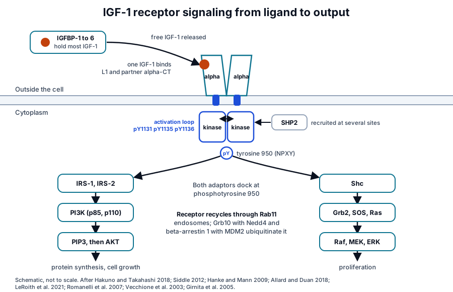
Ligand binding to the alpha subunits changes the conformation of the beta subunits and activates the kinase, which phosphorylates the insulin receptor substrates, IRS-1 and IRS-2, and the adaptor Shc.[19] Both dock at the phosphorylated tyrosine 950 in the juxtamembrane NPXY motif.[6]
Phosphorylated IRS proteins recruit the p85 subunit of PI3K, which generates PIP3 and activates AKT; Shc recruits Grb2 and SOS, which activate Ras and the Raf, MEK and ERK cascade (Fig. 5).[19] Hanke and Mann mapped the phosphotyrosine interactome of this family by quantitative proteomics and found binding partners for 52 of 109 phosphorylation sites, including multisite recruitment of the phosphatase SHP2 by IGF-1R.[20]
The two routes serve different outputs: AKT regulates protein synthesis through mTORC1, and the Ras/MAPK cascade leads to cell proliferation, migration and survival.[6] Siddle concludes that cellular context, rather than intrinsic receptor activity, appears to decide which of these responses dominates in a given tissue.[16]
Internalization, recycling and ubiquitination set the duration of the signal
Romanelli and colleagues followed the receptor after stimulation in glial progenitor cells. IGF-1 caused receptors to leave the cell surface and then return; blocking internalization inhibited AKT phosphorylation, and blocking recycling prevented the surface recovery and the sustained AKT signal.[21] The receptor localized with the transferrin receptor and Rab11-positive endosomes after ligand stimulation, the signature of a recycling route.[21]
Ubiquitination controls how long the receptor lasts. Vecchione and colleagues showed that the adaptor Grb10 brings the HECT ligase Nedd4 to the receptor, that ubiquitination likely occurs at the plasma membrane before endocytic vesicles form, and that both the proteasome and the lysosome then degrade the receptor.[22] Girnita and colleagues found a second route in which beta-arrestin 1 serves as the adaptor for the ligase MDM2, and ligand stimulation accelerated this ubiquitination.[23]
Six IGF binding proteins decide how much ligand reaches the receptor
Most IGF-1 and IGF-2 in blood and in tissue fluid is carried by one of six IGF binding proteins, IGFBP-1 to IGFBP-6, which bind the growth factors with equal or greater affinity than IGF-1R itself.[24] That places the binding proteins in control of how much free ligand the receptor sees.[24]
Clemmons has reviewed how each binding protein is regulated independently, how they govern access of IGF-1 and IGF-2 to the receptor, and how several also act through routes that do not involve the IGFs.[25] LeRoith, Holly and Forbes describe the whole system of three ligands, three receptors and six binding proteins as a unit.[6]
IGF-1R signaling is a major driver of growth in development
Mouse genetics established the receptor’s role. Liu and colleagues found that newborn mice lacking Igf1r were 45 percent of normal size, with smaller organs including muscle and delayed ossification, and did not survive birth; mice lacking Igf1 were 60 percent of normal size and some reached adulthood.[26]
Baker and colleagues timed the effects: between embryonic days 11 and 12.5 the receptor carries only the signal of IGF-2, and from day 13.5 it serves both IGF-1 and IGF-2, while placental growth runs through IGF-2 and a different receptor.[27] Igf1 mutants that survived kept growing more slowly and reached 30 percent of normal adult weight.[27] Yakar, Werner and Rosen have reviewed the receptor’s actions on bone, where IGF-1 signaling supports both longitudinal growth and bone mass.[28]
The growth hormone and IGF-1 axis works through both circulating and local IGF-1
The original somatomedin hypothesis held that growth hormone from the pituitary acts by raising liver-derived IGF-1 in the blood. Le Roith and colleagues reviewed how the hypothesis was revised once IGF-1 was found to be made by most, if not all, tissues, giving autocrine and paracrine IGF-1 a role of its own.[29]
Yakar and colleagues deleted Igf1 in the liver alone: circulating IGF-1 fell sharply, yet body weight, body length and femur length matched wild-type littermates, direct evidence for the importance of locally made IGF-1.[30] Lupu and colleagues compared mice lacking the growth hormone receptor, IGF-1, or both, and found that the two act through both shared and independent functions; the double mutants reached about 17 percent of normal body weight.[31]
Kaplan and Cohen proposed an augmentative and counteractive modification of the hypothesis that takes into account the integrated actions of growth hormone and IGF-1.[32] The growth hormone secretagogue reference describes how pituitary growth hormone release is regulated upstream of this axis.
Advancing the research question
The growth factor receptors overview places IGF-1R beside the other receptor tyrosine kinases that platelet granules and growth factors engage, and the surface receptor chemistry page explains the phosphotyrosine docking modules those receptors share. The PDGFR-beta and EGFR pages show how single-chain receptors form the dimer that IGF-1R already has when it reaches the cell surface.
Referencias comentadas
Numeradas en el orden en que se citan en el texto. Cada fuente lleva una nota sobre lo que respalda en esta página; los artículos revisados por pares se listan con revista, volumen, número y páginas, y las fuentes primarias son regulaciones, documentos de agencias y bases de datos de referencia.
- 1.Peer-reviewedXu Y, Kong GK, Menting JG, et al. How ligand binds to the type 1 insulin-like growth factor receptor. Nature Communications. 2018;9(1):821. https://pubmed.ncbi.nlm.nih.gov/29483580/Annotation: Crystal structures of the IGF-1R ectodomain, apo and with IGF-1 (PDB 5U8R and 5U8Q, Fig. 2 and Fig. 3). Cited for the primary binding site at L1 and the partner alpha-CT, and for domain separation by induced fit.
- 2.Peer-reviewedLi J, Choi E, Yu H, Bai XC. Structural basis of the activation of type 1 insulin-like growth factor receptor. Nature Communications. 2019;10(1):4567. https://pubmed.ncbi.nlm.nih.gov/31594955/Annotation: Cryo-EM structure of full-length IGF-1R with IGF-1 (PDB 6PYH, Fig. 4). Cited for the single ligand on the asymmetric dimer, the alpha-CT beam that explains negative cooperativity, and the L1 to FnIII-2 contact.
- 3.Peer-reviewedHubbard SR. The insulin receptor: both a prototypical and atypical receptor tyrosine kinase. Cold Spring Harbor Perspectives in Biology. 2013;5(3):a008946. https://pubmed.ncbi.nlm.nih.gov/23457259/Annotation: Review of the insulin receptor family architecture. Cited for the preformed, covalently linked tetramer that sets this family apart from single-chain receptor tyrosine kinases.
- 4.DatabaseNCBI Gene 3480, IGF1R insulin like growth factor 1 receptor (Homo sapiens). Accessed 8 November 2026. https://www.ncbi.nlm.nih.gov/gene/3480
- 5.Peer-reviewedUllrich A, Gray A, Tam AW, et al. Insulin-like growth factor I receptor primary structure: comparison with insulin receptor suggests structural determinants that define functional specificity. The EMBO Journal. 1986;5(10):2503-2512. https://pubmed.ncbi.nlm.nih.gov/2877871/Annotation: The cloned human IGF-1R sequence. Cited for the 1,367-residue precursor, the 30-residue signal peptide, the Arg-Lys-Arg-Arg cleavage site and the sizes of the alpha and beta subunits.
- 6.Peer-reviewedLeRoith D, Holly JMP, Forbes BE. Insulin-like growth factors: ligands, binding proteins, and receptors. Molecular Metabolism. 2021;52:101245. https://pubmed.ncbi.nlm.nih.gov/33962049/Annotation: Overview of the IGF system. Cited for the division of labor between alpha and beta subunits, the activation loop tyrosines 1131, 1135 and 1136, tyrosine 950 in the NPXY motif as the IRS and Shc docking site, the low affinity of insulin for IGF-1R, AKT control of protein synthesis through mTORC1 and Ras/MAPK outputs, and the three ligands, three receptors and six binding proteins.
- 7.Peer-reviewedLawrence MC. Understanding insulin and its receptor from their three-dimensional structures. Molecular Metabolism. 2021;52:101255. https://pubmed.ncbi.nlm.nih.gov/33992784/Annotation: Structural review of the insulin receptor family. Cited for the L1, CR, L2 and FnIII-1 to FnIII-3 domain order and the insert that carries alpha-CT (Fig. 1).
- 8.Peer-reviewedGarrett TP, McKern NM, Lou M, et al. Crystal structure of the first three domains of the type-1 insulin-like growth factor receptor. Nature. 1998;394(6691):395-399. https://pubmed.ncbi.nlm.nih.gov/9690478/Annotation: The L1-CR-L2 fragment at 2.6 Å. Cited for the beta-helix L domains, the eight-module cysteine-rich region and the central space that outlines the ligand site.
- 9.Peer-reviewedXu Y, Margetts MB, Venugopal H, et al. How insulin-like growth factor I binds to a hybrid insulin receptor type 1 insulin-like growth factor receptor. Structure. 2022;30(8):1098-1108.e6. https://pubmed.ncbi.nlm.nih.gov/35660159/Annotation: Cryo-EM of an insulin receptor and IGF-1R hybrid with IGF-1. Cited for the ligand engaging the L1 and cysteine-rich region of the IGF-1R half, and for the binding determinants in that region.
- 10.Peer-reviewedZhang X, Yu D, Sun J, et al. Visualization of ligand-bound ectodomain assembly in the full-length human IGF-1 receptor by cryo-EM single-particle analysis. Structure. 2020;28(5):555-561.e4. https://pubmed.ncbi.nlm.nih.gov/32275863/Annotation: Cryo-EM of the full-length receptor with insulin (4.7 Å) and IGF-1 (7.7 Å). Cited for one ligand being enough to bind and activate the receptor.
- 11.Peer-reviewedKavran JM, McCabe JM, Byrne PO, et al. How IGF-1 activates its receptor. eLife. 2014;3:e03772. https://pubmed.ncbi.nlm.nih.gov/25255214/Annotation: Biochemical and biophysical assays of receptor activation. Cited for the autoinhibited ectodomain that holds the transmembrane helices apart and for autophosphorylation as the step the ligand triggers.
- 12.Peer-reviewedXu Y, Kirk NS, Venugopal H, et al. How IGF-II binds to the human type 1 insulin-like growth factor receptor. Structure. 2020;28(7):786-798.e6. https://pubmed.ncbi.nlm.nih.gov/32459985/Annotation: Cryo-EM of IGF-2 bound to a stabilized IGF-1R ectodomain. Cited for the two conformations and the different C-domain contacts of IGF-2 and IGF-1.
- 13.Peer-reviewedAlvino CL, Ong SC, McNeil KA, et al. Understanding the mechanism of insulin and insulin-like growth factor (IGF) receptor activation by IGF-II. PLoS One. 2011;6(11):e27488. https://pubmed.ncbi.nlm.nih.gov/22140443/Annotation: Mutagenesis of IGF-2 Glu12. Cited for that residue bridging the two receptor halves during activation.
- 14.Peer-reviewedBailyes EM, Navé BT, Soos MA, et al. Insulin receptor/IGF-I receptor hybrids are widely distributed in mammalian tissues: quantification of individual receptor species by selective immunoprecipitation and immunoblotting. Biochemical Journal. 1997;327 ( Pt 1)(Pt 1):209-215. https://pubmed.ncbi.nlm.nih.gov/9355755/Annotation: Quantification of hybrid receptors in tissue membranes. Cited for the hybrid fractions in liver, spleen, placenta, skeletal muscle and heart, and for random assembly in placenta.
- 15.Peer-reviewedPandini G, Frasca F, Mineo R, et al. Insulin/insulin-like growth factor I hybrid receptors have different biological characteristics depending on the insulin receptor isoform involved. Journal of Biological Chemistry. 2002;277(42):39684-39695. https://pubmed.ncbi.nlm.nih.gov/12138094/Annotation: Binding and activation of hybrids built with IR-A or IR-B. Cited for the ligand preferences of each hybrid type.
- 16.Peer-reviewedSiddle K. Molecular basis of signaling specificity of insulin and IGF receptors: neglected corners and recent advances. Frontiers in Endocrinology. 2012;3:34. https://pubmed.ncbi.nlm.nih.gov/22649417/Annotation: Review of how insulin and IGF receptors reach distinct responses through shared PI3K/AKT and Ras/ERK pathways. Cited for tissue-level specificity and the conclusion that cellular context, rather than intrinsic receptor activity, sets which responses dominate.
- 17.Peer-reviewedFavelyukis S, Till JH, Hubbard SR, Miller WT. Structure and autoregulation of the insulin-like growth factor 1 receptor kinase. Nature Structural Biology. 2001;8(12):1058-1063. https://pubmed.ncbi.nlm.nih.gov/11694888/Annotation: Kinetics and the 2.1 Å structure of the tris-phosphorylated kinase. Cited for the three activation loop tyrosines, the effect of each phosphorylation on turnover and Km, and substrate recognition at P+1 and P+3.
- 18.Peer-reviewedCabail MZ, Li S, Lemmon E, et al. The insulin and IGF1 receptor kinase domains are functional dimers in the activated state. Nature Communications. 2015;6:6406. https://pubmed.ncbi.nlm.nih.gov/25758790/Annotation: Crystallographic and biochemical evidence for a dimer of phosphorylated kinase domains. Cited for the juxtamembrane exchange that stabilizes helix alphaC in its active position.
- 19.Peer-reviewedHakuno F, Takahashi SI. IGF1 receptor signaling pathways. Journal of Molecular Endocrinology. 2018;61(1):T69-T86. https://pubmed.ncbi.nlm.nih.gov/29535161/Annotation: Review of IGF-1R signaling. Cited for the alpha to beta conformational relay, IRS and Shc phosphorylation, and the PI3K/AKT and Ras/MAPK routes through p85, Grb2 and SOS (Fig. 5).
- 20.Peer-reviewedHanke S, Mann M. The phosphotyrosine interactome of the insulin receptor family and its substrates IRS-1 and IRS-2. Molecular and Cellular Proteomics. 2009;8(3):519-534. https://pubmed.ncbi.nlm.nih.gov/19001411/Annotation: Quantitative interaction proteomics of the family’s phosphotyrosine sites. Cited for partners at 52 of 109 sites and the multisite recruitment of SHP2 by IGF-1R.
- 21.Peer-reviewedRomanelli RJ, LeBeau AP, Fulmer CG, et al. Insulin-like growth factor type-I receptor internalization and recycling mediate the sustained phosphorylation of Akt. Journal of Biological Chemistry. 2007;282(31):22513-22524. https://pubmed.ncbi.nlm.nih.gov/17545147/Annotation: Receptor trafficking in glial progenitor cells. Cited for ligand-induced internalization, colocalization with Rab11-positive endosomes, and the dependence of sustained AKT phosphorylation on both.
- 22.Peer-reviewedVecchione A, Marchese A, Henry P, et al. The Grb10/Nedd4 complex regulates ligand-induced ubiquitination and stability of the insulin-like growth factor I receptor. Molecular and Cellular Biology. 2003;23(9):3363-3372. https://pubmed.ncbi.nlm.nih.gov/12697834/Annotation: Ubiquitination studies in mouse embryo fibroblasts. Cited for Grb10 bridging Nedd4 to the receptor, ubiquitination likely at the plasma membrane, and degradation by both proteasome and lysosome.
- 23.Peer-reviewedGirnita L, Shenoy SK, Sehat B, et al. Beta-arrestin is crucial for ubiquitination and down-regulation of the insulin-like growth factor-1 receptor by acting as adaptor for the MDM2 E3 ligase. Journal of Biological Chemistry. 2005;280(26):24412-24419. https://pubmed.ncbi.nlm.nih.gov/15878855/Annotation: Co-immunoprecipitation and ubiquitination assays. Cited for beta-arrestin 1 as the adaptor that brings MDM2 to the receptor and for ligand accelerating ubiquitination.
- 24.Peer-reviewedAllard JB, Duan C. IGF-binding proteins: why do they exist and why are there so many? Frontiers in Endocrinology. 2018;9:117. https://pubmed.ncbi.nlm.nih.gov/29686648/Annotation: Review of the six IGF binding proteins. Cited for their affinity equal to or greater than the receptor’s and their control of free ligand in blood and tissue.
- 25.Peer-reviewedClemmons DR. Role of IGF-binding proteins in regulating IGF responses to changes in metabolism. Journal of Molecular Endocrinology. 2018;61(1):T139-T169. https://pubmed.ncbi.nlm.nih.gov/29563157/Annotation: Review of IGFBP regulation. Cited for independent regulation of each binding protein, control of receptor access, and IGF-independent actions.
- 26.Peer-reviewedLiu JP, Baker J, Perkins AS, Robertson EJ, Efstratiadis A. Mice carrying null mutations of the genes encoding insulin-like growth factor I (Igf-1) and type 1 IGF receptor (Igf1r). Cell. 1993;75(1):59-72. https://pubmed.ncbi.nlm.nih.gov/8402901/Annotation: Igf1 and Igf1r null mice. Cited for the 45 and 60 percent birth sizes, organ hypoplasia and delayed ossification.
- 27.Peer-reviewedBaker J, Liu JP, Robertson EJ, Efstratiadis A. Role of insulin-like growth factors in embryonic and postnatal growth. Cell. 1993;75(1):73-82. https://pubmed.ncbi.nlm.nih.gov/8402902/Annotation: Growth kinetics of the IGF mutants. Cited for the timing of IGF-2 and IGF-1 signaling through the receptor, placental growth through a different receptor, and 30 percent adult weight in Igf1 mutants.
- 28.Peer-reviewedYakar S, Werner H, Rosen CJ. Insulin-like growth factors: actions on the skeleton. Journal of Molecular Endocrinology. 2018;61(1):T115-T137. https://pubmed.ncbi.nlm.nih.gov/29626053/Annotation: Review of IGF actions on bone. Cited for the receptor’s support of longitudinal growth and bone mass.
- 29.Peer-reviewedLe Roith D, Bondy C, Yakar S, Liu JL, Butler A. The somatomedin hypothesis: 2001. Endocrine Reviews. 2001;22(1):53-74. https://pubmed.ncbi.nlm.nih.gov/11159816/Annotation: Revision of the somatomedin hypothesis. Cited for IGF-1 production by most tissues and the autocrine and paracrine role.
- 30.Peer-reviewedYakar S, Liu JL, Stannard B, et al. Normal growth and development in the absence of hepatic insulin-like growth factor I. Proceedings of the National Academy of Sciences of the United States of America. 1999;96(13):7324-7329. https://pubmed.ncbi.nlm.nih.gov/10377413/Annotation: Liver-specific Igf1 deletion. Cited for the sharp fall in circulating IGF-1 with normal body weight, length and femur length.
- 31.Peer-reviewedLupu F, Terwilliger JD, Lee K, Segre GV, Efstratiadis A. Roles of growth hormone and insulin-like growth factor 1 in mouse postnatal growth. Developmental Biology. 2001;229(1):141-162. https://pubmed.ncbi.nlm.nih.gov/11133160/Annotation: Ghr, Igf1 and double mutant growth curves. Cited for the shared and independent functions of growth hormone and IGF-1 and the 17 percent body weight of double mutants.
- 32.Peer-reviewedKaplan SA, Cohen P. The somatomedin hypothesis 2007: 50 years later. Journal of Clinical Endocrinology and Metabolism. 2007;92(12):4529-4535. https://pubmed.ncbi.nlm.nih.gov/17986643/Annotation: An augmentative and counteractive modification of the somatomedin hypothesis. Cited for the integrated actions of growth hormone and IGF-1.
Cite this page
ExaVeyra Sciences Editorial Team. IGF-1 Receptor: Structure and Signaling. ExaVeyra Sciences; 9 November 2026. Available from: https://exaveyra.com/science/surface-receptors/igf-1-receptor. Accessed [date accessed].
Fuentes de las figuras
- 33.PDB 5U8R, Human IGF-1R ectodomain, apo. ExaVeyra Sciences, rendered in PyMOL from PDB 5U8R. Structure data: RCSB Protein Data Bank (rcsb.org), CC0. Publicación de la estructura: Xu Y, Kong GK, Menting JG, et al. How ligand binds to the type 1 insulin-like growth factor receptor. Nat Commun. 2018;9(1):821.
- 34.PDB 5U8Q, Human IGF-1R ectodomain in complex with IGF-1. ExaVeyra Sciences, rendered in PyMOL from PDB 5U8Q. Structure data: RCSB Protein Data Bank (rcsb.org), CC0. Publicación de la estructura: Xu Y, Kong GK, Menting JG, et al. How ligand binds to the type 1 insulin-like growth factor receptor. Nat Commun. 2018;9(1):821.
- 35.PDB 6PYH, Full-length IGF-1R in complex with IGF-1. ExaVeyra Sciences, rendered in PyMOL from PDB 6PYH. Structure data: RCSB Protein Data Bank (rcsb.org), CC0. Publicación de la estructura: Li J, Choi E, Yu H, Bai XC. Structural basis of the activation of type 1 insulin-like growth factor receptor. Nat Commun. 2019;10(1):4567.
Créditos de imágenes
- Fig. 1ExaVeyra Sciences, figura original. After Ullrich et al., EMBO J 1986; Lawrence, Mol Metab 2021; Hubbard, Cold Spring Harb Perspect Biol 2013; Favelyukis et al., Nat Struct Biol 2001. ↩
- Fig. 4ExaVeyra Sciences, figura original. After Hakuno and Takahashi, J Mol Endocrinol 2018; Siddle, Front Endocrinol 2012; Hanke and Mann, Mol Cell Proteomics 2009; Allard and Duan, Front Endocrinol 2018; LeRoith et al., Mol Metab 2021; Romanelli et al., J Biol Chem 2007; Vecchione et al., Mol Cell Biol 2003; Girnita et al., J Biol Chem 2005. ↩