Esta referencia está disponible actualmente en inglés. La versión en español está en preparación.
Growth Factor Receptors: PDGF, TGF-Beta, VEGF, EGF and IGF-1
Por Benn Bluestein-Veyra, M.Sc. en Química Orgánica · Actualizado el 21 de octubre de 2026
ExaVeyra Sciences es un proveedor B2B con sede en Miami que suministra exosomas para uso estético tópico a clínicas con licencia, y remite a los prescriptores con licencia a una farmacia de preparados compuestos 503A que prepara formulaciones según prescripciones individuales.
| Pregunta | Respuesta |
|---|---|
| Qué cubre esta página | The receptors that read platelet-derived growth factors: receptor tyrosine kinases for PDGF, VEGF, EGF and IGF-1, and serine/threonine kinase receptors for TGF-beta. |
| Quién puede hacer pedidos | Clínicas y profesionales con licencia profesional activa y NPI verificados por ExaVeyra. Los pedidos de exosomas requieren además un reconocimiento firmado de uso profesional. |
| Qué se envía y cómo | El material de exosomas para uso estético tópico, caracterizado por lote, se envía en cadena de frío por exprés nocturno. La entrega puede tardar 1 a 3 días hábiles. Se emite un certificado de análisis para cada lote y se entrega a solicitud. |
Growth factor receptors are the cell-surface proteins through which cells read polypeptide growth factors such as PDGF, VEGF, EGF, IGF-1 and TGF-beta. The first four act on receptor tyrosine kinases, single-pass transmembrane receptors that direct growth, differentiation, metabolism and motility.[1][2] TGF-beta acts on a pair of transmembrane serine/threonine kinases, receptors I and II.[3]
Platelets store many of these ligands. Blair and Flaumenhaft list VEGF, PDGF, EGF and IGF among the growth factors held in alpha granules, which fuse with the plasma membrane on activation and release hundreds of bioactive proteins.[4] Platelets also release TGF-beta1 in a latent form.[5]
This reference follows each receptor family from ligand binding to its first signaling steps. It closes with the binding, phosphorylation and reporter assays that laboratories use to measure receptor activation.
Understanding receptor tyrosine kinase architecture and ligand-induced dimerization
Each receptor tyrosine kinase has a ligand-binding extracellular region, one transmembrane helix and an intracellular tyrosine kinase domain (Fig. 1).[1][6] Ligand binding brings two receptors together, and dimerization leads to autophosphorylation on specific tyrosine residues.[6]
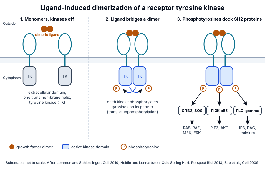
Lemmon and Schlessinger note that the strategies by which ligands induce dimerization are surprisingly diverse, as are the mechanisms that couple dimerization to kinase activation.[2] PDGF bridges two receptors with its own dimeric body, and EGF changes the receptor shape so that the receptors contact each other directly.[7][8]
Autophosphorylation raises kinase activity and creates docking sites for signaling proteins that carry SH2 domains.[6] Hubbard and Miller describe these receptors as essential components of the pathways that mediate cell-to-cell communication.[1]
PDGF dimers clamp PDGFR-beta at their dimerization seam
PDGFs and their receptors have served as prototypes for growth factor and receptor tyrosine kinase function for more than 25 years.[9] In animal development, PDGFR-alpha signaling takes part in gastrulation and in forming the neural crest, lung, intestine, skin and skeleton, and PDGFR-beta signaling in blood vessel formation and early hematopoiesis.[9]
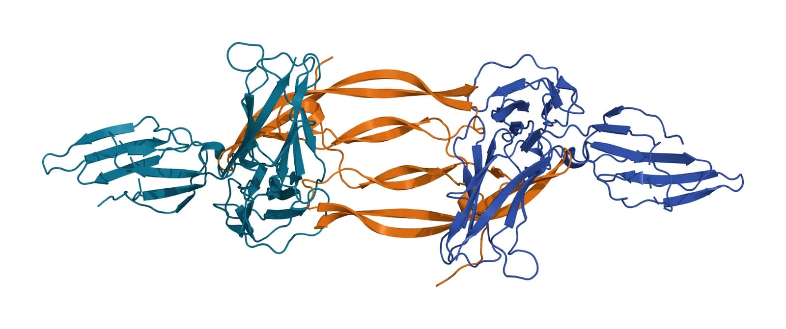
PDGFR-alpha and PDGFR-beta are type III receptor tyrosine kinases, with five Ig-like domains outside the cell and a split kinase domain inside.[6] The human PDGFRB gene maps to chromosome 5q32.[10]
Shim and colleagues solved PDGF-B bound to the first three Ig domains of PDGFR-beta and found that the two PDGF-B protomers clamp the receptor at their dimerization seam (Fig. 2).[7] The interface is predominantly hydrophobic, and the PDGF-B L1 loop, disordered in the free growth factor, adopts a defined conformation to contact the third Ig domain.[7]
In the same study, PDGF propeptides occupied positions on the mature growth factor that overlap the receptor sites, which the authors read as a way to cover hydrophobic surfaces during secretion.[7] Calorimetry showed that the membrane-proximal contact between PDGFR-alpha molecules, required for activation, contributes negatively to ligand binding.[7]
EGF binding exposes a dimerization arm on EGFR
Ogiso and colleagues solved a 2:2 complex of human EGF with the EGFR extracellular region at 3.3 Å resolution. Domains I to III form a C shape, with EGF docked between domains I and III (Fig. 3).[8] The human EGFR gene maps to chromosome 7p11.2.[11]
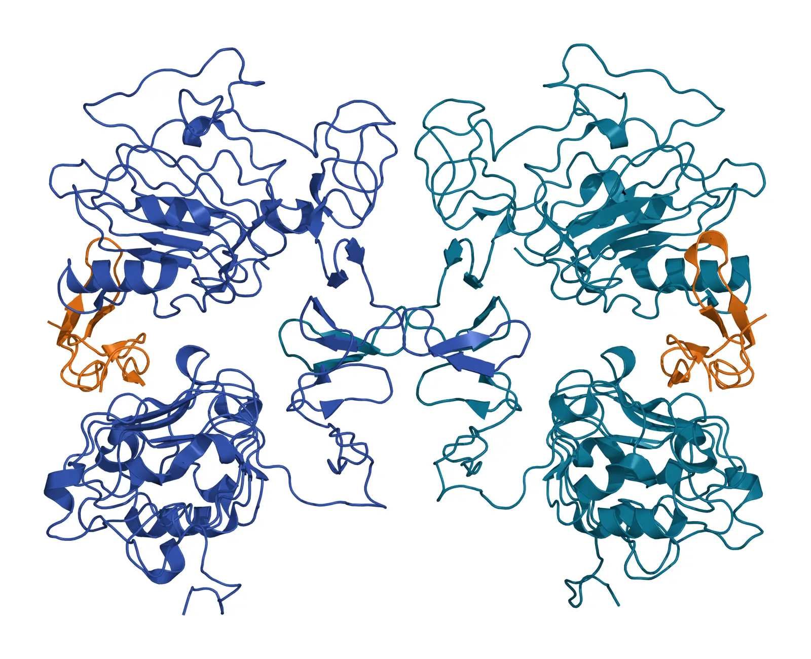
The 1:1 complexes dimerize through a direct receptor contact: a protruding beta-hairpin arm of each domain II holds the body of the other, and mutagenesis confirmed this receptor-mediated dimerization.[8] Lemmon, Schlessinger and Ferguson describe it as distinctive to the EGFR family, with ligand binding inducing a dramatic conformational change that exposes the arm.[12]
An asymmetric kinase dimer switches the EGFR kinase on
Zhang and colleagues found that the EGFR kinase domain is intrinsically autoinhibited, in a conformation that resembles Src and the cyclin-dependent kinases.[13] Activation comes from an asymmetric dimer in which the C-terminal lobe of one kinase domain plays the role that cyclin plays in an active CDK and cyclin complex.[13]
In this family, allosteric changes in the asymmetric dimer drive kinase regulation, and the intracellular juxtamembrane segment activates the receptor.[12] A long carboxy-terminal tail carries the autophosphorylation sites and serves an autoregulatory function.[12]
VEGF-A signals through VEGFR-1, VEGFR-2 and neuropilin coreceptors
Five mammalian VEGF ligands act through three receptor tyrosine kinases, and coreceptors such as neuropilins and integrins associate with the ligand and receptor complex to modulate its output.[14] VEGF-A binds two of these receptors, KDR (VEGFR-2) and Flt-1 (VEGFR-1).[15]
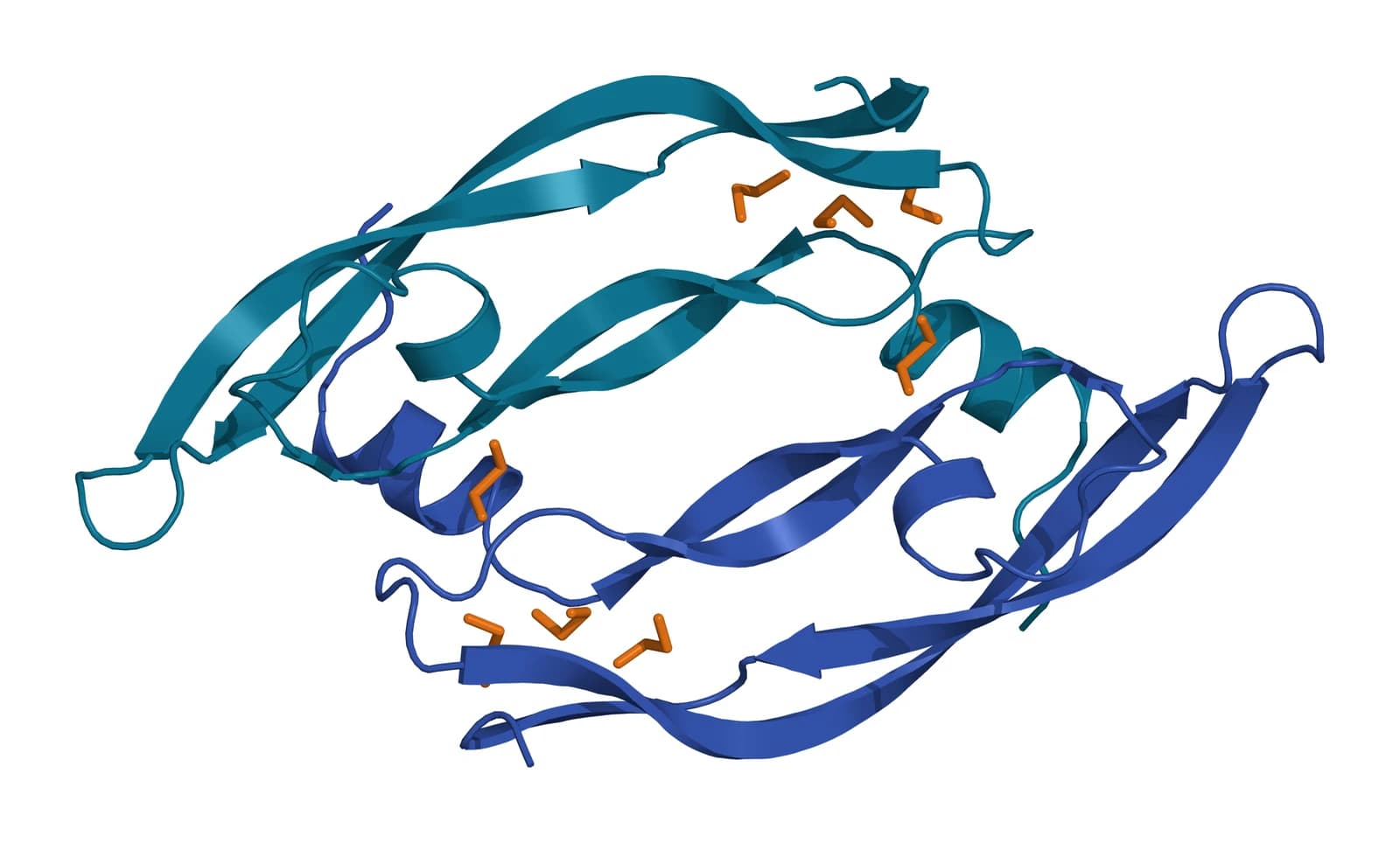
VEGF-A is a homodimeric cystine knot growth factor whose closest relative is PDGF. Muller and colleagues refined its receptor-binding domain to 1.93 Å with eight copies in the asymmetric unit, and mutagenesis placed one flexible loop in the receptor-binding site (Fig. 4).[15]
The VEGF receptor extracellular region holds seven immunoglobulin homology domains, and ligand binding promotes receptor dimerization.[16] Leppänen and colleagues located the high-affinity VEGFR-2 binding site in domains D2 and D3, and VEGFR-1 and VEGFR-2 chimeras defined the VEGFR-2 residues critical for binding VEGF-A and VEGF-C.[16] The human KDR gene maps to chromosome 4q12.[17]
Simons, Gordon and Claesson-Welsh describe VEGFR-2 signaling as tightly regulated at several levels: by neuropilins, proteoglycans, integrins and protein tyrosine phosphatases, and by receptor endocytosis and trafficking.[18] VEGFs and their receptors balance the formation of new blood vessels with the upkeep of existing ones, in development and in adult tissues.[18]
Platelets sort VEGF into its own granules. Italiano and colleagues found VEGF and endostatin in separate alpha-granule subpopulations, and a selective PAR4 agonist released the endostatin-containing granules while the VEGF-containing granules stayed in the platelet.[19]
IGF-1 binding releases an autoinhibited IGF-1R homodimer
The type 1 insulin-like growth factor receptor is a homodimeric receptor tyrosine kinase that signals into pathways directing normal cellular growth, differentiation and proliferation.[20] The human IGF1R gene maps to chromosome 15q26.3.[21]
Xu and colleagues solved crystal structures of the IGF-1R ectodomain alone and bound to IGF-I. The structures show the growth factor at the primary ligand-binding site and indicate that binding separates receptor domains by induced fit.[20]
Kavran and colleagues found that the IGF1R ectodomain maintains an autoinhibited state with the transmembrane helices held apart. Ligand binding releases this constraint, letting the transmembrane segments associate and the intracellular regions autophosphorylate.[22]
Phosphorylated IGF1R was fully active independent of ligand and of the extracellular and transmembrane regions, so the authors identify autophosphorylation as the key step that ligand binding triggers.[22] The active receptor then phosphorylates insulin receptor substrates and SHC.[23]
TGF-beta is stored in latent form and signals through serine/threonine kinase receptors
Assoian and colleagues identified platelets in 1983 as a major storage site of TGF-beta, a 25,000-dalton protein of two disulfide-linked 12,500-dalton subunits. The platelet extract had a specific activity 100-fold greater than the other normal tissues tested.[24]
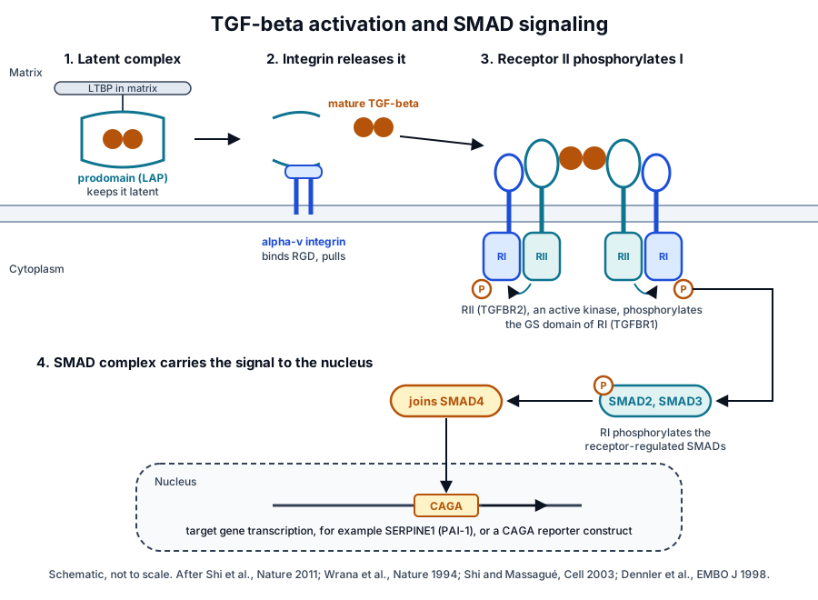
TGF-beta is stored in the extracellular matrix as a latent complex with its prodomain, held there by latent TGF-beta binding proteins (Fig. 5).[25] Robertson and Rifkin review the molecules that set its bioavailability and note that TGF-beta must be activated before it can bind its receptors.[26]
Shi and colleagues solved porcine proTGF-beta1 as a ring-shaped dimer in which the prodomain shields the growth factor from receptors. Activation required alpha-v integrin binding to an RGD sequence in the prodomain and force that unfastens a straitjacket around each growth factor monomer.[25]
Platelets carry an activation route of their own. Blakytny and colleagues found that thrombin-stimulated platelets activated a significant proportion of the latent TGF-beta they released, through a released furin-like proprotein convertase.[5]
Receptor II recruits and phosphorylates receptor I
Wrana and colleagues showed in 1994 that TGF-beta binds directly to receptor II, a constitutively active kinase. Receptor I then recognizes the bound ligand, joins the complex and is phosphorylated by receptor II, which lets it pass the signal to downstream substrates.[3]
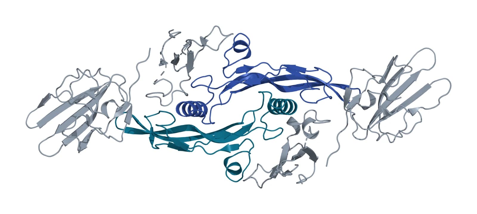
The genes encoding the two receptors, TGFBR1 and TGFBR2, map to chromosomes 9q22.33 and 3p24.1.[27][28] Heldin and Moustakas describe the active signaling unit as a heterotetrameric complex of type I and type II receptors.[29]
Radaev and colleagues solved human TGF-beta1 with the extracellular domains of both receptors at 3.0 Å (Fig. 6). Binding of all three TGF-beta isoforms to the type I receptor rose considerably in the presence of the type II receptor.[30]
Hinck, Mueller and Springer trace the growth factor domain of the TGF-beta family to a common cystine knot ancestor. They describe how ternary complexes show TGF-beta growth factor dimers binding type I and type II receptors at the cell surface.[31]
SMAD proteins carry the signal to the nucleus
After receptor activation, SMAD2 and SMAD3 are phosphorylated and form complexes with SMAD4, which move to the nucleus and control expression of target genes.[32][33] Shi and Massagué review this route from ligand binding to transcriptional regulation and the control of SMAD activity and degradation.[33]
TGF-beta receptors also signal through non-SMAD pathways, including the ERK1/2, JNK and p38 MAP kinases, Src, PI3K and Rho GTPases.[29] Massagué frames the central question of the field as how context determines the cellular response to TGF-beta.[34]
Phosphotyrosine docking sites route signals to the MAPK, PI3K and PLC-gamma pathways
SH2 domains recognize phosphotyrosine within particular sequence motifs. Bae and colleagues noted that the modest affinity of SH2 domains for phosphotyrosine peptides likely gives an oversimplified account of how cells select signaling pathways.[35]
Their crystal structure of the activated FGFR1 kinase with a phospholipase C-gamma fragment showed a secondary binding site on an SH2 domain contacting the kinase domain. This phosphorylation-independent contact set the selectivity of phospholipase C-gamma binding and signaling in cultured cells.[35]
At IGF-1R, phosphotyrosines on IRS proteins and SHC recruit the p85 subunit of PI3K, GRB2 and SHP2, which lead into the PI3K and Ras-MAP kinase pathways.[23] Hakuno and Takahashi note that these pathways are required for IGF-induced cell proliferation, differentiation and survival.[23]
PDGF receptors use the same logic: their SH2-docking pathways promote cell growth, survival and migration.[6] The table summarizes the receptors on this page.
| Receptor | Ligand on this page | Receptor class | Structure |
|---|---|---|---|
| PDGFR-alpha, PDGFR-beta | PDGF dimers | Type III receptor tyrosine kinase, five Ig-like domains | PDGF-B with PDGFR-beta D1 to D3, PDB 3MJG |
| EGFR | EGF | EGFR family receptor tyrosine kinase, domains I to IV | EGF with the EGFR extracellular region, PDB 1IVO |
| VEGFR-1, VEGFR-2 | VEGF-A | Receptor tyrosine kinase, seven Ig homology domains | VEGF-A receptor-binding domain, PDB 2VPF |
| IGF-1R | IGF-1 | Homodimeric receptor tyrosine kinase | Ectodomain crystal structures, Xu et al. 2018 |
| TGFBR2 and TGFBR1 | TGF-beta1, 2 and 3 | Serine/threonine kinase receptors, type II and type I | TGF-beta1 ternary complex, PDB 3KFD |
Binding, phosphorylation and reporter assays measure receptor activation
Binding studies measure the first step. Shim and colleagues used calorimetry on PDGF and its receptor, and Radaev and colleagues used quantitative binding studies to compare receptor kinetics across the TGF-beta isoforms.[7][30]
Receptor chimeras map specificity: Leppänen and colleagues swapped regions between VEGFR-1 and VEGFR-2 to find the residues that bind VEGF-A.[16] Kinase activation is read as phosphorylation, and Kavran and colleagues measured it in full-length IGF1R and in kinase-containing fragments.[22]
Reporter constructs read the transcriptional output. Dennler and colleagues identified CAGA boxes in the human PAI-1 promoter that bind SMAD3 and SMAD4, and these boxes conferred TGF-beta and activin responsiveness on a heterologous promoter reporter construct.[32]
On the ligand side, mass spectrometry defines what platelets store. Maynard and colleagues identified 284 proteins in a fraction enriched for alpha granules, 44 of them newly assigned to the granule, and confirmed several by immunoelectron microscopy.[36]
Where to go from here
The platelet granule biology reference describes how alpha granules form, what they hold and how activation releases them. The PRP preparation variables reference explains how spin settings, anticoagulant and activation shape the growth factor content of a preparation, and the platelet-rich fibrin reference follows the fibrin matrix that holds and releases these proteins.
The BMAC and SVF composition reference and the platelet study reporting reference complete the cluster, and the MSC secretome reference covers the same growth factors from stromal cells. Our research methods, regulatory and quality references support study design and documentation, and our team is glad to discuss the methods described on this page.
Referencias comentadas
Numeradas en el orden en que se citan en el texto. Cada fuente lleva una nota sobre lo que respalda en esta página; los artículos revisados por pares se listan con revista, volumen, número y páginas, y las fuentes primarias son regulaciones, documentos de agencias y bases de datos de referencia.
- 1.Peer-reviewedHubbard SR, Miller WT. Receptor tyrosine kinases: mechanisms of activation and signaling. Current Opinion in Cell Biology. 2007. https://pubmed.ncbi.nlm.nih.gov/17306972/Annotation: Review of receptor tyrosine kinases. Cited for single-pass receptors that bind growth factors and direct growth, differentiation, metabolism and motility in cell-to-cell communication.
- 2.Peer-reviewedLemmon MA, Schlessinger J. Cell signaling by receptor tyrosine kinases. Cell. 2010. https://pubmed.ncbi.nlm.nih.gov/20602996/Annotation: Review of receptor tyrosine kinase activation. Cited for the diversity of ligand-induced dimerization strategies and of the mechanisms that couple dimerization to kinase activation.
- 3.Peer-reviewedWrana JL, Attisano L, Wieser R, et al. Mechanism of activation of the TGF-beta receptor. Nature. 1994. https://pubmed.ncbi.nlm.nih.gov/8047140/Annotation: Biochemical study of the two TGF-beta receptors. Cited for ligand binding to the constitutively active receptor II and the recruitment and phosphorylation of receptor I.
- 4.Peer-reviewedBlair P, Flaumenhaft R. Platelet alpha-granules: basic biology and clinical correlates. Blood Reviews. 2009. https://pubmed.ncbi.nlm.nih.gov/19450911/Annotation: Review of alpha-granule biology. Cited for VEGF, PDGF, EGF and IGF among stored growth factors (full text) and for granule fusion and release of hundreds of bioactive proteins on activation.
- 5.Peer-reviewedBlakytny R, Ludlow A, Martin GE, et al. Latent TGF-beta1 activation by platelets. Journal of Cellular Physiology. 2004. https://pubmed.ncbi.nlm.nih.gov/14978736/Annotation: Study of thrombin-stimulated human platelets and their releasates. Cited for latent TGF-beta1 release and its activation by a released furin-like proprotein convertase.
- 6.Peer-reviewedHeldin CH, Lennartsson J. Structural and functional properties of platelet-derived growth factor and stem cell factor receptors. Cold Spring Harbor Perspectives in Biology. 2013. https://pubmed.ncbi.nlm.nih.gov/23906712/Annotation: Review of the type III receptor tyrosine kinases. Cited for five Ig-like domains, the split kinase domain, dimerization-driven autophosphorylation and SH2 docking pathways for growth, survival and migration.
- 7.Peer-reviewedShim AH, Liu H, Focia PJ, et al. Structures of a platelet-derived growth factor/propeptide complex and a platelet-derived growth factor/receptor complex. Proceedings of the National Academy of Sciences of the United States of America. 2010. https://pubmed.ncbi.nlm.nih.gov/20534510/Annotation: Crystal structures of PDGF-A with its propeptide and PDGF-B with PDGFR-beta D1 to D3 (PDB 3MJG). Cited for the clamp at the dimerization seam, the hydrophobic interface, the L1 loop, propeptide overlap and calorimetry.
- 8.Peer-reviewedOgiso H, Ishitani R, Nureki O, et al. Crystal structure of the complex of human epidermal growth factor and receptor extracellular domains. Cell. 2002. https://pubmed.ncbi.nlm.nih.gov/12297050/Annotation: Crystal structure of the 2:2 EGF and EGFR extracellular region complex at 3.3 Å (PDB 1IVO). Cited for the C-shaped domains I to III, EGF docking and receptor-mediated dimerization through the domain II arm.
- 9.Peer-reviewedAndrae J, Gallini R, Betsholtz C. Role of platelet-derived growth factors in physiology and medicine. Genes & Development. 2008. https://pubmed.ncbi.nlm.nih.gov/18483217/Annotation: Review of PDGF ligands and receptors. Cited for PDGF as a prototype growth factor and for the developmental roles of PDGFR-alpha and PDGFR-beta signaling.
- 10.DatabaseNCBI Gene 5159, PDGFRB platelet derived growth factor receptor beta (Homo sapiens). Accessed 21 October 2026. https://www.ncbi.nlm.nih.gov/gene/5159
- 11.DatabaseNCBI Gene 1956, EGFR epidermal growth factor receptor (Homo sapiens). Accessed 21 October 2026. https://www.ncbi.nlm.nih.gov/gene/1956
- 12.Peer-reviewedLemmon MA, Schlessinger J, Ferguson KM. The EGFR family: not so prototypical receptor tyrosine kinases. Cold Spring Harbor Perspectives in Biology. 2014. https://pubmed.ncbi.nlm.nih.gov/24691965/Annotation: Review of EGFR family mechanisms. Cited for the exposed dimerization arm, allosteric activation in the asymmetric kinase dimer, the activating juxtamembrane segment and the autoregulatory tail.
- 13.Peer-reviewedZhang X, Gureasko J, Shen K, et al. An allosteric mechanism for activation of the kinase domain of epidermal growth factor receptor. Cell. 2006. https://pubmed.ncbi.nlm.nih.gov/16777603/Annotation: Mutational and crystallographic study of the EGFR kinase domain. Cited for intrinsic autoinhibition resembling Src and CDKs and for activation through an asymmetric, cyclin-like kinase dimer.
- 14.Peer-reviewedKoch S, Claesson-Welsh L. Signal transduction by vascular endothelial growth factor receptors. Cold Spring Harbor Perspectives in Medicine. 2012. https://pubmed.ncbi.nlm.nih.gov/22762016/Annotation: Review of VEGF signal transduction. Cited for five mammalian VEGF ligands acting through three receptor tyrosine kinases, with neuropilin and integrin coreceptors modulating the output.
- 15.Peer-reviewedMuller YA, Christinger HW, Keyt BA, et al. The crystal structure of vascular endothelial growth factor (VEGF) refined to 1.93 A resolution: multiple copy flexibility and receptor binding. Structure. 1997;5(10):1325-1338. https://pubmed.ncbi.nlm.nih.gov/9351807/Annotation: Crystal structure of the VEGF receptor-binding domain (PDB 2VPF). Cited for the cystine knot homodimer related to PDGF, binding to KDR and Flt-1, eight copies in the asymmetric unit and the flexible receptor-binding loop.
- 16.Peer-reviewedLeppänen VM, Prota AE, Jeltsch M, et al. Structural determinants of growth factor binding and specificity by VEGF receptor 2. Proceedings of the National Academy of Sciences of the United States of America. 2010. https://pubmed.ncbi.nlm.nih.gov/20145116/Annotation: Crystal structure of VEGF-C with VEGFR-2 D2 and D3 plus VEGFR-1 and VEGFR-2 chimeras. Cited for seven Ig homology domains, the D2 and D3 high-affinity site and residues that bind VEGF-A and VEGF-C.
- 17.DatabaseNCBI Gene 3791, KDR kinase insert domain receptor (Homo sapiens). Accessed 21 October 2026. https://www.ncbi.nlm.nih.gov/gene/3791
- 18.Peer-reviewedSimons M, Gordon E, Claesson-Welsh L. Mechanisms and regulation of endothelial VEGF receptor signalling. Nature Reviews Molecular Cell Biology. 2016. https://pubmed.ncbi.nlm.nih.gov/27461391/Annotation: Review of VEGFR-2 signaling. Cited for regulation by neuropilins, proteoglycans, integrins, phosphatases, endocytosis and trafficking, and for the balance of new and existing blood vessels.
- 19.Peer-reviewedItaliano JE Jr, Richardson JL, Patel-Hett S, et al. Angiogenesis is regulated by a novel mechanism: pro- and antiangiogenic proteins are organized into separate platelet alpha granules and differentially released. Blood. 2008. https://pubmed.ncbi.nlm.nih.gov/17962514/Annotation: Immunofluorescence and immunoelectron microscopy of platelets and megakaryocytes. Cited for VEGF and endostatin in separate alpha-granule subpopulations and selective release by a PAR4 agonist.
- 20.Peer-reviewedXu Y, Kong GK, Menting JG, et al. How ligand binds to the type 1 insulin-like growth factor receptor. Nature Communications. 2018. https://pubmed.ncbi.nlm.nih.gov/29483580/Annotation: Crystal structures of the IGF-1R ectodomain, apo and with IGF-I. Cited for the homodimeric receptor, the primary ligand-binding site and domain separation by induced fit.
- 21.DatabaseNCBI Gene 3480, IGF1R insulin like growth factor 1 receptor (Homo sapiens). Accessed 21 October 2026. https://www.ncbi.nlm.nih.gov/gene/3480
- 22.Peer-reviewedKavran JM, McCabe JM, Byrne PO, et al. How IGF-1 activates its receptor. eLife. 2014. https://pubmed.ncbi.nlm.nih.gov/25255214/Annotation: Biochemical, biophysical and enzymatic study of IGF1R. Cited for the autoinhibited ectodomain holding the transmembrane helices apart and for autophosphorylation as the key ligand-triggered step.
- 23.Peer-reviewedHakuno F, Takahashi SI. IGF1 receptor signaling pathways. Journal of Molecular Endocrinology. 2018. https://pubmed.ncbi.nlm.nih.gov/29535161/Annotation: Review of IGF1 receptor signaling. Cited for IRS and SHC substrates, recruitment of p85, GRB2 and SHP2, and the PI3K and Ras-MAP kinase pathways required for IGF bioactivities.
- 24.Peer-reviewedAssoian RK, Komoriya A, Meyers CA, et al. Transforming growth factor-beta in human platelets. Identification of a major storage site, purification, and characterization. The Journal of Biological Chemistry. 1983. https://pubmed.ncbi.nlm.nih.gov/6602130/Annotation: Purification of TGF-beta from human platelets. Cited for platelets as a major storage site, the 25,000-dalton disulfide-linked dimer and the 100-fold specific activity of the platelet extract.
- 25.Peer-reviewedShi M, Zhu J, Wang R, et al. Latent TGF-β structure and activation. Nature. 2011. https://pubmed.ncbi.nlm.nih.gov/21677751/Annotation: Crystal structure of porcine proTGF-beta1. Cited for matrix storage with LTBPs, the ring-shaped latent complex, integrin binding to RGD and force-dependent release of the growth factor.
- 26.Peer-reviewedRobertson IB, Rifkin DB. Regulation of the Bioavailability of TGF-β and TGF-β-Related Proteins. Cold Spring Harbor Perspectives in Biology. 2016. https://pubmed.ncbi.nlm.nih.gov/27252363/Annotation: Review of TGF-beta bioavailability. Cited for matrix sequestration of latent TGF-beta and the requirement for activation before receptor binding.
- 27.DatabaseNCBI Gene 7046, TGFBR1 transforming growth factor beta receptor 1 (Homo sapiens). Accessed 21 October 2026. https://www.ncbi.nlm.nih.gov/gene/7046
- 28.DatabaseNCBI Gene 7048, TGFBR2 transforming growth factor beta receptor 2 (Homo sapiens). Accessed 21 October 2026. https://www.ncbi.nlm.nih.gov/gene/7048
- 29.Peer-reviewedHeldin CH, Moustakas A. Signaling Receptors for TGF-β Family Members. Cold Spring Harbor Perspectives in Biology. 2016. https://pubmed.ncbi.nlm.nih.gov/27481709/Annotation: Review of TGF-beta family receptors. Cited for heterotetrameric type I and type II receptor complexes and for the non-SMAD pathways: ERK1/2, JNK, p38, Src, PI3K and Rho GTPases.
- 30.Peer-reviewedRadaev S, Zou Z, Huang T, et al. Ternary complex of transforming growth factor-beta1 reveals isoform-specific ligand recognition and receptor recruitment in the superfamily. The Journal of Biological Chemistry. 2010;285(19):14806-14814. https://pubmed.ncbi.nlm.nih.gov/20207738/Annotation: Crystal structure of TGF-beta1 with TbetaRI and TbetaRII extracellular domains at 3.0 Å (PDB 3KFD) and binding kinetics. Cited for type II receptor enhancement of type I receptor binding.
- 31.Peer-reviewedHinck AP, Mueller TD, Springer TA. Structural Biology and Evolution of the TGF-β Family. Cold Spring Harbor Perspectives in Biology. 2016. https://pubmed.ncbi.nlm.nih.gov/27638177/Annotation: Review of TGF-beta family structure and evolution. Cited for the cystine knot origin of the growth factor domain and ternary complexes with type I and type II receptors.
- 32.Peer-reviewedDennler S, Itoh S, Vivien D, et al. Direct binding of Smad3 and Smad4 to critical TGF beta-inducible elements in the promoter of human plasminogen activator inhibitor-type 1 gene. The EMBO Journal. 1998. https://pubmed.ncbi.nlm.nih.gov/9606191/Annotation: Promoter and binding study of the PAI-1 gene. Cited for SMAD2 and SMAD3 phosphorylation and SMAD4 complexes, and for CAGA boxes that confer TGF-beta responsiveness on a reporter construct.
- 33.Peer-reviewedShi Y, Massagué J. Mechanisms of TGF-beta signaling from cell membrane to the nucleus. Cell. 2003. https://pubmed.ncbi.nlm.nih.gov/12809600/Annotation: Review of TGF-beta signaling mechanisms. Cited for receptor activation, SMAD phosphorylation, transcriptional regulation and the control of SMAD activity and degradation.
- 34.Peer-reviewedMassagué J. TGFβ signalling in context. Nature Reviews Molecular Cell Biology. 2012. https://pubmed.ncbi.nlm.nih.gov/22992590/Annotation: Review of TGF-beta signaling. Cited for the question of how cellular context determines the response to TGF-beta.
- 35.Peer-reviewedBae JH, Lew ED, Yuzawa S, et al. The selectivity of receptor tyrosine kinase signaling is controlled by a secondary SH2 domain binding site. Cell. 2009. https://pubmed.ncbi.nlm.nih.gov/19665973/Annotation: Crystal structure of activated FGFR1 kinase with a phospholipase C-gamma fragment, with biochemistry and cultured cells. Cited for SH2 recognition of phosphotyrosine and the secondary binding site that sets selectivity.
- 36.Peer-reviewedMaynard DM, Heijnen HF, Horne MK, et al. Proteomic analysis of platelet alpha-granules using mass spectrometry. Journal of Thrombosis and Haemostasis. 2007. https://pubmed.ncbi.nlm.nih.gov/17723134/Annotation: Mass spectrometry of an alpha-granule-enriched platelet fraction. Cited for 284 identified proteins, 44 newly assigned to the granule, and confirmation by immunoelectron microscopy.
Cite this page
ExaVeyra Sciences Editorial Team. Growth Factor Receptors: PDGF, TGF-Beta, VEGF, EGF and IGF-1. ExaVeyra Sciences; 21 October 2026. Available from: https://exaveyra.com/science/platelet-biology/growth-factor-receptors. Accessed [date accessed].
Fuentes de las figuras
- 37.PDB 3MJG, PDGF-B in complex with PDGFR-beta domains 1 to 3. ExaVeyra Sciences, rendered in PyMOL from PDB 3MJG. Structure data: RCSB Protein Data Bank (rcsb.org), CC0. Publicación de la estructura: Shim AH, Liu H, Focia PJ, Chen X, Lin PC, He X. Structures of a platelet-derived growth factor/propeptide complex and a platelet-derived growth factor/receptor complex. Proc Natl Acad Sci U S A. 2010;107(25):11307-11312.
- 38.PDB 1IVO, Human EGF in complex with the EGFR extracellular domains. ExaVeyra Sciences, rendered in PyMOL from PDB 1IVO. Structure data: RCSB Protein Data Bank (rcsb.org), CC0. Publicación de la estructura: Ogiso H, Ishitani R, Nureki O, et al. Crystal structure of the complex of human epidermal growth factor and receptor extracellular domains. Cell. 2002;110(6):775-787.
- 39.PDB 2VPF, VEGF-A receptor-binding domain homodimer. ExaVeyra Sciences, rendered in PyMOL from PDB 2VPF. Structure data: RCSB Protein Data Bank (rcsb.org), CC0. Publicación de la estructura: Muller YA, Christinger HW, Keyt BA, de Vos AM. The crystal structure of vascular endothelial growth factor (VEGF) refined to 1.93 A resolution: multiple copy flexibility and receptor binding. Structure. 1997;5(10):1325-1338.
- 40.PDB 3KFD, TGF-beta1 ternary complex with TGFBR2 and TGFBR1. ExaVeyra Sciences, rendered in PyMOL from PDB 3KFD. Structure data: RCSB Protein Data Bank (rcsb.org), CC0. Publicación de la estructura: Radaev S, Zou Z, Huang T, Lafer EM, Hinck AP, Sun PD. Ternary complex of transforming growth factor-beta1 reveals isoform-specific ligand recognition and receptor recruitment in the superfamily. J Biol Chem. 2010;285(19):14806-14814.