Esta referencia está disponible actualmente en inglés. La versión en español está en preparación.
VEGFR-2 and FGFR: Two Receptor Families Behind Vessel and Tissue Growth
Por Benn Bluestein-Veyra, M.Sc. en Química Orgánica · Actualizado el 9 de noviembre de 2026
ExaVeyra Sciences es un proveedor B2B con sede en Miami que suministra exosomas para uso estético tópico a clínicas con licencia, y trabaja con una farmacia de preparados compuestos 503A que prepara formulaciones para prescriptores con licencia según prescripciones individuales.
| Pregunta | Respuesta |
|---|---|
| Qué cubre esta página | How VEGFR-2 binds VEGF-A through domains 2 and 3, dimerizes through domain 7 and signals from Y1175, and how FGFRs use heparan sulfate, spliced domain 3 and FRS2. |
| Quién puede hacer pedidos | Clínicas y profesionales con licencia profesional activa y NPI verificados por ExaVeyra. Los pedidos de exosomas requieren además un reconocimiento firmado de uso profesional. |
| Qué se envía y cómo | El material de exosomas para uso estético tópico, caracterizado por lote, se envía en cadena de frío por exprés nocturno. La entrega puede tardar 1 a 3 días hábiles. Se emite un certificado de análisis para cada lote y se entrega a solicitud. |
VEGFR-2 is the receptor tyrosine kinase through which vascular endothelial growth factor A directs the growth of blood vessels, and the four FGF receptors are the tyrosine kinases through which the eighteen secreted fibroblast growth factors direct the growth of limbs, skeleton and many other tissues.[1][2] Both bind a growth factor outside the cell, pair up, and phosphorylate each other on tyrosines that recruit signaling proteins.
The two families solve the pairing problem differently. VEGFR-2 is brought together by a dimeric ligand and then locked by contacts between its own membrane-proximal domains, while FGF receptors recruit a sugar chain from the cell surface, heparan sulfate, to hold the complex together.[3][4] This page follows each family from ligand binding through internalization and into normal development.
VEGFR-2 binds VEGF-A through its second and third Ig-like domains
Five mammalian VEGF ligands act through three receptor tyrosine kinases, VEGFR-1, VEGFR-2 and VEGFR-3, with co-receptors such as neuropilins and integrins associating with the complex and shaping its output.[1] The extracellular region of each receptor is built from seven immunoglobulin homology domains, D1 to D7, and ligand binding promotes dimerization of two receptor chains.[5] The human VEGFR-2 gene, KDR, maps to chromosome 4q12.[6]
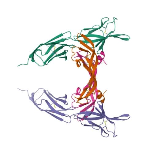
Leppänen and colleagues solved the structure of a VEGF ligand bound to the high-affinity site of VEGFR-2, which consists of D2 and D3. The complex is a symmetrical 2:2 assembly in which the twisted receptor domains wrap around the twofold axis of the ligand dimer, and an N-terminal helix plus three loops of the ligand set receptor specificity.[5] Brozzo and colleagues then solved VEGF-A bound to D2 and D3 (Fig. 1) and found a comparable binding surface, with differences in the twist angle between the two receptor domains.[7]
The same study measured binding thermodynamics. VEGF-A binding is entropy driven, and binding to the full extracellular domain is 1.0 to 1.7 kcal/mol less favorable than binding to D2 and D3 alone, a difference the authors linked to homotypic contacts within domains 4 to 7 that cost energy to form and may keep the receptor from activating without ligand.[7]
Membrane-proximal domain contacts turn ligand binding into kinase activation
Electron microscopy first showed how the signal crosses the membrane. Ruch and colleagues found that the monomeric VEGFR-2 extracellular domain is flexible, that VEGF binding to the membrane-distal domains dimerizes it, and that dimerization promotes a further contact between the membrane-proximal domain 7 of the two chains.[3]
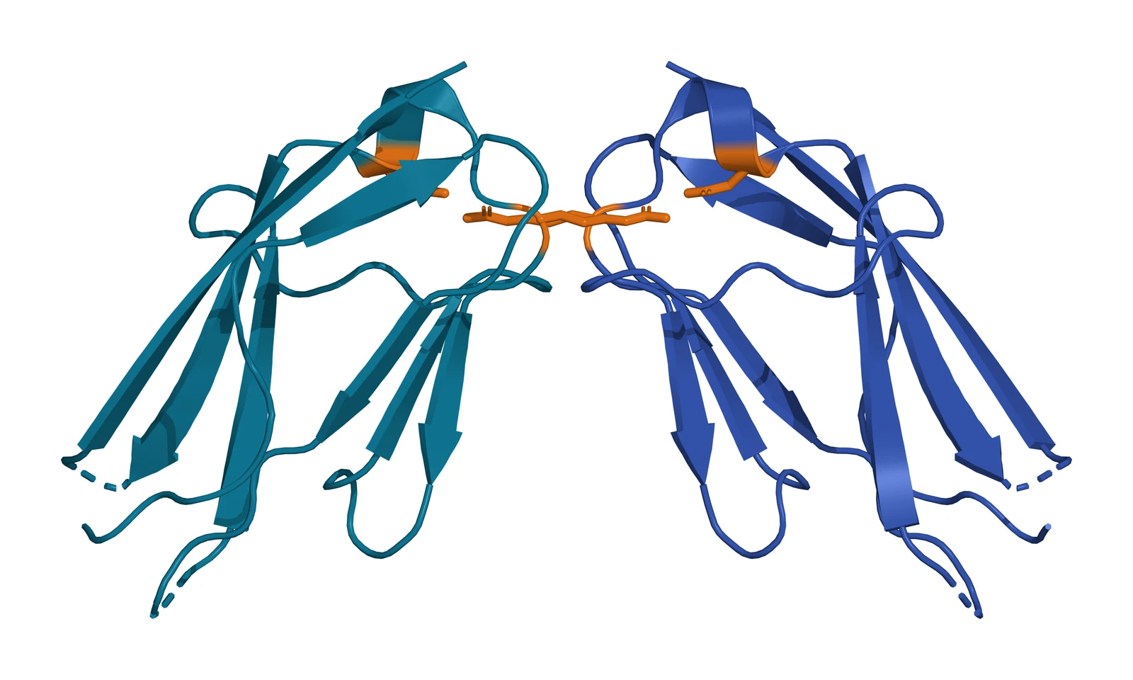
Yang and colleagues then solved domain 7 at 2.7 Å (Fig. 2). Two copies meet through salt bridges between Arg726 of one protomer and Asp731 of the other, and receptors mutated at either residue show strongly impaired autophosphorylation and signaling. The D7 dimer resembles the D4 dimer of the related receptor KIT, which points to a shared activation mechanism.[8]
Hyde and colleagues confirmed that D4 and D7 are indispensable with designed ankyrin repeat proteins. Binders directed at D2 and D3 blocked ligand binding and dimerization, while binders directed at D4 or D7 left both intact and still blocked kinase activation, so these domains regulate the receptor allosterically.[9] Sarabipour and colleagues added that VEGFR-2 expressed at physiological levels forms dimers before ligand arrives, and that ligand binding changes the conformation of the transmembrane helices to raise kinase phosphorylation.[10]
Inside the cell, the kinase domain is split by a 68-residue insert, as in the PDGF receptors. McTigue and colleagues solved it at 2.4 Å with Y1059 of the activation loop autophosphorylated and the ends of the insert folded into a small beta structure against the C terminus.[11] Like the FGF receptors, VEGFR-2 is also held in check by a molecular brake, a triad of residues in the hinge region of the kinase that activating changes release.[12]
Autophosphorylation at tyrosine 1175 recruits PLC-gamma
Takahashi and colleagues mapped the autophosphorylation sites of human VEGFR-2 and found Y1175 and Y1214 to be the two major sites phosphorylated in response to VEGF-A. A receptor with Y1175 changed to phenylalanine lost the ability to phosphorylate PLC-gamma and showed reduced MAP kinase activation and DNA synthesis in endothelial cells.[13]
The same site is essential in the embryo. Sakurai and colleagues made knock-in mice carrying phenylalanine at the equivalent residue, Y1173 of mouse Flk-1, and the homozygous embryos formed no organized blood vessels or yolk sac blood islands, closely resembling embryos that lack the receptor altogether. Mice carrying the Y1212 substitution were viable and fertile.[14] Shalaby and colleagues had shown that Flk-1 null embryos lack blood islands and organized vessels and have severely reduced hematopoietic progenitors, which identified VEGFR-2 as the receptor behind the first steps of vascular development.[15]
PLC-gamma at Y1175 therefore anchors the main output of VEGFR-2 (Fig. 3). Simons, Gordon and Claesson-Welsh describe the layers that regulate that output, from neuropilins, proteoglycans, integrins and phosphatases to endocytosis and trafficking, all serving to balance the formation of new vessels with the maintenance of existing ones.[16]
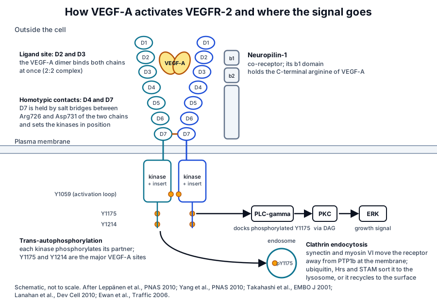
Endocytosis moves VEGFR-2 signaling into endosomes
VEGFR-2 cycles between the plasma membrane and early endosomes even in resting cells. Ewan and colleagues found constitutive, clathrin-linked endocytosis and recycling in quiescent endothelial cells, and showed that ligand-activated receptor codistributes with the Hrs and STAM sorting complex and is degraded in lysosomes, a route that requires the receptor’s own kinase activity.[17]
Internalization is part of signaling. Lanahan and colleagues showed that a complex of synectin and myosin VI carries VEGFR-2 endosomes away from the plasma membrane, where the phosphatase PTP1b would otherwise remove the phosphate from Y1175; when trafficking is delayed, Y1175 signaling falls and arterial morphogenesis is reduced in mice and zebrafish.[18]
Nakayama and colleagues found that sprouting endothelial cells in the developing retina take up VEGF and turn over its receptor at high rates, and that the clathrin-associated sorting protein Dab2, with ephrin-B2 and the polarity protein PAR-3, enables receptor endocytosis and downstream signaling. Atypical protein kinase C phosphorylates Dab2 in mature vessels and slows the process, so vessel growth is tuned locally by the rate of receptor internalization.[19]
VEGFR-1, VEGFR-3 and neuropilins shape the VEGFR-2 response
VEGFR-1, encoded by FLT1, binds VEGF-A with higher affinity than VEGFR-2 but has weaker kinase activity.[20][21] Fong and colleagues found that embryos lacking Flt-1 form endothelial cells but assemble them into disorganized channels,[22] and Hiratsuka and colleagues showed that mice whose Flt-1 lacks the kinase domain develop normal vessels, which identifies the ligand-binding extracellular region as the part of VEGFR-1 that matters for vascular development.[20]
Markovic-Mueller and colleagues solved the full-length VEGFR-1 extracellular domain bound to VEGF-A at 4 Å and found homotypic receptor contacts in domains 4, 5 and 7.[23] VEGFR-3, encoded by FLT4, uses the same logic with its own ligand VEGF-C: Leppänen and colleagues found a conserved ligand site in D2 and homotypic contacts in D5 and D7 that are required for activation.[24][25]
Neuropilin-1 is the best studied co-receptor. Parker and colleagues solved its b1 domain bound to the heparin-binding domain of VEGF-A and showed that the C-terminal arginine encoded by exon 8 is essential for the interaction, with exon 7 residues adding further contacts.[26] Gelfand and colleagues then made mice whose neuropilin-1 cannot bind VEGF and found normal vasculature, while vessels lacking neuropilin-1 had less VEGFR-2 at the cell surface, so the co-receptor supports VEGFR-2 through a mechanism independent of ligand binding.[27]
FGF receptors use heparan sulfate to assemble a 2:2:2 complex
The FGF family is encoded by 22 genes in mammals, eighteen of which are secreted signaling proteins, and they act on alternatively spliced forms of four receptor tyrosine kinases, FGFR1 to FGFR4.[2][28] Each receptor has an extracellular region of three Ig-like domains, D1 to D3, a single transmembrane helix and a cytoplasmic tyrosine kinase domain.[29][30][31][32][33] Unlike most growth factors, FGFs act in concert with heparin or heparan sulfate proteoglycans to activate their receptors.[34]
Yayon and colleagues established the requirement in 1991. Cells unable to make heparan sulfate could not bind basic FGF even when they expressed the receptor, and adding free heparin or heparan sulfate restored binding, so the low-affinity sugar site is an obligatory accessory to the high-affinity receptor.[35]
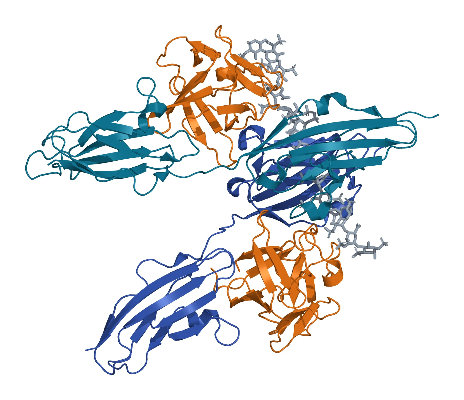
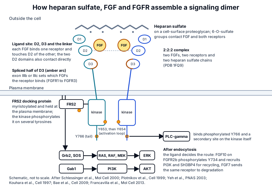
Plotnikov and colleagues solved FGF2 bound to D2 and D3 of FGFR1 at 2.8 Å. Each FGF contacts D2, D3 and the linker, two complexes form a twofold symmetric dimer held by contacts between FGF2 and the neighboring D2 and by a direct D2 to D2 contact, and a canyon of basic residues likely marks the heparin site.[36] Schlessinger and colleagues then solved the ternary complex with heparin (Fig. 4): a 2:2:2 assembly in which each heparin chain contacts its own FGF and receptor, augmenting their binding, and reaches across to the receptor of the adjoining half to promote dimerization, with the 6-O-sulfate groups pivotal to both roles.[4]
Pellegrini and colleagues reported a second architecture the same year, with FGF1, FGFR2 and a heparin decasaccharide arranged around a single central heparin chain that links two ligands and contacts only one receptor.[37] Mohammadi, Olsen and Ibrahimi review how the two models differ in stoichiometry and quaternary arrangement and how later biochemistry weighed them.[29] Goetz and Mohammadi show how splicing, ligand dimerization, proteolysis and co-receptors generate distinct FGF outputs.[38]
Endocrine FGFs need a different partner. Chen and colleagues solved the 1:1:1 complex of the shed extracellular domain of alpha-Klotho with the FGFR1c ligand-binding region and FGF23, in which Klotho tethers the receptor by its D3 domain and the hormone by its C-terminal tail; dimerization and activation of the stabilized complex still depend on heparan sulfate.[39]
Alternative splicing of domain 3 sets FGFR ligand specificity
Two mutually exclusive exons, IIIb and IIIc, encode the second half of D3 in FGFR1, FGFR2 and FGFR3, and the choice between them decides which FGFs a receptor binds. FGF7 and FGF10, for example, activate only the IIIb isoform of FGFR2.[40] Zhang and colleagues used cell lines expressing each of the seven principal receptor isoforms to map the specificity of every mammalian FGF.[28]
Yeh and colleagues solved FGF10 bound to the ligand-binding portion of FGFR2b and found contacts between divergent regions of FGF10 and two IIIb-specific loops in D3. FGF10 binding also rotates D2 into new contacts, so both domains contribute to the exceptional specificity of this pair.[40] Because epithelial cells tend to express IIIb isoforms and mesenchymal cells IIIc isoforms, splicing lets the two tissue layers signal to each other with ligands the sender itself cannot read.[2]
FRS2 docking links FGFR kinase activity to RAS and MAPK
Mohammadi, Schlessinger and Hubbard solved the FGFR1 kinase domain at 2.0 Å and found that activation loop residues interfere with substrate binding without blocking ATP, an autoinhibitory mechanism the authors proposed may be general among receptor tyrosine kinases.[41] Furdui and colleagues then showed that autophosphorylation of FGFR1 proceeds in a strictly ordered sequence: phosphorylation of Y653 in the activation loop raises substrate turnover 50- to 100-fold, and phosphorylation of Y654 raises it 500- to 1,000-fold.[42]
The main output runs through a docking protein. Kouhara and colleagues purified FRS2, a myristoylated membrane-anchored protein that is tyrosine phosphorylated in response to FGF and recruits Grb2 and SOS, linking the receptor to the RAS and MAP kinase cascade.[43] Eswarakumar, Lax and Schlessinger summarize the wider set of substrates, including Shc, PLC-gamma, STAT1 and Gab1, with FRS2 proteins as the major mediators of the RAS and MAPK and PI3K routes.[34]
PLC-gamma binds the receptor directly, at phosphorylated Y766 in the tail.[44] Bae and colleagues solved the activated FGFR1 kinase bound to a PLC-gamma fragment and found that a secondary site on the SH2 domain contacts the kinase itself, independent of phosphorylation, and that this contact sets the selectivity of PLC-gamma signaling in cells.[45] Brewer, Mazot and Soriano review the mouse genetics and find an important role for ERK1 and ERK2 as mediators of FGF signaling in many processes, with strong evidence for additional routes.[44]
The ligand also decides what happens to the receptor after endocytosis (Fig. 5). Francavilla and colleagues found that FGF7 sends FGFR2b to degradation, whereas FGF10 induces rapid phosphorylation of Y734, recruits PI3K and SH3BP4, and returns the receptor to the surface by recycling, a switch that supports epithelial branching in lung explants.[46]
FGFR signaling patterns limbs and skeleton
The limb bud is the classic FGF system. Sekine and colleagues found that mice lacking Fgf10 initiate limb buds that never grow out, with no apical ectodermal ridge or zone of polarizing activity, and that pulmonary branching fails after the trachea forms, so FGF10 is an essential regulator of both limb and lung formation.[47]
Once the ridge forms, it answers with its own FGFs. Sun, Mariani and Martin inactivated Fgf4 and Fgf8 in ridge cells and found that the two ligands regulate cell number in the nascent bud and the survival of cells far from the ridge, ensuring enough progenitors for the full set of skeletal elements; without both, limb development fails.[48]
In the skeleton, FGF and FGFR signaling regulates chondrocyte proliferation and differentiation, osteoblast activity and the maintenance of adult bone, as Su, Jin and Chen summarize from knock-in, knockout and transgenic mouse models.[49] Members of the family act from the earliest stages of embryonic development through organogenesis, where they maintain progenitor cells and mediate their growth, differentiation and patterning, and continue to regulate metabolism and tissue upkeep in the adult.[2]
Advancing the research question
The growth factor receptor overview sets VEGFR-2 beside PDGFR-beta, EGFR, IGF-1R and the TGF-beta receptors, and the surface receptor chemistry page explains the Ig-like fold and membrane anchoring they share. The PDGFR-beta page describes the closest relative of VEGFR-2, and the EGFR page shows a receptor that dimerizes through its own extracellular arm.
The platelet granule biology page describes where VEGF and FGF are stored and released, and the extracellular vesicle uptake page covers how target cells take in the vesicles that carry them.
Referencias comentadas
Numeradas en el orden en que se citan en el texto. Cada fuente lleva una nota sobre lo que respalda en esta página; los artículos revisados por pares se listan con revista, volumen, número y páginas, y las fuentes primarias son regulaciones, documentos de agencias y bases de datos de referencia.
- 1.Peer-reviewedKoch S, Claesson-Welsh L. Signal transduction by vascular endothelial growth factor receptors. Cold Spring Harbor Perspectives in Medicine. 2012;2(7):a006502. https://pubmed.ncbi.nlm.nih.gov/22762016/Annotation: Review of VEGF signal transduction. Cited for five mammalian ligands acting through three receptor tyrosine kinases and for neuropilin and integrin co-receptors.
- 2.Peer-reviewedOrnitz DM, Itoh N. The Fibroblast Growth Factor signaling pathway. Wiley Interdisciplinary Reviews: Developmental Biology. 2015;4(3):215-266. https://pubmed.ncbi.nlm.nih.gov/25772309/Annotation: Review of the FGF signaling pathway. Cited for eighteen secreted FGFs acting on four receptors, the epithelial and mesenchymal distribution of IIIb and IIIc isoforms, and FGF roles from early development to adult tissue upkeep.
- 3.Peer-reviewedRuch C, Skiniotis G, Steinmetz MO, et al. Structure of a VEGF-VEGF receptor complex determined by electron microscopy. Nature Structural & Molecular Biology. 2007;14(3):249-250. https://pubmed.ncbi.nlm.nih.gov/17293873/Annotation: Electron microscopy of the VEGF and VEGFR-2 extracellular complex. Cited for the flexible monomer and the domain 7 contact that follows ligand-induced dimerization.
- 4.Peer-reviewedSchlessinger J, Plotnikov AN, Ibrahimi OA, et al. Crystal structure of a ternary FGF-FGFR-heparin complex reveals a dual role for heparin in FGFR binding and dimerization. Molecular Cell. 2000;6(3):743-750. https://pubmed.ncbi.nlm.nih.gov/11030354/Annotation: Crystal structure of the 2:2:2 FGF2, FGFR1 and heparin complex (PDB 1FQ9, Fig. 4). Cited for heparin augmenting binding within each half and bridging to the neighboring receptor, and for the pivotal 6-O-sulfate.
- 5.Peer-reviewedLeppänen VM, Prota AE, Jeltsch M, et al. Structural determinants of growth factor binding and specificity by VEGF receptor 2. Proceedings of the National Academy of Sciences of the United States of America. 2010;107(6):2425-2430. https://pubmed.ncbi.nlm.nih.gov/20145116/Annotation: Crystal structure of VEGF-C bound to VEGFR-2 D2 and D3. Cited for the seven Ig homology domains, the symmetrical 2:2 complex, and the N-terminal helix and three loops that set receptor specificity.
- 6.DatabaseNCBI Gene 3791, KDR kinase insert domain receptor (Homo sapiens). Accessed 8 November 2026. https://www.ncbi.nlm.nih.gov/gene/3791
- 7.Peer-reviewedBrozzo MS, Bjelić S, Kisko K, et al. Thermodynamic and structural description of allosterically regulated VEGFR-2 dimerization. Blood. 2012;119(7):1781-1788. https://pubmed.ncbi.nlm.nih.gov/22207738/Annotation: Crystal structures of VEGF-A and VEGF-E with VEGFR-2 D2 and D3 (PDB 3V2A, Fig. 1) and calorimetry. Cited for the entropy-driven binding, the 1.0 to 1.7 kcal/mol penalty of the full ectodomain and the proposed role of D4 to D7 homotypic contacts.
- 8.Peer-reviewedYang Y, Xie P, Opatowsky Y, et al. Direct contacts between extracellular membrane-proximal domains are required for VEGF receptor activation and cell signaling. Proceedings of the National Academy of Sciences of the United States of America. 2010;107(5):1906-1911. https://pubmed.ncbi.nlm.nih.gov/20080685/Annotation: Crystal structure of VEGFR-2 domain 7 (PDB 3KVQ, Fig. 2) with mutagenesis. Cited for the Arg726 and Asp731 salt bridges, impaired autophosphorylation in the mutants and the resemblance to KIT domain 4.
- 9.Peer-reviewedHyde CA, Giese A, Stuttfeld E, et al. Targeting extracellular domains D4 and D7 of vascular endothelial growth factor receptor 2 reveals allosteric receptor regulatory sites. Molecular and Cellular Biology. 2012;32(19):3802-3813. https://pubmed.ncbi.nlm.nih.gov/22801374/Annotation: Designed ankyrin repeat proteins against VEGFR-2 D2 and D3, D4 and D7. Cited for D4 and D7 binders that leave ligand binding and dimerization intact yet block kinase activation.
- 10.Peer-reviewedSarabipour S, Ballmer-Hofer K, Hristova K. VEGFR-2 conformational switch in response to ligand binding. eLife. 2016;5:e13876. https://pubmed.ncbi.nlm.nih.gov/27052508/Annotation: Quantitative FRET and biochemistry in cells. Cited for unliganded VEGFR-2 dimers at physiological expression and the transmembrane conformational change on ligand binding.
- 11.Peer-reviewedMcTigue MA, Wickersham JA, Pinko C, et al. Crystal structure of the kinase domain of human vascular endothelial growth factor receptor 2: a key enzyme in angiogenesis. Structure. 1999;7(3):319-330. https://pubmed.ncbi.nlm.nih.gov/10368301/Annotation: Crystal structure of the VEGFR-2 kinase domain at 2.4 Å. Cited for the 68-residue kinase insert, autophosphorylated Y1059 in the activation loop and the beta structure formed by the ends of the insert.
- 12.Peer-reviewedChen H, Ma J, Li W, et al. A molecular brake in the kinase hinge region regulates the activity of receptor tyrosine kinases. Molecular Cell. 2007;27(5):717-730. https://pubmed.ncbi.nlm.nih.gov/17803937/Annotation: Crystal structures of FGFR2 kinase domains in several states. Cited for the autoinhibitory hinge-region triad shared by FGFRs, VEGFRs, PDGFRs and KIT.
- 13.Peer-reviewedTakahashi T, Yamaguchi S, Chida K, et al. A single autophosphorylation site on KDR/Flk-1 is essential for VEGF-A-dependent activation of PLC-gamma and DNA synthesis in vascular endothelial cells. The EMBO Journal. 2001;20(11):2768-2778. https://pubmed.ncbi.nlm.nih.gov/11387210/Annotation: Phosphopeptide mapping and mutant receptors in endothelial cells. Cited for Y1175 and Y1214 as the major VEGF-A autophosphorylation sites and for Y1175 docking PLC-gamma.
- 14.Peer-reviewedSakurai Y, Ohgimoto K, Kataoka Y, et al. Essential role of Flk-1 (VEGF receptor 2) tyrosine residue 1173 in vasculogenesis in mice. Proceedings of the National Academy of Sciences of the United States of America. 2005;102(4):1076-1081. https://pubmed.ncbi.nlm.nih.gov/15644447/Annotation: Knock-in mice with phenylalanine at Y1173 or Y1212 of Flk-1. Cited for the Y1173F embryos that form no vessels or blood islands and the viable Y1212F mice.
- 15.Peer-reviewedShalaby F, Rossant J, Yamaguchi TP, et al. Failure of blood-island formation and vasculogenesis in Flk-1-deficient mice. Nature. 1995;376(6535):62-66. https://pubmed.ncbi.nlm.nih.gov/7596435/Annotation: Targeted disruption of Flk-1 in mice. Cited for the absence of yolk sac blood islands and organized vessels and the reduction of hematopoietic progenitors in null embryos.
- 16.Peer-reviewedSimons M, Gordon E, Claesson-Welsh L. Mechanisms and regulation of endothelial VEGF receptor signalling. Nature Reviews Molecular Cell Biology. 2016;17(10):611-625. https://pubmed.ncbi.nlm.nih.gov/27461391/Annotation: Review of VEGFR-2 regulation. Cited for the roles of neuropilins, proteoglycans, integrins, phosphatases and trafficking in balancing new vessel formation with maintenance of existing vessels.
- 17.Peer-reviewedEwan LC, Jopling HM, Jia H, et al. Intrinsic tyrosine kinase activity is required for vascular endothelial growth factor receptor 2 ubiquitination, sorting and degradation in endothelial cells. Traffic. 2006;7(9):1270-1282. https://pubmed.ncbi.nlm.nih.gov/17004325/Annotation: Trafficking studies in human endothelial cells. Cited for constitutive clathrin-linked endocytosis and recycling, codistribution of activated receptor with Hrs and STAM, and kinase-dependent lysosomal degradation.
- 18.Peer-reviewedLanahan AA, Hermans K, Claes F, et al. VEGF receptor 2 endocytic trafficking regulates arterial morphogenesis. Developmental Cell. 2010;18(5):713-724. https://pubmed.ncbi.nlm.nih.gov/20434959/Annotation: Synectin and myosin VI mutant mice and zebrafish. Cited for endosomal trafficking that protects Y1175 from PTP1b and for its requirement in arterial morphogenesis.
- 19.Peer-reviewedNakayama M, Nakayama A, van Lessen M, et al. Spatial regulation of VEGF receptor endocytosis in angiogenesis. Nature Cell Biology. 2013;15(3):249-260. https://pubmed.ncbi.nlm.nih.gov/23354168/Annotation: Mouse retina studies of receptor uptake. Cited for high VEGF receptor endocytosis in sprouting cells, the Dab2, ephrin-B2 and PAR-3 route and its restraint by atypical PKC in mature vessels.
- 20.Peer-reviewedHiratsuka S, Minowa O, Kuno J, et al. Flt-1 lacking the tyrosine kinase domain is sufficient for normal development and angiogenesis in mice. Proceedings of the National Academy of Sciences of the United States of America. 1998;95(16):9349-9354. https://pubmed.ncbi.nlm.nih.gov/9689083/Annotation: Kinase-deleted Flt-1 mice. Cited for the higher affinity and lower kinase activity of VEGFR-1 and for normal vessel development when only its ligand-binding region remains.
- 21.DatabaseNCBI Gene 2321, FLT1 fms related receptor tyrosine kinase 1 (Homo sapiens). Accessed 8 November 2026. https://www.ncbi.nlm.nih.gov/gene/2321
- 22.Peer-reviewedFong GH, Rossant J, Gertsenstein M, et al. Role of the Flt-1 receptor tyrosine kinase in regulating the assembly of vascular endothelium. Nature. 1995;376(6535):66-70. https://pubmed.ncbi.nlm.nih.gov/7596436/Annotation: Targeted Flt-1 mutation in mice. Cited for endothelial cells that form but assemble into disorganized channels without VEGFR-1.
- 23.Peer-reviewedMarkovic-Mueller S, Stuttfeld E, Asthana M, et al. Structure of the Full-length VEGFR-1 Extracellular Domain in Complex with VEGF-A. Structure. 2017;25(2):341-352. https://pubmed.ncbi.nlm.nih.gov/28111021/Annotation: Hybrid structure of the full VEGFR-1 ectodomain with VEGF-A at 4 Å. Cited for homotypic receptor contacts in domains 4, 5 and 7.
- 24.Peer-reviewedLeppänen VM, Tvorogov D, Kisko K, et al. Structural and mechanistic insights into VEGF receptor 3 ligand binding and activation. Proceedings of the National Academy of Sciences of the United States of America. 2013;110(32):12960-12965. https://pubmed.ncbi.nlm.nih.gov/23878260/Annotation: Crystal structures of VEGF-C with VEGFR-3 D1 and D2 and of the VEGFR-3 D4 and D5 homodimer. Cited for the conserved D2 ligand site and the homotypic D5 and D7 contacts required for activation.
- 25.DatabaseNCBI Gene 2324, FLT4 fms related receptor tyrosine kinase 4 (Homo sapiens). Accessed 8 November 2026. https://www.ncbi.nlm.nih.gov/gene/2324
- 26.Peer-reviewedParker MW, Xu P, Li X, et al. Structural basis for selective vascular endothelial growth factor-A (VEGF-A) binding to neuropilin-1. The Journal of Biological Chemistry. 2012;287(14):11082-11089. https://pubmed.ncbi.nlm.nih.gov/22318724/Annotation: Crystal structure of the VEGF-A heparin-binding domain with the neuropilin-1 b1 domain. Cited for the essential exon 8 C-terminal arginine and the additional exon 7 contacts.
- 27.Peer-reviewedGelfand MV, Hagan N, Tata A, et al. Neuropilin-1 functions as a VEGFR2 co-receptor to guide developmental angiogenesis independent of ligand binding. eLife. 2014;3:e03720. https://pubmed.ncbi.nlm.nih.gov/25244320/Annotation: Mice with a neuropilin-1 point mutation that abolishes VEGF binding. Cited for their normal vasculature and for reduced VEGFR-2 surface levels in vessels lacking neuropilin-1.
- 28.Peer-reviewedZhang X, Ibrahimi OA, Olsen SK, et al. Receptor specificity of the fibroblast growth factor family. The complete mammalian FGF family. The Journal of Biological Chemistry. 2006;281(23):15694-15700. https://pubmed.ncbi.nlm.nih.gov/16597617/Annotation: Mitogenesis assays in BaF3 lines expressing the seven principal FGFR isoforms. Cited for the 22 mammalian FGF genes and the complete receptor specificity table.
- 29.Peer-reviewedMohammadi M, Olsen SK, Ibrahimi OA. Structural basis for fibroblast growth factor receptor activation. Cytokine & Growth Factor Reviews. 2005;16(2):107-137. https://pubmed.ncbi.nlm.nih.gov/15863029/Annotation: Review of FGF and FGFR structural biology. Cited for the receptor architecture and for the two crystallographic models of heparin-mediated dimerization and how they were weighed.
- 30.DatabaseNCBI Gene 2260, FGFR1 fibroblast growth factor receptor 1 (Homo sapiens). Accessed 8 November 2026. https://www.ncbi.nlm.nih.gov/gene/2260
- 31.DatabaseNCBI Gene 2263, FGFR2 fibroblast growth factor receptor 2 (Homo sapiens). Accessed 8 November 2026. https://www.ncbi.nlm.nih.gov/gene/2263
- 32.DatabaseNCBI Gene 2261, FGFR3 fibroblast growth factor receptor 3 (Homo sapiens). Accessed 8 November 2026. https://www.ncbi.nlm.nih.gov/gene/2261
- 33.DatabaseNCBI Gene 2264, FGFR4 fibroblast growth factor receptor 4 (Homo sapiens). Accessed 8 November 2026. https://www.ncbi.nlm.nih.gov/gene/2264
- 34.Peer-reviewedEswarakumar VP, Lax I, Schlessinger J. Cellular signaling by fibroblast growth factor receptors. Cytokine & Growth Factor Reviews. 2005;16(2):139-149. https://pubmed.ncbi.nlm.nih.gov/15863030/Annotation: Review of FGFR signaling. Cited for FGFs acting in concert with heparan sulfate proteoglycans and for the substrate set, including Shc, PLC-gamma, STAT1, Gab1 and the FRS2 proteins.
- 35.Peer-reviewedYayon A, Klagsbrun M, Esko JD, et al. Cell surface, heparin-like molecules are required for binding of basic fibroblast growth factor to its high affinity receptor. Cell. 1991;64(4):841-848. https://pubmed.ncbi.nlm.nih.gov/1847668/Annotation: Binding studies in heparan sulfate-deficient CHO cells. Cited for the obligatory role of heparan sulfate in high-affinity FGF2 binding.
- 36.Peer-reviewedPlotnikov AN, Schlessinger J, Hubbard SR, et al. Structural basis for FGF receptor dimerization and activation. Cell. 1999;98(5):641-650. https://pubmed.ncbi.nlm.nih.gov/10490103/Annotation: Crystal structure of FGF2 with FGFR1 D2 and D3 at 2.8 Å. Cited for ligand contacts with D2, D3 and the linker, the twofold symmetric dimer, the direct D2 to D2 contact and the basic canyon.
- 37.Peer-reviewedPellegrini L, Burke DF, von Delft F, et al. Crystal structure of fibroblast growth factor receptor ectodomain bound to ligand and heparin. Nature. 2000;407(6807):1029-1034. https://pubmed.ncbi.nlm.nih.gov/11069186/Annotation: Crystal structure of FGF1, FGFR2 and a heparin decasaccharide. Cited for the alternative architecture built around a single central heparin chain contacting one receptor.
- 38.Peer-reviewedGoetz R, Mohammadi M. Exploring mechanisms of FGF signalling through the lens of structural biology. Nature Reviews Molecular Cell Biology. 2013;14(3):166-180. https://pubmed.ncbi.nlm.nih.gov/23403721/Annotation: Structural review of FGF signaling. Cited for alternative splicing, ligand homodimerization, proteolytic processing and heparan sulfate and Klotho co-receptors as sources of distinct outputs.
- 39.Peer-reviewedChen G, Liu Y, Goetz R, et al. α-Klotho is a non-enzymatic molecular scaffold for FGF23 hormone signalling. Nature. 2018;553(7689):461-466. https://pubmed.ncbi.nlm.nih.gov/29342138/Annotation: Crystal structure of the alpha-Klotho, FGFR1c and FGF23 ternary complex. Cited for Klotho tethering receptor D3 and the hormone tail, with dimerization still dependent on heparan sulfate.
- 40.Peer-reviewedYeh BK, Igarashi M, Eliseenkova AV, et al. Structural basis by which alternative splicing confers specificity in fibroblast growth factor receptors. Proceedings of the National Academy of Sciences of the United States of America. 2003;100(5):2266-2271. https://pubmed.ncbi.nlm.nih.gov/12591959/Annotation: Crystal structure of FGF10 with FGFR2b. Cited for the IIIb and IIIc exons encoding the second half of D3, the IIIb-specific loop contacts and the rotation of D2 on FGF10 binding.
- 41.Peer-reviewedMohammadi M, Schlessinger J, Hubbard SR. Structure of the FGF receptor tyrosine kinase domain reveals a novel autoinhibitory mechanism. Cell. 1996;86(4):577-587. https://pubmed.ncbi.nlm.nih.gov/8752212/Annotation: Crystal structure of the FGFR1 kinase domain at 2.0 Å. Cited for activation loop residues that interfere with substrate peptide binding while leaving ATP binding intact, proposed as a general autoinhibitory mechanism.
- 42.Peer-reviewedFurdui CM, Lew ED, Schlessinger J, et al. Autophosphorylation of FGFR1 kinase is mediated by a sequential and precisely ordered reaction. Molecular Cell. 2006;21(5):711-717. https://pubmed.ncbi.nlm.nih.gov/16507368/Annotation: Rapid quench kinetics and mass spectrometry of FGFR1 autophosphorylation. Cited for the ordered reaction and the 50- to 100-fold and 500- to 1,000-fold rate increases after Y653 and Y654.
- 43.Peer-reviewedKouhara H, Hadari YR, Spivak-Kroizman T, et al. A lipid-anchored Grb2-binding protein that links FGF-receptor activation to the Ras/MAPK signaling pathway. Cell. 1997;89(5):693-702. https://pubmed.ncbi.nlm.nih.gov/9182757/Annotation: Purification and cloning of FRS2. Cited for its myristoylation, membrane anchoring, tyrosine phosphorylation and recruitment of Grb2 and SOS.
- 44.Peer-reviewedBrewer JR, Mazot P, Soriano P. Genetic insights into the mechanisms of Fgf signaling. Genes & Development. 2016;30(7):751-771. https://pubmed.ncbi.nlm.nih.gov/27036966/Annotation: Review of mouse genetic analyses of FGF signaling. Cited for an important in vivo role of ERK1 and ERK2 with strong evidence for additional routes, and for Y766 as the PLC-gamma binding site of FGFR1.
- 45.Peer-reviewedBae JH, Lew ED, Yuzawa S, et al. The selectivity of receptor tyrosine kinase signaling is controlled by a secondary SH2 domain binding site. Cell. 2009;138(3):514-524. https://pubmed.ncbi.nlm.nih.gov/19665973/Annotation: Crystal structure of the activated FGFR1 kinase with a PLC-gamma fragment. Cited for the secondary SH2 domain contact with the kinase that sets PLC-gamma selectivity.
- 46.Peer-reviewedFrancavilla C, Rigbolt KT, Emdal KB, et al. Functional proteomics defines the molecular switch underlying FGF receptor trafficking and cellular outputs. Molecular Cell. 2013;51(6):707-722. https://pubmed.ncbi.nlm.nih.gov/24011590/Annotation: Quantitative proteomics and imaging of FGFR2b trafficking. Cited for FGF7 directing the receptor to degradation and FGF10 phosphorylating Y734 to recruit PI3K and SH3BP4 for recycling, supporting epithelial branching in lung explants.
- 47.Peer-reviewedSekine K, Ohuchi H, Fujiwara M, et al. Fgf10 is essential for limb and lung formation. Nature Genetics. 1999;21(1):138-141. https://pubmed.ncbi.nlm.nih.gov/9916808/Annotation: Fgf10-deficient mice. Cited for limb buds that initiate without outgrowth, the missing apical ectodermal ridge and zone of polarizing activity, and the failure of pulmonary branching after the trachea forms.
- 48.Peer-reviewedSun X, Mariani FV, Martin GR. Functions of FGF signalling from the apical ectodermal ridge in limb development. Nature. 2002;418(6897):501-508. https://pubmed.ncbi.nlm.nih.gov/12152071/Annotation: Conditional inactivation of Fgf4 and Fgf8 in the mouse apical ectodermal ridge. Cited for the regulation of limb bud cell number and survival and for the failure of limb development without both ligands.
- 49.Peer-reviewedSu N, Jin M, Chen L. Role of FGF/FGFR signaling in skeletal development and homeostasis: learning from mouse models. Bone Research. 2014;2:14003. https://pubmed.ncbi.nlm.nih.gov/26273516/Annotation: Review of mouse models of FGF and FGFR signaling in bone. Cited for the regulation of chondrocyte and osteoblast biology during skeletal development and in adult bone maintenance.
Cite this page
ExaVeyra Sciences Editorial Team. VEGFR-2 and FGFR: Angiogenic Receptor Tyrosine Kinases. ExaVeyra Sciences; 9 November 2026. Available from: https://exaveyra.com/science/surface-receptors/vegfr-2-and-fgfr. Accessed [date accessed].
Fuentes de las figuras
- 50.PDB 3V2A, VEGFR-2 bound to VEGF-A. RCSB Protein Data Bank (rcsb.org). Image released under CC0. Publicación de la estructura: Brozzo MS, Bjelić S, Kisko K, et al. Thermodynamic and structural description of allosterically regulated VEGFR-2 dimerization. Blood. 2012;119(7):1781-1788.
- 51.PDB 3KVQ, VEGFR-2 extracellular domain D7. ExaVeyra Sciences, rendered in PyMOL from PDB 3KVQ. Structure data: RCSB Protein Data Bank (rcsb.org), CC0. Publicación de la estructura: Yang Y, Xie P, Opatowsky Y, Schlessinger J. Direct contacts between extracellular membrane-proximal domains are required for VEGF receptor activation and cell signaling. Proc Natl Acad Sci U S A. 2010;107(5):1906-1911.
- 52.PDB 1FQ9, ternary FGF2, FGFR1 and heparin complex. ExaVeyra Sciences, rendered in PyMOL from PDB 1FQ9. Structure data: RCSB Protein Data Bank (rcsb.org), CC0. Publicación de la estructura: Schlessinger J, Plotnikov AN, Ibrahimi OA, et al. Crystal structure of a ternary FGF-FGFR-heparin complex reveals a dual role for heparin in FGFR binding and dimerization. Mol Cell. 2000;6(3):743-750.
Créditos de imágenes
- Fig. 3ExaVeyra Sciences, figura original. After Leppänen et al., Proc Natl Acad Sci U S A 2010; Yang et al., Proc Natl Acad Sci U S A 2010; Takahashi et al., EMBO J 2001; Lanahan et al., Dev Cell 2010; Ewan et al., Traffic 2006. ↩
- Fig. 5ExaVeyra Sciences, figura original. After Schlessinger et al., Mol Cell 2000; Plotnikov et al., Cell 1999; Yeh et al., Proc Natl Acad Sci U S A 2003; Kouhara et al., Cell 1997; Bae et al., Cell 2009; Francavilla et al., Mol Cell 2013. ↩