Esta referencia está disponible actualmente en inglés. La versión en español está en preparación.
PDGFR-beta: How the Receptor Is Built and What It Signals
Por Benn Bluestein-Veyra, M.Sc. en Química Orgánica · Actualizado el 9 de noviembre de 2026
ExaVeyra Sciences es un proveedor B2B con sede en Miami que suministra exosomas para uso estético tópico a clínicas con licencia, y trabaja con una farmacia de preparados compuestos 503A que prepara formulaciones para prescriptores con licencia según prescripciones individuales.
| Pregunta | Respuesta |
|---|---|
| Qué cubre esta página | PDGFR-beta structure: five Ig-like domains and a split kinase, activation by PDGF-B and PDGF-D, phosphotyrosine docking, and pericyte recruitment. |
| Quién puede hacer pedidos | Clínicas y profesionales con licencia profesional activa y NPI verificados por ExaVeyra. Los pedidos de exosomas requieren además un reconocimiento firmado de uso profesional. |
| Qué se envía y cómo | El material de exosomas para uso estético tópico, caracterizado por lote, se envía en cadena de frío por exprés nocturno. La entrega puede tardar 1 a 3 días hábiles. Se emite un certificado de análisis para cada lote y se entrega a solicitud. |
PDGFR-beta is the cell surface receptor tyrosine kinase that reads the B and D chains of platelet-derived growth factor, a single-pass membrane protein of 1,106 amino acids encoded by the PDGFRB gene on chromosome 5q32.[1][2] It belongs to the type III class of receptor tyrosine kinases, defined by five immunoglobulin-like domains outside the cell and a kinase domain split in two by an insert inside it.[3]
Andrae and colleagues described PDGFs and their receptors in 2008 as prototypes for growth factor and receptor tyrosine kinase function for more than 25 years.[4] This page follows PDGFR-beta from its domain architecture to ligand binding, kinase activation, the docking of signaling proteins, its trafficking after activation, and its roles in blood vessel development and on mesenchymal stromal cells.
Understanding the structure of PDGFR-beta
The receptor reads from its N terminus as five Ig-like domains, D1 to D5, followed by one transmembrane helix, a juxtamembrane segment, the two lobes of the kinase and a C-terminal tail. The kinase insert separates the lobes and carries several of the tyrosines described below.[3][5]
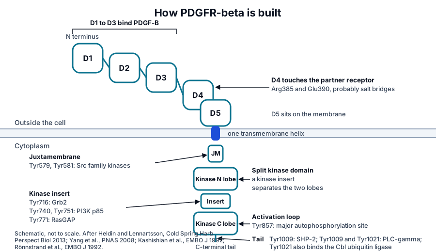
The first three Ig domains hold the ligand.[6] The fourth makes a direct contact with the fourth domain of the partner receptor: Yang and colleagues found that mutating Arg385 or Glu390 in D4 compromised activation, and proposed that salt bridges between these residues hold the two membrane-proximal regions together.[7]
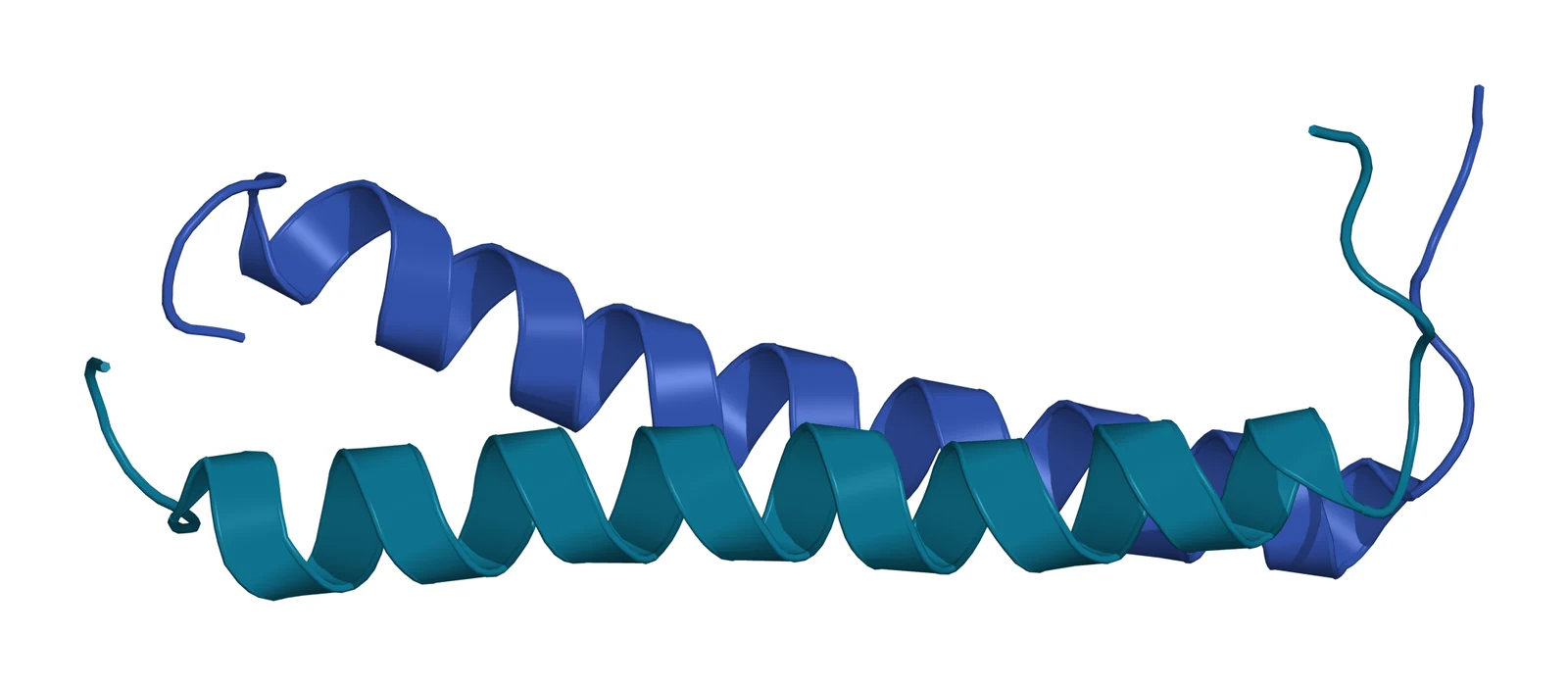
The transmembrane segment has its own structure. Muhle-Goll and colleagues solved it by solution NMR in micelles and found a stable left-handed helical dimer whose tilt in a bilayer rose from 10 to 30 degrees as the bilayer thinned, which they read as a way for membrane thickness to influence where the receptor sits and how it activates.[8]
Chen, Unger and He then imaged the full-length human receptor bound to PDGF-B by negative-stain electron microscopy at 27 Å. Two receptors formed a dimer through an interface running the full length of the protein, from the membrane-proximal domains through the transmembrane helix to the kinase domains, and the two kinases sat in an asymmetric arrangement.[9]
PDGF-B and PDGF-D are the ligands that activate PDGFR-beta
The PDGF family has four chains, A, B, C and D, secreted as disulfide-linked dimers. PDGFR-alpha binds the A, B and C chains, while PDGFR-beta binds PDGF-B and PDGF-D.[10][11] The identity of the chains in a dimer decides which receptor pair forms: PDGF-BB and PDGF-DD assemble PDGFR-beta homodimers, and PDGF-AB and PDGF-BB can also assemble alpha and beta heterodimers.[1][5]
Bergsten and colleagues identified PDGF-D as the first ligand specific to PDGFR-beta. It is secreted as a two-domain homodimer that becomes an active agonist only after limited proteolysis removes its N-terminal CUB domain.[5][10] PDGF-B carries a different kind of control: a C-terminal retention motif that holds the secreted factor within the pericellular space.[12]
All four PDGFs share a cystine-knot growth factor domain and a conserved prodomain region. Shim and colleagues showed that the propeptide can stay noncovalently bound to the mature growth factor after processing and covers the same hydrophobic surfaces that the receptor later binds.[6]
PDGF-B clamps two receptors at its dimerization seam
The 2.3 Å crystal structure of PDGF-B bound to PDGFR-beta D1 to D3 shows how a dimeric ligand brings two receptors together. The two PDGF-B protomers clamp each receptor at their dimerization seam, so one growth factor dimer holds two receptor chains in a 2:2 complex (Fig. 1).[6]
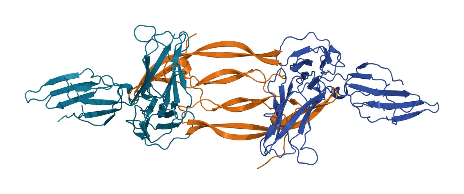
The interface is predominantly hydrophobic. On binding, the PDGF-B L1 loop, disordered in the free growth factor, adopts a defined conformation and packs against the third Ig domain of the receptor.[6] Chen, Chen and He review these structures as a template for how the whole family recognizes its receptors.[5]
Dimerization by itself is necessary but insufficient. Yang and colleagues cross-linked PDGF-stimulated cells and found that a Glu390Ala receptor still dimerized on ligand binding yet stayed inactive, which places the D4 homotypic contact after dimerization as the step that commits the pair to activation.[7] Calorimetry in the Shim study showed that the membrane-proximal homotypic contact of the closely related PDGFR-alpha, although required for activation, contributes negatively to ligand binding.[6]
Lemmon and Schlessinger place PDGFR-beta within the wider family: ligands induce dimerization by surprisingly diverse strategies, and the mechanisms that couple dimerization to kinase activation are equally varied.[13] In PDGF signaling, the ligand dimer itself forms the bridge between the two receptors.[6]
Dimerization releases a split kinase domain that phosphorylates itself
Once two receptors are held together, each kinase domain phosphorylates tyrosines on its partner. Autophosphorylation raises catalytic activity and, at the same time, creates docking sites for proteins that carry SH2 domains.[3][13]
Kazlauskas and Cooper mapped the first two sites in 1989. Tyr857, in the activation loop of the kinase, is the major site and corresponds to the conserved activation loop tyrosine of many kinases. Tyr751, in the kinase insert, is a second site in cells and the major site in vitro.[14]
Kazlauskas later described ligand-dependent de-repression of the kinase as the main mode of PDGFR activation, with the dimer relieving an autoinhibited state of the unliganded receptor.[11] The asymmetric kinase arrangement seen in the full-length complex is consistent with that reading.[9]
Phosphotyrosines dock PI3K, PLC-gamma, SHP-2 and Grb2
The autophosphorylated receptor is a platform. Kashishian and colleagues identified three phosphorylation sites in the kinase insert, Tyr740, Tyr751 and Tyr771, and showed that Tyr740 and Tyr751 bind the p85 subunit of PI3K while Tyr771 binds RasGAP, so one receptor signals to different pathways through different tyrosines.[15]
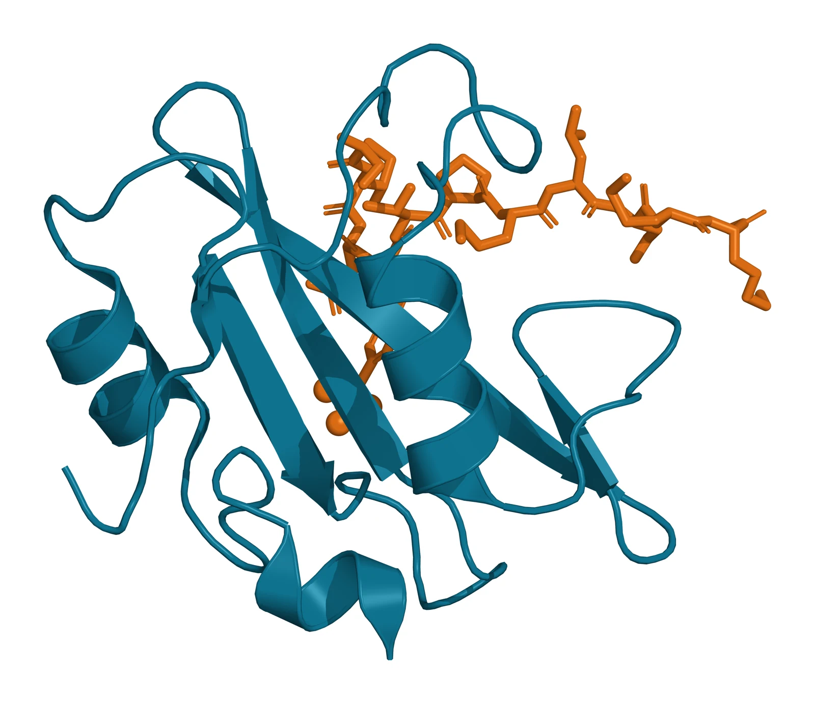
The crystal structure of the p85-alpha N-terminal SH2 domain bound to a PDGFR phosphopeptide shows how that recognition works: the phosphotyrosine sits in one pocket and a methionine three residues downstream in a second, hydrophobic pocket that forms when a tyrosine in the BG loop swings aside (Fig. 2).[16]
Grb2 binds Tyr716, a further site in the kinase insert, and through it the receptor activates Ras; a phosphopeptide spanning Tyr716 blocked that interaction and lowered Ras-GTP in cells.[17] Two tail sites, Tyr1009 and Tyr1021, are needed for PLC-gamma to associate with the receptor and become phosphorylated.[18]
Tyr1009 also recruits the SH2-containing tyrosine phosphatase SHP-2, then called Syp, whose stable association with the receptor depends on that single phosphotyrosine.[19] Two juxtamembrane tyrosines, Tyr579 and Tyr581, are docking sites for Src family kinases.[20] Heldin and Lennartsson summarize the outcome: the pathways these docking proteins start promote cell growth, survival and migration.[3]
| Phosphotyrosine | Location | Docking protein | Pathway |
|---|---|---|---|
| Tyr579, Tyr581 | Juxtamembrane segment | Src family kinases | Src signaling |
| Tyr716 | Kinase insert | Grb2 | Ras activation |
| Tyr740, Tyr751 | Kinase insert | PI3K p85 subunit | PI3K and AKT |
| Tyr771 | Kinase insert | RasGAP | Ras regulation |
| Tyr857 | Activation loop | None; raises kinase activity | Catalysis |
| Tyr1009 | C-terminal tail | SHP-2, PLC-gamma | Phosphatase signaling, PLC-gamma |
| Tyr1021 | C-terminal tail | PLC-gamma, Cbl | Calcium and PKC; ubiquitination |
Ubiquitination and endocytosis decide how long the receptor signals
The ligand-bound receptor leaves the surface quickly: in human fibroblasts, labeled PDGF-BB reached early endosomes within 20 minutes.[21] Sadowski and colleagues tracked this labeled PDGF-BB and found that both dynamin-dependent and dynamin-independent routes take it in, with both ending in lysosomal degradation of the ligand.[21]
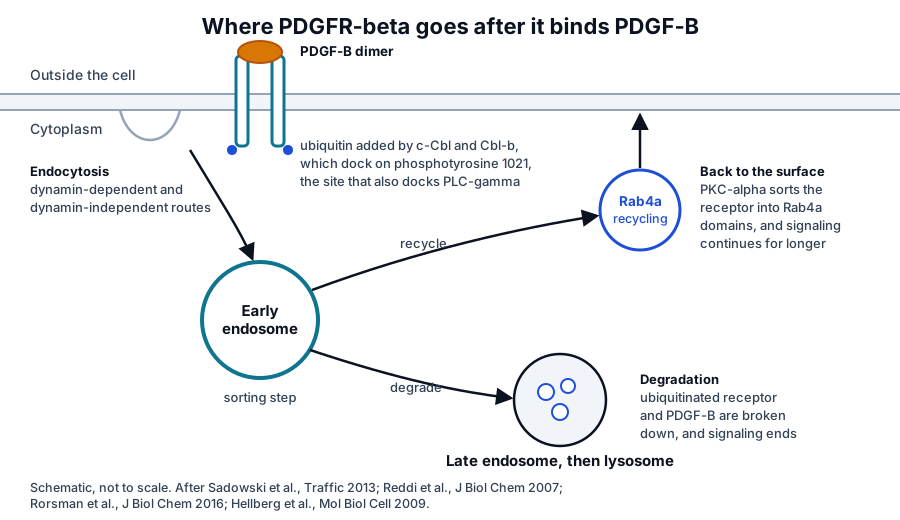
Ubiquitin marks the receptor for that fate. Reddi and colleagues showed that the ligase Cbl binds phosphotyrosine 1021 through its tyrosine kinase binding domain, which both drives ubiquitin-dependent sorting to the lysosome and competes with PLC-gamma for the same site.[22] Rorsman and colleagues found that c-Cbl and Cbl-b act together, with Tyr1021 as the primary Cbl-b site, and that cells depleted of both kept more receptor at the surface and phosphorylated Src and PLC-gamma more strongly.[20]
Sorting at the early endosome offers a second exit. Hellberg and colleagues showed that PKC-alpha activity routes the ligand-bound receptor into Rab4a-positive recycling domains and back to the surface, and that blocking this sorting raised the rate of receptor degradation, so the sorting step sets the duration of signaling.[23]
Endocytosis also shapes which signals are sent. In the Sadowski study, acute dynamin inhibition left most PDGF uptake intact but specifically lowered STAT3 signaling and entry into S phase, which the authors read as support for the view that the endocytic machinery can selectively regulate particular signaling outputs.[21]
PDGFR-beta recruits pericytes and shapes mesenchymal development
In animal development, PDGFR-beta signaling has established roles in blood vessel formation and early hematopoiesis, while PDGFR-alpha directs gastrulation, neural crest and organ mesenchyme.[4] Hellström and colleagues showed that PDGFR-beta positive progenitors of vascular smooth muscle cells and pericytes proliferate at sites where immature capillary endothelium and growing arteries express PDGF-B, and proposed a model in which these progenitors co-migrate along sprouting vessels.[24]
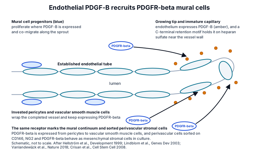
The retention motif on PDGF-B makes that gradient local. Lindblom and colleagues deleted it in mice and found that pericytes invested the microvessel wall improperly and that the glomerular mesangium formed late, so PDGF-B needs to stay near the endothelium that makes it for pericyte recruitment to work.[12]
Pericyte biology has since become a field of its own. Armulik, Genové and Betsholtz review pericytes as regulators of vascular morphogenesis and homeostasis and note that some pericytes are probably mesenchymal stem or progenitor cells able to give rise to adipocytes, cartilage, bone and muscle.[25] Kazlauskas frames PDGFR signaling as essential for the migration and proliferation of pericytes that build and maintain vessels.[11]
The level of signaling matters. Olson and Soriano generated mice with activating PDGFR-beta mutations and found that increased signaling drove proliferation and lowered differentiation genes in aortic smooth muscle, induced immune response genes in pericytes and mesenchymal cells, and inhibited white adipocyte differentiation, identifying PDGFR-beta as a regulator of mural cell progenitor potential.[26] Tokunaga and colleagues reached a complementary result in bone marrow stromal cells, where deleting PDGFR-beta lowered mitogenic and migratory responses and enhanced osteogenic differentiation.[27]
PDGFR-beta marks perivascular and mesenchymal stromal cells
Single-cell transcriptomics of the adult mouse brain vasculature places PDGFR-beta across the mural cell lineage, which Vanlandewijck and colleagues describe as a punctuated continuum from pericytes to vascular smooth muscle cells, distinct from the seamless continuum of endothelial cells.[28] He and colleagues used Pdgfrb-eGFP together with NG2-DsRed to isolate brain mural cells and cataloged 260 transcripts enriched in them.[29]
PDGFR-beta is also the receptor that links pericytes to mesenchymal stromal cells. Crisan and colleagues sorted perivascular cells from human skeletal muscle, pancreas, adipose tissue and placenta on CD146, NG2 and PDGFR-beta expression, and the sorted cells were myogenic, osteogenic, chondrogenic and adipogenic in culture and expressed MSC markers.[30]
The ISCT minimal criteria that define cultured MSCs rely on plastic adherence, trilineage differentiation and a panel of CD105, CD73 and CD90 with absence of hematopoietic markers.[31] PDGFR-beta is outside that panel; studies such as Crisan and colleagues use it alongside other markers to identify perivascular cells.[30][31]
Lineage tracing qualifies the in vivo picture. Guimarães-Camboa and colleagues found that Tbx18-marked pericytes and smooth muscle cells of adult mice behaved as MSCs after sorting and culture yet kept their identity in the animal, and the authors suggest that the multipotency seen in culture arises from manipulation of the cells ex vivo.[32]
Advancing the research question
The growth factor receptors overview compares PDGFR-beta with EGFR, VEGFR-2, IGF-1R and the TGF-beta receptors, and the sibling pages in this cluster take each of those receptors in the same depth. The surface receptor chemistry page explains the Ig-like fold and the glycosylation that PDGFR-beta shares with other receptors.
The platelet granule biology reference describes where PDGF-B is stored and released, and the extracellular vesicle uptake reference covers the endocytic machinery that this receptor also uses. Our team is glad to discuss the structural and signaling literature summarized here.
Referencias comentadas
Numeradas en el orden en que se citan en el texto. Cada fuente lleva una nota sobre lo que respalda en esta página; los artículos revisados por pares se listan con revista, volumen, número y páginas, y las fuentes primarias son regulaciones, documentos de agencias y bases de datos de referencia.
- 1.DatabaseNCBI Gene 5159, PDGFRB platelet derived growth factor receptor beta (Homo sapiens). Accessed 8 November 2026. https://www.ncbi.nlm.nih.gov/gene/5159
- 2.DatabaseNCBI Protein NP_002600.1, platelet-derived growth factor receptor beta isoform 1 precursor (Homo sapiens), 1106 aa. Accessed 8 November 2026. https://www.ncbi.nlm.nih.gov/protein/NP_002600.1
- 3.Peer-reviewedHeldin CH, Lennartsson J. Structural and functional properties of platelet-derived growth factor and stem cell factor receptors. Cold Spring Harbor Perspectives in Biology. 2013;5(8):a009100. https://pubmed.ncbi.nlm.nih.gov/23906712/Annotation: Review of the type III receptor tyrosine kinases. Cited for the five Ig-like domains, the split kinase domain, dimerization-driven autophosphorylation and the SH2 docking pathways for growth, survival and migration.
- 4.Peer-reviewedAndrae J, Gallini R, Betsholtz C. Role of platelet-derived growth factors in physiology and medicine. Genes & Development. 2008;22(10):1276-1312. https://pubmed.ncbi.nlm.nih.gov/18483217/Annotation: Review of PDGF ligands and receptors. Cited for PDGF as a prototype system and for the developmental roles of PDGFR-beta in blood vessel formation and early hematopoiesis.
- 5.Peer-reviewedChen PH, Chen X, He X. Platelet-derived growth factors and their receptors: structural and functional perspectives. Biochimica et Biophysica Acta. 2013;1834(10):2176-2186. https://pubmed.ncbi.nlm.nih.gov/23137658/Annotation: Structural review of the PDGF system. Cited for the domain layout of the receptors, the PDGF-B complex as a template for receptor recognition, the N-terminal CUB domain of PDGF-D, and PDGF-BB and PDGF-AB as inducers of alpha and beta heterodimers.
- 6.Peer-reviewedShim AH, Liu H, Focia PJ, et al. Structures of a platelet-derived growth factor/propeptide complex and a platelet-derived growth factor/receptor complex. Proceedings of the National Academy of Sciences of the United States of America. 2010;107(25):11307-11312. https://pubmed.ncbi.nlm.nih.gov/20534510/Annotation: Crystal structure of PDGF-B with PDGFR-beta D1 to D3 (PDB 3MJG, Fig. 1). Cited for the clamp at the dimerization seam, the hydrophobic interface, the L1 loop, propeptide overlap and the calorimetry on the PDGFR-alpha membrane-proximal contact.
- 7.Peer-reviewedYang Y, Yuzawa S, Schlessinger J. Contacts between membrane proximal regions of the PDGF receptor ectodomain are required for receptor activation but not for receptor dimerization. Proceedings of the National Academy of Sciences of the United States of America. 2008;105(22):7681-7686. https://pubmed.ncbi.nlm.nih.gov/18505839/Annotation: Mutagenesis and cross-linking of PDGFR-beta D4. Cited for the Arg385 and Glu390 homotypic contact and for dimerization being necessary but insufficient for activation.
- 8.Peer-reviewedMuhle-Goll C, Hoffmann S, Afonin S, et al. Hydrophobic matching controls the tilt and stability of the dimeric platelet-derived growth factor receptor (PDGFR) β transmembrane segment. Journal of Biological Chemistry. 2012;287(31):26178-26186. https://pubmed.ncbi.nlm.nih.gov/22619173/Annotation: NMR structure of the transmembrane segment (PDB 2L6W). Cited for the left-handed helical dimer and the dependence of its tilt on bilayer thickness.
- 9.Peer-reviewedChen PH, Unger V, He X. Structure of Full-Length Human PDGFRβ Bound to Its Activating Ligand PDGF-B as Determined by Negative-Stain Electron Microscopy. Journal of Molecular Biology. 2015;427(24):3921-3934. https://pubmed.ncbi.nlm.nih.gov/26463591/Annotation: Negative-stain structure of the full-length receptor with PDGF-B at 27 Å. Cited for the dimer interface running the length of the receptor and the asymmetric arrangement of the kinase domains.
- 10.Peer-reviewedBergsten E, Uutela M, Li X, et al. PDGF-D is a specific, protease-activated ligand for the PDGF beta-receptor. Nature Cell Biology. 2001;3(5):512-516. https://pubmed.ncbi.nlm.nih.gov/11331881/Annotation: Identification of PDGF-D. Cited for the receptor specificity of the four PDGF chains and for proteolytic activation of PDGF-DD as a PDGFR-beta specific agonist.
- 11.Peer-reviewedKazlauskas A. PDGFs and their receptors. Gene. 2017;614:1-7. https://pubmed.ncbi.nlm.nih.gov/28267575/Annotation: Short review of the PDGF system. Cited for ligand-dependent de-repression of the kinase as the main activation mode and for the role of PDGFR signaling in pericyte migration and proliferation.
- 12.Peer-reviewedLindblom P, Gerhardt H, Liebner S, et al. Endothelial PDGF-B retention is required for proper investment of pericytes in the microvessel wall. Genes & Development. 2003;17(15):1835-1840. https://pubmed.ncbi.nlm.nih.gov/12897053/Annotation: Deletion of the PDGF-B retention motif in mice. Cited for the C-terminal motif that keeps PDGF-B in the pericellular space and its requirement for pericyte investment and mesangium formation.
- 13.Peer-reviewedLemmon MA, Schlessinger J. Cell signaling by receptor tyrosine kinases. Cell. 2010;141(7):1117-1134. https://pubmed.ncbi.nlm.nih.gov/20602996/Annotation: Review of receptor tyrosine kinase activation. Cited for the diversity of dimerization strategies and of the mechanisms that couple dimerization to kinase activation.
- 14.Peer-reviewedKazlauskas A, Cooper JA. Autophosphorylation of the PDGF receptor in the kinase insert region regulates interactions with cell proteins. Cell. 1989;58(6):1121-1133. https://pubmed.ncbi.nlm.nih.gov/2550144/Annotation: First mapping of PDGFR-beta autophosphorylation sites. Cited for Tyr857 as the major activation loop site and Tyr751 in the kinase insert as the site that recruits a PI 3-kinase activity.
- 15.Peer-reviewedKashishian A, Kazlauskas A, Cooper JA. Phosphorylation sites in the PDGF receptor with different specificities for binding GAP and PI3 kinase in vivo. EMBO Journal. 1992;11(4):1373-1382. https://pubmed.ncbi.nlm.nih.gov/1314164/Annotation: Mutagenesis of the kinase insert tyrosines. Cited for Tyr740 and Tyr751 binding PI3K and Tyr771 binding RasGAP, and for one receptor signaling to different pathways through different tyrosines.
- 16.Peer-reviewedNolte RT, Eck MJ, Schlessinger J, Shoelson SE, Harrison SC. Crystal structure of the PI 3-kinase p85 amino-terminal SH2 domain and its phosphopeptide complexes. Nature Structural Biology. 1996;3(4):364-374. https://pubmed.ncbi.nlm.nih.gov/8599763/Annotation: Crystal structures of the p85 N-terminal SH2 domain with PDGFR phosphopeptides (PDB 2IUI, Fig. 2). Cited for the two-pronged binding mode and the methionine pocket formed by the BG loop.
- 17.Peer-reviewedArvidsson AK, Rupp E, Nånberg E, et al. Tyr-716 in the platelet-derived growth factor beta-receptor kinase insert is involved in GRB2 binding and Ras activation. Molecular and Cellular Biology. 1994;14(10):6715-6726. https://pubmed.ncbi.nlm.nih.gov/7935391/Annotation: Mutant receptor and phosphopeptide studies. Cited for Tyr716 as the Grb2 binding site and its contribution to Ras activation.
- 18.Peer-reviewedRönnstrand L, Mori S, Arvidsson AK, et al. Identification of two C-terminal autophosphorylation sites in the PDGF beta-receptor: involvement in the interaction with phospholipase C-gamma. EMBO Journal. 1992;11(11):3911-3919. https://pubmed.ncbi.nlm.nih.gov/1396585/Annotation: Mapping of the tail autophosphorylation sites. Cited for Tyr1009 and Tyr1021 being required for PLC-gamma association and phosphorylation.
- 19.Peer-reviewedKazlauskas A, Feng GS, Pawson T, Valius M. The 64-kDa protein that associates with the platelet-derived growth factor receptor beta subunit via Tyr-1009 is the SH2-containing phosphotyrosine phosphatase Syp. Proceedings of the National Academy of Sciences of the United States of America. 1993;90(15):6939-6943. https://pubmed.ncbi.nlm.nih.gov/7688466/Annotation: Identification of the Tyr1009 binding partner. Cited for SHP-2 (Syp) docking on phosphotyrosine 1009.
- 20.Peer-reviewedRorsman C, Tsioumpekou M, Heldin CH, Lennartsson J. The Ubiquitin Ligases c-Cbl and Cbl-b Negatively Regulate Platelet-derived Growth Factor (PDGF) BB-induced Chemotaxis by Affecting PDGF Receptor β (PDGFRβ) Internalization and Signaling. Journal of Biological Chemistry. 2016;291(22):11608-11618. https://pubmed.ncbi.nlm.nih.gov/27048651/Annotation: Depletion of c-Cbl and Cbl-b. Cited for cooperative ubiquitination of PDGFR-beta, Tyr1021 as the primary Cbl-b site, Tyr579 and Tyr581 as Src docking sites, and reduced receptor clearance with stronger Src and PLC-gamma signaling after depletion.
- 21.Peer-reviewedSadowski Ł, Jastrzębski K, Kalaidzidis Y, et al. Dynamin inhibitors impair endocytosis and mitogenic signaling of PDGF. Traffic. 2013;14(6):725-736. https://pubmed.ncbi.nlm.nih.gov/23425318/Annotation: Quantitative imaging of PDGF-BB uptake in human fibroblasts. Cited for arrival of labeled PDGF-BB in early endosomes within 20 minutes, dynamin-dependent and dynamin-independent internalization ending in lysosomal degradation, and dynamin activity selectively supporting STAT3 signaling.
- 22.Peer-reviewedReddi AL, Ying G, Duan L, et al. Binding of Cbl to a phospholipase Cgamma1-docking site on platelet-derived growth factor receptor beta provides a dual mechanism of negative regulation. Journal of Biological Chemistry. 2007;282(40):29336-29347. https://pubmed.ncbi.nlm.nih.gov/17620338/Annotation: Cbl binding and ubiquitination studies. Cited for Cbl docking on phosphotyrosine 1021, ubiquitin-dependent lysosomal sorting and competition with PLC-gamma.
- 23.Peer-reviewedHellberg C, Schmees C, Karlsson S, Ahgren A, Heldin CH. Activation of protein kinase C alpha is necessary for sorting the PDGF beta-receptor to Rab4a-dependent recycling. Molecular Biology of the Cell. 2009;20(12):2856-2863. https://pubmed.ncbi.nlm.nih.gov/19369415/Annotation: Endosomal sorting studies in fibroblasts. Cited for PKC-alpha and Rab4a routing the receptor to recycling and for sorting at the early endosome setting the duration of signaling.
- 24.Peer-reviewedHellström M, Kalén M, Lindahl P, Abramsson A, Betsholtz C. Role of PDGF-B and PDGFR-beta in recruitment of vascular smooth muscle cells and pericytes during embryonic blood vessel formation in the mouse. Development. 1999;126(14):3047-3055. https://pubmed.ncbi.nlm.nih.gov/10375497/Annotation: Analysis of mural cell development in PDGF-B and PDGFR-beta mutant embryos. Cited for PDGFR-beta positive progenitors proliferating at sites of endothelial PDGF-B expression and for the proposed model of co-migration along growing vessels.
- 25.Peer-reviewedArmulik A, Genové G, Betsholtz C. Pericytes: developmental, physiological, and pathological perspectives, problems, and promises. Developmental Cell. 2011;21(2):193-215. https://pubmed.ncbi.nlm.nih.gov/21839917/Annotation: Review of pericyte biology. Cited for pericytes as regulators of vascular morphogenesis and homeostasis and for the view that some are probably mesenchymal stem or progenitor cells.
- 26.Peer-reviewedOlson LE, Soriano P. PDGFRβ signaling regulates mural cell plasticity and inhibits fat development. Developmental Cell. 2011;20(6):815-826. https://pubmed.ncbi.nlm.nih.gov/21664579/Annotation: Conditional knock-in mice with activating PDGFR-beta mutations. Cited for increased signaling driving smooth muscle proliferation, lowering differentiation genes, inducing immune response genes in pericytes and mesenchymal cells, and inhibiting white adipocyte differentiation.
- 27.Peer-reviewedTokunaga A, Oya T, Ishii Y, et al. PDGF receptor beta is a potent regulator of mesenchymal stromal cell function. Journal of Bone and Mineral Research. 2008;23(9):1519-1528. https://pubmed.ncbi.nlm.nih.gov/18410236/Annotation: Cre-loxP deletion of PDGFR-beta in mouse marrow stromal cells. Cited for reduced mitogenic and migratory responses and enhanced osteogenic differentiation without the receptor.
- 28.Peer-reviewedVanlandewijck M, He L, Mäe MA, et al. A molecular atlas of cell types and zonation in the brain vasculature. Nature. 2018;554(7693):475-480. https://pubmed.ncbi.nlm.nih.gov/29443965/Annotation: Single-cell transcriptomics of the adult mouse brain vasculature. Cited for the punctuated continuum of mural cells from pericytes to vascular smooth muscle cells.
- 29.Peer-reviewedHe L, Vanlandewijck M, Raschperger E, et al. Analysis of the brain mural cell transcriptome. Scientific Reports. 2016;6:35108. https://pubmed.ncbi.nlm.nih.gov/27725773/Annotation: RNA sequencing of Pdgfrb-eGFP and NG2-DsRed double-positive mural cells. Cited for Pdgfrb as a mural cell reporter and the catalog of 260 mural cell-enriched transcripts.
- 30.Peer-reviewedCrisan M, Yap S, Casteilla L, et al. A perivascular origin for mesenchymal stem cells in multiple human organs. Cell Stem Cell. 2008;3(3):301-313. https://pubmed.ncbi.nlm.nih.gov/18786417/Annotation: Prospective sorting of human perivascular cells on CD146, NG2 and PDGFR-beta. Cited for the sorted cells showing myogenic, osteogenic, chondrogenic and adipogenic potential and MSC markers in culture.
- 31.Peer-reviewedDominici M, Le Blanc K, Mueller I, et al. Minimal criteria for defining multipotent mesenchymal stromal cells. The International Society for Cellular Therapy position statement. Cytotherapy. 2006;8(4):315-317. https://pubmed.ncbi.nlm.nih.gov/16923606/Annotation: The ISCT minimal criteria for MSCs. Cited for the CD105, CD73 and CD90 panel and the absence of PDGFR-beta from it.
- 32.Peer-reviewedGuimarães-Camboa N, Cattaneo P, Sun Y, et al. Pericytes of Multiple Organs Do Not Behave as Mesenchymal Stem Cells In Vivo. Cell Stem Cell. 2017;20(3):345-359.e5. https://pubmed.ncbi.nlm.nih.gov/28111199/Annotation: Tbx18 lineage tracing of pericytes in adult mice. Cited for sorted pericytes behaving as MSCs in culture while keeping their identity in the animal.
Cite this page
ExaVeyra Sciences Editorial Team. PDGFR-beta: Structure, Ligands and Signaling. ExaVeyra Sciences; 9 November 2026. Available from: https://exaveyra.com/science/surface-receptors/pdgfr-beta. Accessed [date accessed].
Fuentes de las figuras
- 33.PDB 2L6W, PDGFR-beta transmembrane segment dimer. ExaVeyra Sciences, rendered in PyMOL from PDB 2L6W. Structure data: RCSB Protein Data Bank (rcsb.org), CC0. Publicación de la estructura: Muhle-Goll C, Hoffmann S, Afonin S, et al. Hydrophobic matching controls the tilt and stability of the dimeric platelet-derived growth factor receptor (PDGFR) β transmembrane segment. J Biol Chem. 2012;287(31):26178-26186.
- 34.PDB 3MJG, PDGF-B in complex with PDGFR-beta domains 1 to 3. ExaVeyra Sciences, rendered in PyMOL from PDB 3MJG. Structure data: RCSB Protein Data Bank (rcsb.org), CC0. Publicación de la estructura: Shim AH, Liu H, Focia PJ, Chen X, Lin PC, He X. Structures of a platelet-derived growth factor/propeptide complex and a platelet-derived growth factor/receptor complex. Proc Natl Acad Sci U S A. 2010;107(25):11307-11312.
- 35.PDB 2IUI, PI3K p85 N-terminal SH2 domain with a PDGFR phosphotyrosyl peptide. ExaVeyra Sciences, rendered in PyMOL from PDB 2IUI. Structure data: RCSB Protein Data Bank (rcsb.org), CC0. Publicación de la estructura: Nolte RT, Eck MJ, Schlessinger J, Shoelson SE, Harrison SC. Crystal structure of the PI 3-kinase p85 amino-terminal SH2 domain and its phosphopeptide complexes. Nat Struct Biol. 1996;3(4):364-374.
Créditos de imágenes
- Fig. 1ExaVeyra Sciences, figura original. After Heldin and Lennartsson, Cold Spring Harb Perspect Biol 2013; Yang et al., PNAS 2008; Kashishian et al., EMBO J 1992; Rönnstrand et al., EMBO J 1992. ↩
- Fig. 5ExaVeyra Sciences, figura original. After Sadowski et al., Traffic 2013; Reddi et al., J Biol Chem 2007; Rorsman et al., J Biol Chem 2016; Hellberg et al., Mol Biol Cell 2009. ↩
- Fig. 6ExaVeyra Sciences, figura original. After Hellström et al., Development 1999; Lindblom et al., Genes Dev 2003; Vanlandewijck et al., Nature 2018; Crisan et al., Cell Stem Cell 2008. ↩