Esta referencia está disponible actualmente en inglés. La versión en español está en preparación.
TGF-beta Receptors: How Type I and Type II Receptors Work Together
Por Benn Bluestein-Veyra, M.Sc. en Química Orgánica · Actualizado el 9 de noviembre de 2026
ExaVeyra Sciences es un proveedor B2B con sede en Miami que suministra exosomas para uso estético tópico a clínicas con licencia, y trabaja con una farmacia de preparados compuestos 503A que prepara formulaciones para prescriptores con licencia según prescripciones individuales.
| Pregunta | Respuesta |
|---|---|
| Qué cubre esta página | How TGFBR2 binds TGF-beta and activates TGFBR1 through its GS domain, SMAD2/3 and SMAD4 signaling, SMAD7 turnover, co-receptors and latent TGF-beta activation. |
| Quién puede hacer pedidos | Clínicas y profesionales con licencia profesional activa y NPI verificados por ExaVeyra. Los pedidos de exosomas requieren además un reconocimiento firmado de uso profesional. |
| Qué se envía y cómo | El material de exosomas para uso estético tópico, caracterizado por lote, se envía en cadena de frío por exprés nocturno. La entrega puede tardar 1 a 3 días hábiles. Se emite un certificado de análisis para cada lote y se entrega a solicitud. |
The TGF-beta receptors are transmembrane serine/threonine kinases that work as a pair: the type II receptor binds the growth factor and carries a constitutively active kinase, and the type I receptor is recruited to the bound ligand, phosphorylated by the type II receptor, and then propagates the signal to SMAD proteins inside the cell.[1] Wrana and colleagues established that division of labor in 1994, and later crystal structures of the receptor complexes are consistent with it.[2][3]
This page follows the signal from the surface to the nucleus. It covers the receptor family, the ligand-binding type II receptor, the ternary complex, the GS domain switch on the type I receptor, the SMAD cycle, the non-SMAD branches, the co-receptors betaglycan and endoglin, the release of TGF-beta from its latent complex, receptor endocytosis and turnover, and the roles of these receptors in vascular and mesenchymal development.
The TGF-beta receptor family signals through serine/threonine kinases
The TGF-beta family comprises 33 secreted dimeric ligands in mammals, including the three TGF-beta isoforms, the activins, the bone morphogenetic proteins (BMPs), growth and differentiation factors, nodal and anti-Müllerian hormone.[4] All of them signal through heterotetrameric complexes of two type I and two type II receptors, each a transmembrane serine/threonine kinase with an extracellular ligand-binding domain and a cytoplasmic kinase domain.[2][5][6]
The type I receptors are known as activin receptor-like kinases (ALKs). TGF-beta itself uses TGFBR2 as its type II receptor and TGFBR1, also called ALK5, as its type I receptor.[5][7][8] In endothelial cells TGF-beta can also engage ALK1, which phosphorylates SMAD1 and SMAD5 instead of SMAD2.[9]
The kinase domains belong to the serine/threonine kinase class, yet their catalytic centers carry several features otherwise seen in tyrosine kinases, and both receptors can autophosphorylate on tyrosine as well as serine and threonine.[5][10]
TGFBR2 binds the ligand with a constitutively active kinase
TGF-beta initiates receptor assembly by binding TGFBR2 with high affinity. Hart and colleagues solved the 2.15 Å structure of the TGFBR2 extracellular domain bound to TGF-beta3 and found that the receptor grips identical finger segments at the two opposite ends of the growth factor dimer.[6]
The TGFBR2 kinase needs no ligand to be active. It is a constitutively active kinase, and its job on ligand binding is to recruit the type I receptor and phosphorylate it.[1] TGFBR2 is therefore the receptor that reads whether ligand is present, and TGFBR1 is the receptor that reads whether TGFBR2 has acted on it.
Among the three isoforms, TGF-beta1 and TGF-beta3 bind TGFBR2 directly with high affinity, while TGF-beta2 binds it weakly and depends on the co-receptor betaglycan to reach the signaling receptors.[3][11] That difference in affinity is one of the clearest examples of a co-receptor setting the potency of a ligand.
The ternary complex brings TGFBR1 to the ligand-bound TGFBR2
Groppe and colleagues solved the first ternary complex, TGF-beta3 with the extracellular domains of both receptor pairs, and showed how the type I receptor is recruited.[2] TGFBR1 docks against a composite surface made of the ligand and the already-bound TGFBR2, and it is tethered there by extensions unique to the TGF-beta branch. Disrupting those receptor-to-receptor contacts abolished assembly of the signaling complex and SMAD activation.[2]
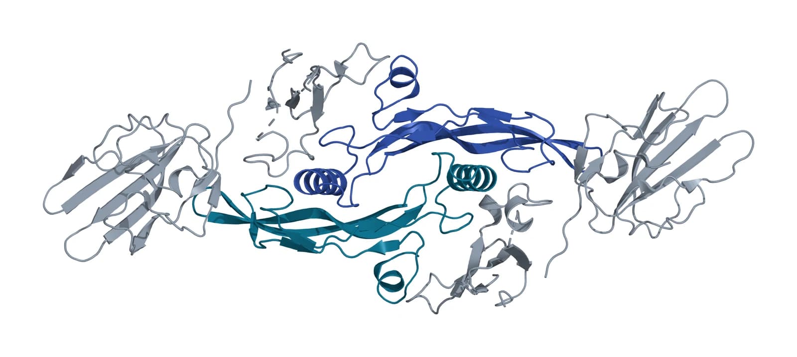
Radaev and colleagues then solved the TGF-beta1 ternary complex at 3.0 Å (PDB 3KFD, Fig. 1). The assembly matches the TGF-beta3 complex apart from a 10 degree rotation in the docking orientation of TGFBR1, and quantitative binding studies showed that TGFBR1 binds every isoform far better once TGFBR2 is already in place.[3]
This cooperative assembly distinguishes the TGF-beta branch from the BMP branch, whose receptors bind the ligand independently and gain avidity from membrane localization.[2] Hinck, Mueller and Springer place these complexes in the evolutionary context of the cystine knot growth factor fold shared by the whole family.[12]
The GS domain switches TGFBR1 from inhibited to active
TGFBR1 carries a conserved 30-residue segment, the GS domain, between its transmembrane helix and its kinase domain. Wieser, Wrana and Massagué showed that TGFBR2 phosphorylates a cluster of serines and threonines in the motif TTSGSGSG at the center of the GS domain, that mutating these residues impairs signaling, and that replacing Thr204 with aspartate produces a receptor that signals without ligand or TGFBR2.[13]
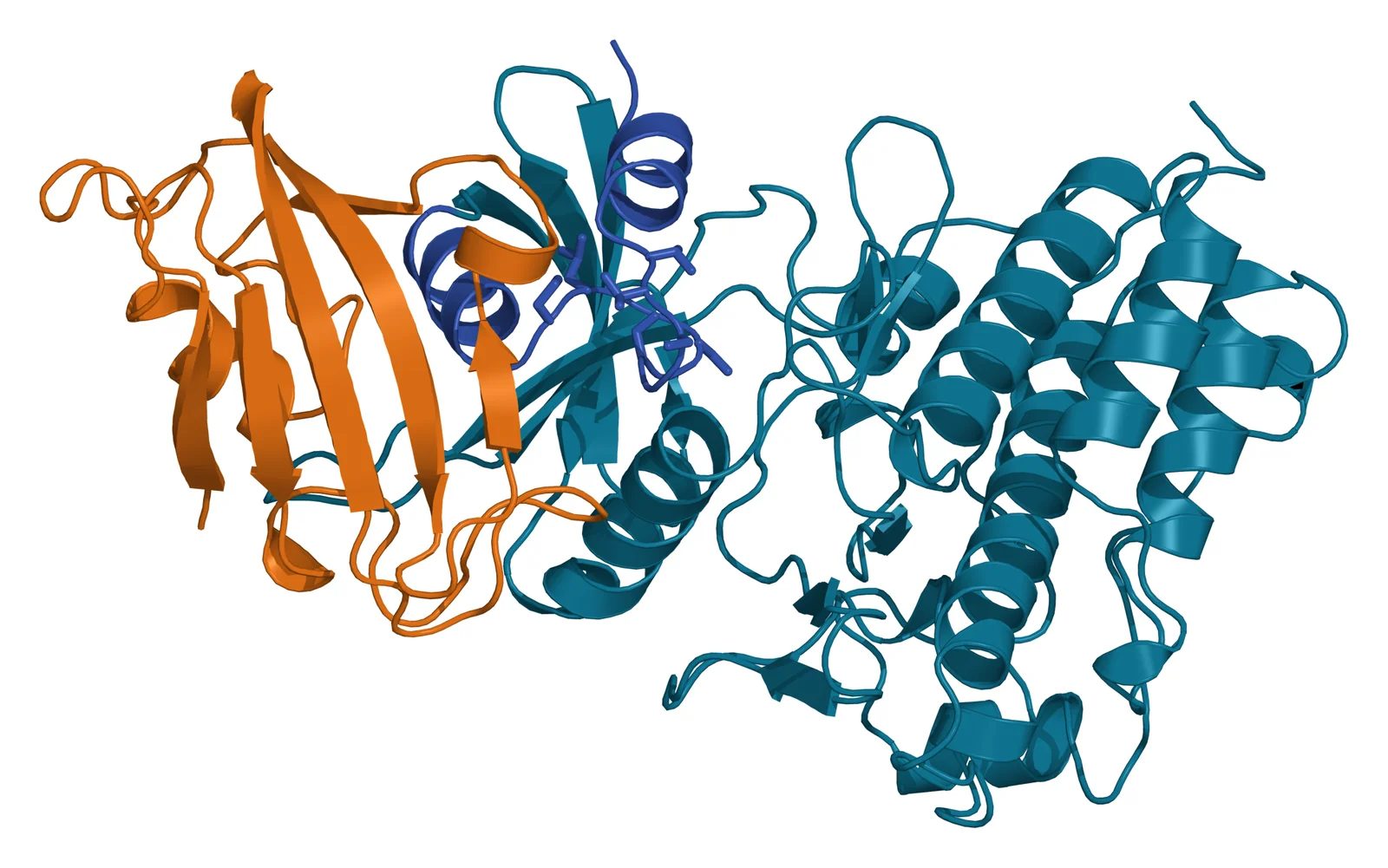
Huse and colleagues solved the cytoplasmic domain of unphosphorylated TGFBR1 in complex with FKBP12 (PDB 1B6C, Fig. 2). The receptor adopts an inactive conformation that the unphosphorylated GS domain holds in place, and FKBP12 binds the GS domain and caps the TGFBR2 phosphorylation sites, stabilizing that inactive state further.[10]
The same group then prepared a homogeneously tetraphosphorylated TGFBR1 by protein semisynthesis. Phosphorylation of the GS domain sharply increased the specificity of the kinase for the C-terminal serines of SMAD2, created a phosphorylation-dependent binding site for SMAD2, and ended recognition by FKBP12. The GS domain therefore acts as a switch that converts a binding surface for an inhibitor into a binding surface for the substrate.[14]
Chaikuad and Bullock review the structural allostery of the receptors and SMADs, including the FKBP12, Ski and SMURF1 complexes that mark the down-regulated state.[15]
Receptor-regulated SMADs carry the signal and SMAD7 shuts it off
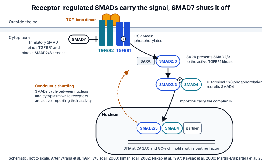
The SMAD family has three classes. Receptor-regulated SMADs (R-SMADs) are direct substrates of the type I receptors: SMAD2 and SMAD3 for ALK4, ALK5 (TGFBR1) and ALK7, and SMAD1, SMAD5 and SMAD8 for the BMP-type receptors. SMAD4 is the common mediator that partners with every phosphorylated R-SMAD, and SMAD6 and SMAD7 are inhibitory SMADs.[16][17][18][19]
SMAD2 is delivered to the receptor by SARA, the SMAD anchor for receptor activation. Wu and colleagues solved the SMAD2 MH2 domain bound to the SMAD-binding domain of SARA at 2.2 Å and showed how a rigid coil of SARA reads the beta sheet of SMAD2, a recognition that also suggested how type I receptors tell one R-SMAD from another.[20]
Activated TGFBR1 phosphorylates the two C-terminal serines of SMAD2 and SMAD3, and the phosphorylated SMADs form heteromeric complexes with SMAD4 that accumulate in the nucleus.[14][21][22] Martin-Malpartida and colleagues showed that these complexes recognize both the classical CAGAC motif and a 5 base pair GC-rich motif on DNA, which explains how SMAD2, SMAD3 and SMAD4 reach GC-rich regulatory elements genome-wide.[18]
The SMADs never settle in one compartment. Inman, Nicolás and Hill showed that receptors in epithelial cells remain active for at least three to four hours after stimulation, that continuous receptor activity is needed to keep active SMADs in the nucleus, and that SMAD2, SMAD3 and SMAD4 shuttle continuously between nucleus and cytoplasm so that the nuclear concentration of active SMADs reports the amount of active receptor at every moment.[22]
SMAD7 closes the loop. TGF-beta rapidly induces its expression, and it associates stably with the activated receptor complex and inhibits phosphorylation of SMAD2 and SMAD3.[19] SMAD7 also recruits the HECT ubiquitin ligase SMURF2, which leaves the nucleus on binding SMAD7 and targets the receptor complex for proteasomal and lysosomal degradation.[23] Hata and Chen review how posttranslational modifications and modulatory partners of the receptors and SMADs make the output context dependent.[17]
Non-SMAD routes branch from the same receptors
The ligand-occupied receptor complex also activates pathways that do not run through SMADs, including the ERK, JNK and p38 MAP kinases, PI3K and AKT, the Src tyrosine kinase and Rho family GTPases.[5][24] These branches reinforce, attenuate or redirect the SMAD response, and Zhang reviews how each is engaged directly by the receptors.[24]
The best characterized example is the TRAF6 to TAK1 route. Sorrentino and colleagues found that the ubiquitin ligase TRAF6 binds a consensus motif in TGFBR1, that TGF-beta induces TRAF6 autoubiquitination and lysine 63-linked polyubiquitination of TAK1, and that this activates TAK1 and the p38 and JNK kinases independently of the TGFBR1 kinase activity that SMAD2 requires.[25]
Derynck and Budi summarize how the receptors are presented and activated, how SMAD signaling draws on crosstalk with other pathways, and how non-SMAD mechanisms complement it, across the 33-ligand family.[4]
Betaglycan and endoglin present the ligand as co-receptors
Two membrane co-receptors, the proteoglycan betaglycan and the glycoprotein endoglin, are sometimes called type III receptors. Betaglycan binds all three TGF-beta isoforms, presents them to TGFBR2, and sets the potency of the ligands on their target cells; it also binds inhibin and takes part in the trafficking of the signaling receptors.[11][26]
Endoglin, also known as CD105, is a dimeric membrane glycoprotein that shares sequence with betaglycan and is abundant on vascular endothelial cells. Cheifetz and colleagues showed that it is the major TGF-beta1 binding protein that coexists with TGFBR1 and TGFBR2 on human umbilical vein endothelial cells, binding TGF-beta1 with a dissociation constant near 50 pM, binding TGF-beta3 with high affinity, and showing no measurable binding of TGF-beta2.[26] Saito and colleagues later solved the endoglin ectodomain alone and bound to BMP9, revealing a duplicated orphan domain that holds the ligand in a way compatible with type I receptor binding.[27]
Integrins release TGF-beta from its latent complex
TGF-beta is secreted and stored in the extracellular matrix as a latent complex with its own prodomain, which is in turn tethered to the matrix by latent TGF-beta binding proteins.[28] Shi and colleagues solved the structure of dimeric pro-TGF-beta1 and found a ring-shaped complex in which the prodomain forms a straitjacket around each growth factor monomer, shielding it from its receptors. Binding of an alpha-v integrin to the RGD motif of the prodomain is necessary but insufficient for release; the integrin must also apply force.[29]
Dong and colleagues showed how integrin alpha-v beta-6 binds pro-TGF-beta1 in the orientation needed for force-dependent release and how the prodomain is remodeled by the contact.[30] Campbell and colleagues then used cryo-electron microscopy to show that integrin alpha-v beta-8 can expose the growth factor to its receptors while it remains inside the latent complex, so that release and diffusion of TGF-beta are not required for signaling in that setting.[31]
Robertson and Rifkin review the full set of latent TGF-beta activators and extracellular modulators, with the mouse genetics that establish which ones matter in vivo.[28] Massagué frames this ligand availability, together with receptor levels and SMAD partners, as the context that decides what a cell does in response to TGF-beta.[32]
Endocytosis sorts receptors toward signaling or SMAD7 and SMURF turnover
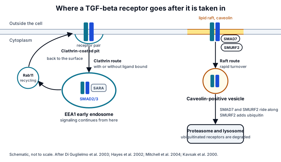
Activated TGF-beta receptors are rapidly internalized. Hayes, Chawla and Corvera showed that type I and type II receptors enter endosomes marked by EEA1, that SARA is enriched there, and that the extent of SMAD2 phosphorylation, nuclear translocation and transcription tracks the extent of internalization, revealing the early endosome as a compartment that amplifies the signal.[33]
Di Guglielmo and colleagues then showed that the receptors use two distinct routes. Clathrin-dependent internalization into EEA1-positive endosomes, where SARA resides, promotes SMAD signaling, while a lipid raft and caveolin-dependent route carries receptors bound by SMAD7 and SMURF2 and is required for rapid receptor turnover.[34] Mitchell and colleagues found that receptors also recycle, with or without ligand, through clathrin-coated pits and Rab11-dependent return to the surface, before activated receptors are diverted toward degradation.[35]
Heldin and Moustakas review the full set of modifications that govern receptor stability, including phosphorylation, ubiquitination, sumoylation and neddylation, along with the surface and cytoplasmic partners that regulate them.[5]
TGF-beta receptors shape vascular and mesenchymal development
Mouse genetics show what the receptors do in a developing animal. Oshima, Oshima and Taketo found that embryos lacking Tgfbr2 die around embryonic day 10.5 with defects in yolk sac hematopoiesis and vasculogenesis, a phenotype matching loss of TGF-beta1 itself.[36] Larsson and colleagues found that embryos lacking Tgfbr1 die at midgestation with severe defects in yolk sac and placental vascular development, while their hematopoietic precursors retain normal potential in culture; endothelial cells from these embryos proliferate more, migrate improperly and produce less fibronectin.[37]
The same receptors direct mesenchymal lineage decisions. Grafe and colleagues review how TGF-beta family signaling through SMAD and non-SMAD routes regulates commitment and differentiation of mesenchymal stem cells into osteoblasts, chondrocytes, myoblasts, adipocytes and tenocytes, both in development and in adult tissue maintenance.[38] Schmierer and Hill examine how the strength and duration of the signal, as well as its presence, are sensed and interpreted in embryos and adult tissues.[39]
Together these studies show a receptor system built for control at every step: ligand availability, co-receptor presentation, cooperative complex assembly, the GS domain switch, a shuttling SMAD readout, and an endocytic choice between signaling and degradation.
Advancing the research question
The growth factor receptors overview places the TGF-beta receptors beside the tyrosine kinase receptors for PDGF, EGF, VEGF and IGF-1, and the sibling pages on PDGFR-beta, EGFR, VEGFR-2 and FGFR, and the IGF-1 receptor cover each of those in the same depth. The surface receptor chemistry page explains the membrane biochemistry the whole cluster shares.
Platelet granule biology explains where platelet-stored TGF-beta comes from, and the extracellular vesicle uptake page describes how vesicle-borne ligands reach a receiving cell.
Referencias comentadas
Numeradas en el orden en que se citan en el texto. Cada fuente lleva una nota sobre lo que respalda en esta página; los artículos revisados por pares se listan con revista, volumen, número y páginas, y las fuentes primarias son regulaciones, documentos de agencias y bases de datos de referencia.
- 1.Peer-reviewedWrana JL, Attisano L, Wieser R, et al. Mechanism of activation of the TGF-beta receptor. Nature. 1994;370(6488):341-347. https://pubmed.ncbi.nlm.nih.gov/8047140/Annotation: Receptor reconstitution and phosphorylation studies. Cited for TGFBR2 as the constitutively active, ligand-binding kinase that recruits and phosphorylates TGFBR1.
- 2.Peer-reviewedGroppe J, Hinck CS, Samavarchi-Tehrani P, et al. Cooperative assembly of TGF-beta superfamily signaling complexes is mediated by two disparate mechanisms and distinct modes of receptor binding. Molecular Cell. 2008;29(2):157-168. https://pubmed.ncbi.nlm.nih.gov/18243111/Annotation: The first TGF-beta ternary complex structure. Cited for TGFBR1 docking at a composite ligand and TGFBR2 interface, the tethering extensions, and the contrast with avidity-driven BMP receptor assembly.
- 3.Peer-reviewedRadaev S, Zou Z, Huang T, et al. Ternary complex of transforming growth factor-beta1 reveals isoform-specific ligand recognition and receptor recruitment in the superfamily. Journal of Biological Chemistry. 2010;285(19):14806-14814. https://pubmed.ncbi.nlm.nih.gov/20207738/Annotation: The PDB 3KFD structure shown in Fig. 1. Cited for the TGF-beta1 ternary complex, the 10 degree difference in TGFBR1 docking, and the isoform-specific binding kinetics of the receptors.
- 4.Peer-reviewedDerynck R, Budi EH. Specificity, versatility, and control of TGF-β family signaling. Science Signaling. 2019;12(570):eaav5183. https://pubmed.ncbi.nlm.nih.gov/30808818/Annotation: Review of the 33-ligand family. Cited for the ligand count, the control of receptor presentation and activation, and the interplay of SMAD and non-SMAD signaling.
- 5.Peer-reviewedHeldin CH, Moustakas A. Signaling Receptors for TGF-β Family Members. Cold Spring Harbor Perspectives in Biology. 2016;8(8):a022053. https://pubmed.ncbi.nlm.nih.gov/27481709/Annotation: Review of the type I and type II receptors. Cited for the heterotetrameric complex, TGFBR2 and TGFBR1 (ALK5) as the TGF-beta receptor pair, dual specificity, the non-SMAD outputs, and the modifications that control receptor stability.
- 6.Peer-reviewedHart PJ, Deep S, Taylor AB, et al. Crystal structure of the human TbetaR2 ectodomain--TGF-beta3 complex. Nature Structural Biology. 2002;9(3):203-208. https://pubmed.ncbi.nlm.nih.gov/11850637/Annotation: The 2.15 Å structure of the TGFBR2 ectodomain bound to TGF-beta3. Cited for TGFBR2 binding identical finger segments at opposite ends of the growth factor dimer.
- 7.DatabaseNCBI Gene 7046, TGFBR1 transforming growth factor beta receptor 1 (Homo sapiens). Accessed 8 November 2026. https://www.ncbi.nlm.nih.gov/gene/7046
- 8.DatabaseNCBI Gene 7048, TGFBR2 transforming growth factor beta receptor 2 (Homo sapiens). Accessed 8 November 2026. https://www.ncbi.nlm.nih.gov/gene/7048
- 9.Peer-reviewedGoumans MJ, Valdimarsdottir G, Itoh S, et al. Balancing the activation state of the endothelium via two distinct TGF-beta type I receptors. EMBO Journal. 2002;21(7):1743-1753. https://pubmed.ncbi.nlm.nih.gov/11927558/Annotation: Endothelial cell signaling studies. Cited for TGF-beta inducing SMAD1/5 phosphorylation through ALK1 and SMAD2 phosphorylation through ALK5 in endothelial cells.
- 10.Peer-reviewedHuse M, Chen YG, Massagué J, Kuriyan J. Crystal structure of the cytoplasmic domain of the type I TGF beta receptor in complex with FKBP12. Cell. 1999;96(3):425-436. https://pubmed.ncbi.nlm.nih.gov/10025408/Annotation: The PDB 1B6C structure shown in Fig. 2. Cited for the inactive conformation held by the unphosphorylated GS domain, FKBP12 capping the phosphorylation sites, and the tyrosine kinase features of the catalytic center.
- 11.Peer-reviewedBilandzic M, Stenvers KL. Betaglycan: a multifunctional accessory. Molecular and Cellular Endocrinology. 2011;339(1-2):180-189. https://pubmed.ncbi.nlm.nih.gov/21550381/Annotation: Review of betaglycan. Cited for its role in presenting all three TGF-beta isoforms, setting ligand potency, binding inhibin and trafficking the signaling receptors.
- 12.Peer-reviewedHinck AP, Mueller TD, Springer TA. Structural Biology and Evolution of the TGF-β Family. Cold Spring Harbor Perspectives in Biology. 2016;8(12):a022103. https://pubmed.ncbi.nlm.nih.gov/27638177/Annotation: Structural review of the family, its prodomains and receptors. Cited for the cystine knot growth factor fold and the ternary complexes that explain receptor specificity.
- 13.Peer-reviewedWieser R, Wrana JL, Massagué J. GS domain mutations that constitutively activate T beta R-I, the downstream signaling component in the TGF-beta receptor complex. EMBO Journal. 1995;14(10):2199-2208. https://pubmed.ncbi.nlm.nih.gov/7774578/Annotation: Mutational analysis of the GS domain. Cited for the TTSGSGSG phosphorylation cluster and the constitutively active Thr204 to aspartate receptor.
- 14.Peer-reviewedHuse M, Muir TW, Xu L, et al. The TGF beta receptor activation process: an inhibitor- to substrate-binding switch. Molecular Cell. 2001;8(3):671-682. https://pubmed.ncbi.nlm.nih.gov/11583628/Annotation: Semisynthetic tetraphosphorylated TGFBR1. Cited for GS domain phosphorylation switching the receptor from an FKBP12-binding to a SMAD2-binding state and sharpening kinase specificity.
- 15.Peer-reviewedChaikuad A, Bullock AN. Structural Basis of Intracellular TGF-β Signaling: Receptors and Smads. Cold Spring Harbor Perspectives in Biology. 2016;8(11):a022111. https://pubmed.ncbi.nlm.nih.gov/27549117/Annotation: Structural review of the intracellular receptor domains and SMAD complexes. Cited for the allosteric mechanisms of activation and for the FKBP12, Ski and SMURF1 complexes that mark down-regulation.
- 16.Peer-reviewedShi Y, Massagué J. Mechanisms of TGF-beta signaling from cell membrane to the nucleus. Cell. 2003;113(6):685-700. https://pubmed.ncbi.nlm.nih.gov/12809600/Annotation: Foundational review of receptor and SMAD activation. Cited for the three SMAD classes and the overall route from membrane to nucleus.
- 17.Peer-reviewedHata A, Chen YG. TGF-β Signaling from Receptors to Smads. Cold Spring Harbor Perspectives in Biology. 2016;8(9):a022061. https://pubmed.ncbi.nlm.nih.gov/27449815/Annotation: Review of receptor to SMAD signaling. Cited for the SMAD classes and for the posttranslational modifications and partners that make the output context dependent.
- 18.Peer-reviewedMartin-Malpartida P, Batet M, Kaczmarska Z, et al. Structural basis for genome wide recognition of 5-bp GC motifs by SMAD transcription factors. Nature Communications. 2017;8(1):2070. https://pubmed.ncbi.nlm.nih.gov/29234012/Annotation: Crystal structures of SMAD3 and SMAD4 bound to GC-rich DNA. Cited for the 5 base pair GC motif that SMAD2, SMAD3 and SMAD4 recognize alongside CAGAC.
- 19.Peer-reviewedNakao A, Afrakhte M, Morén A, et al. Identification of Smad7, a TGFbeta-inducible antagonist of TGF-beta signalling. Nature. 1997;389(6651):631-635. https://pubmed.ncbi.nlm.nih.gov/9335507/Annotation: Identification of SMAD7 in mammalian cells and Xenopus embryos. Cited for SMAD7 as a TGF-beta-inducible inhibitory SMAD that associates stably with the receptor complex and inhibits phosphorylation of SMAD2 and SMAD3 (Fig. 3).
- 20.Peer-reviewedWu G, Chen YG, Ozdamar B, et al. Structural basis of Smad2 recognition by the Smad anchor for receptor activation. Science. 2000;287(5450):92-97. https://pubmed.ncbi.nlm.nih.gov/10615055/Annotation: The 2.2 Å structure of the SMAD2 MH2 domain bound to SARA. Cited for how SARA recognizes SMAD2 and presents it to the receptor.
- 21.Peer-reviewedMacias MJ, Martin-Malpartida P, Massagué J. Structural determinants of Smad function in TGF-β signaling. Trends in Biochemical Sciences. 2015;40(6):296-308. https://pubmed.ncbi.nlm.nih.gov/25935112/Annotation: Structural review of the SMADs. Cited for phosphorylated R-SMADs forming complexes with SMAD4.
- 22.Peer-reviewedInman GJ, Nicolás FJ, Hill CS. Nucleocytoplasmic shuttling of Smads 2, 3, and 4 permits sensing of TGF-beta receptor activity. Molecular Cell. 2002;10(2):283-294. https://pubmed.ncbi.nlm.nih.gov/12191474/Annotation: Live-cell and inhibitor studies of SMAD localization. Cited for continuous shuttling, sustained receptor activity after stimulation, and nuclear SMAD levels reporting receptor activity.
- 23.Peer-reviewedKavsak P, Rasmussen RK, Causing CG, et al. Smad7 binds to Smurf2 to form an E3 ubiquitin ligase that targets the TGF beta receptor for degradation. Molecular Cell. 2000;6(6):1365-1375. https://pubmed.ncbi.nlm.nih.gov/11163210/Annotation: Identification of the SMAD7 and SMURF2 complex. Cited for SMAD7 recruiting SMURF2 from the nucleus to the activated receptor and for proteasomal and lysosomal degradation of the receptors (Figs. 3 and 4).
- 24.Peer-reviewedZhang YE. Non-Smad Signaling Pathways of the TGF-β Family. Cold Spring Harbor Perspectives in Biology. 2017;9(2):a022129. https://pubmed.ncbi.nlm.nih.gov/27864313/Annotation: Review of the non-SMAD branches. Cited for the MAP kinase, PI3K and AKT, and Rho GTPase routes engaged directly by the receptors.
- 25.Peer-reviewedSorrentino A, Thakur N, Grimsby S, et al. The type I TGF-beta receptor engages TRAF6 to activate TAK1 in a receptor kinase-independent manner. Nature Cell Biology. 2008;10(10):1199-1207. https://pubmed.ncbi.nlm.nih.gov/18758450/Annotation: Cell biology of the TRAF6 to TAK1 route. Cited for TRAF6 binding a motif in TGFBR1, lysine 63-linked ubiquitination of TAK1, and p38 and JNK activation independent of the TGFBR1 kinase.
- 26.Peer-reviewedCheifetz S, Bellón T, Calés C, et al. Endoglin is a component of the transforming growth factor-beta receptor system in human endothelial cells. Journal of Biological Chemistry. 1992;267(27):19027-19030. https://pubmed.ncbi.nlm.nih.gov/1326540/Annotation: Affinity labeling of endothelial cells. Cited for endoglin as the major TGF-beta1 binding protein beside TGFBR1 and TGFBR2 on endothelial cells, its 50 pM affinity, and its isoform selectivity.
- 27.Peer-reviewedSaito T, Bokhove M, Croci R, et al. Structural Basis of the Human Endoglin-BMP9 Interaction: Insights into BMP Signaling and HHT1. Cell Reports. 2017;19(9):1917-1928. https://pubmed.ncbi.nlm.nih.gov/28564608/Annotation: Crystal structures of the endoglin ectodomain alone and bound to BMP9. Cited for the duplicated orphan domain and for ligand binding compatible with type I receptor recognition.
- 28.Peer-reviewedRobertson IB, Rifkin DB. Regulation of the Bioavailability of TGF-β and TGF-β-Related Proteins. Cold Spring Harbor Perspectives in Biology. 2016;8(6):a021907. https://pubmed.ncbi.nlm.nih.gov/27252363/Annotation: Review of latent TGF-beta and its activators. Cited for matrix sequestration by latent TGF-beta binding proteins and for the in vivo evidence on each activator.
- 29.Peer-reviewedShi M, Zhu J, Wang R, et al. Latent TGF-β structure and activation. Nature. 2011;474(7351):343-349. https://pubmed.ncbi.nlm.nih.gov/21677751/Annotation: Crystal structure of pro-TGF-beta1. Cited for the ring-shaped latent complex, the prodomain straitjacket, and force-dependent release after alpha-v integrin binding to the RGD motif.
- 30.Peer-reviewedDong X, Zhao B, Iacob RE, et al. Force interacts with macromolecular structure in activation of TGF-β. Nature. 2017;542(7639):55-59. https://pubmed.ncbi.nlm.nih.gov/28117447/Annotation: Structure of integrin alpha-v beta-6 bound to pro-TGF-beta1. Cited for the binding orientation that allows force-dependent release and for remodeling of the prodomain on integrin contact.
- 31.Peer-reviewedCampbell MG, Cormier A, Ito S, et al. Cryo-EM Reveals Integrin-Mediated TGF-β Activation without Release from Latent TGF-β. Cell. 2020;180(3):490-501.e16. https://pubmed.ncbi.nlm.nih.gov/31955848/Annotation: Cryo-EM of integrin alpha-v beta-8 with latent TGF-beta. Cited for signaling by the growth factor while it remains within the latent complex.
- 32.Peer-reviewedMassagué J. TGFβ signalling in context. Nature Reviews Molecular Cell Biology. 2012;13(10):616-630. https://pubmed.ncbi.nlm.nih.gov/22992590/Annotation: Review of how cellular context shapes the response. Cited for ligand availability, receptor levels and SMAD partners as the determinants of the response to TGF-beta.
- 33.Peer-reviewedHayes S, Chawla A, Corvera S. TGF beta receptor internalization into EEA1-enriched early endosomes: role in signaling to Smad2. Journal of Cell Biology. 2002;158(7):1239-1249. https://pubmed.ncbi.nlm.nih.gov/12356868/Annotation: Receptor trafficking and SMAD2 signaling. Cited for internalization into EEA1 endosomes where SARA resides, and for signaling that tracks the extent of internalization (Fig. 4).
- 34.Peer-reviewedDi Guglielmo GM, Le Roy C, Goodfellow AF, Wrana JL. Distinct endocytic pathways regulate TGF-beta receptor signalling and turnover. Nature Cell Biology. 2003;5(5):410-421. https://pubmed.ncbi.nlm.nih.gov/12717440/Annotation: The two endocytic routes drawn in Fig. 4. Cited for clathrin-dependent entry into EEA1 endosomes promoting SMAD signaling and the caveolin route carrying SMAD7 and SMURF2-bound receptors to turnover.
- 35.Peer-reviewedMitchell H, Choudhury A, Pagano RE, Leof EB. Ligand-dependent and -independent transforming growth factor-beta receptor recycling regulated by clathrin-mediated endocytosis and Rab11. Molecular Biology of the Cell. 2004;15(9):4166-4178. https://pubmed.ncbi.nlm.nih.gov/15229286/Annotation: Receptor recycling studies. Cited for ligand-independent recycling through clathrin-coated pits and Rab11-dependent return to the surface (Fig. 4).
- 36.Peer-reviewedOshima M, Oshima H, Taketo MM. TGF-beta receptor type II deficiency results in defects of yolk sac hematopoiesis and vasculogenesis. Developmental Biology. 1996;179(1):297-302. https://pubmed.ncbi.nlm.nih.gov/8873772/Annotation: Tgfbr2 knockout mice. Cited for yolk sac hematopoiesis and vasculogenesis defects with lethality around embryonic day 10.5, matching loss of TGF-beta1.
- 37.Peer-reviewedLarsson J, Goumans MJ, Sjöstrand LJ, et al. Abnormal angiogenesis but intact hematopoietic potential in TGF-beta type I receptor-deficient mice. EMBO Journal. 2001;20(7):1663-1673. https://pubmed.ncbi.nlm.nih.gov/11285230/Annotation: Tgfbr1 knockout mice. Cited for yolk sac and placental vascular defects at midgestation, intact hematopoietic potential, and the behavior of receptor-deficient endothelial cells.
- 38.Peer-reviewedGrafe I, Alexander S, Peterson JR, et al. TGF-β Family Signaling in Mesenchymal Differentiation. Cold Spring Harbor Perspectives in Biology. 2018;10(5):a022202. https://pubmed.ncbi.nlm.nih.gov/28507020/Annotation: Review of TGF-beta family signaling in mesenchymal lineages. Cited for commitment and differentiation of mesenchymal stem cells into osteoblasts, chondrocytes, myoblasts, adipocytes and tenocytes.
- 39.Peer-reviewedSchmierer B, Hill CS. TGFbeta-SMAD signal transduction: molecular specificity and functional flexibility. Nature Reviews Molecular Cell Biology. 2007;8(12):970-982. https://pubmed.ncbi.nlm.nih.gov/18000526/Annotation: Review of SMAD specificity and signal interpretation. Cited for how signal strength and duration are sensed in embryos and adult tissues.
Cite this page
ExaVeyra Sciences Editorial Team. TGF-beta Receptors: Type I, Type II and Signaling. ExaVeyra Sciences; 9 November 2026. Available from: https://exaveyra.com/science/surface-receptors/tgf-beta-receptors. Accessed [date accessed].
Fuentes de las figuras
- 40.PDB 3KFD, TGF-beta1 ternary complex with TGFBR2 and TGFBR1. ExaVeyra Sciences, rendered in PyMOL from PDB 3KFD. Structure data: RCSB Protein Data Bank (rcsb.org), CC0. Publicación de la estructura: Radaev S, Zou Z, Huang T, Lafer EM, Hinck AP, Sun PD. Ternary complex of transforming growth factor-beta1 reveals isoform-specific ligand recognition and receptor recruitment in the superfamily. J Biol Chem. 2010;285(19):14806-14814.
- 41.PDB 1B6C, TGFBR1 cytoplasmic domain in complex with FKBP12. ExaVeyra Sciences, rendered in PyMOL from PDB 1B6C. Structure data: RCSB Protein Data Bank (rcsb.org), CC0. Publicación de la estructura: Huse M, Chen YG, Massagué J, Kuriyan J. Crystal structure of the cytoplasmic domain of the type I TGF beta receptor in complex with FKBP12. Cell. 1999;96(3):425-436.
Créditos de imágenes
- Fig. 3ExaVeyra Sciences, figura original. After Wrana et al., Nature 1994; Wu et al., Science 2000; Inman et al., Mol Cell 2002; Nakao et al., Nature 1997; Kavsak et al., Mol Cell 2000; Martin-Malpartida et al., Nat Commun 2017. ↩
- Fig. 4ExaVeyra Sciences, figura original. After Di Guglielmo et al., Nat Cell Biol 2003; Hayes et al., J Cell Biol 2002; Mitchell et al., Mol Biol Cell 2004; Kavsak et al., Mol Cell 2000. ↩