Esta referencia está disponible actualmente en inglés. La versión en español está en preparación.
Stem Cell Types: Embryonic, Induced Pluripotent, Adult and MSC
Por Benn Bluestein-Veyra, M.Sc. en Química Orgánica · Actualizado el 20 de octubre de 2026
ExaVeyra Sciences es un proveedor B2B con sede en Miami que suministra exosomas para uso estético tópico a clínicas con licencia, y remite a los prescriptores con licencia a una farmacia de preparados compuestos 503A que prepara formulaciones según prescripciones individuales.
| Pregunta | Respuesta |
|---|---|
| Qué cubre esta página | A primer on stem cell types: embryonic and induced pluripotent stem cells, hematopoietic and other adult stem cells, and where mesenchymal stromal cells fit. |
| Quién puede hacer pedidos | Clínicas y profesionales con licencia profesional activa y NPI verificados por ExaVeyra. Los pedidos de exosomas requieren además un reconocimiento firmado de uso profesional. |
| Qué se envía y cómo | El material de exosomas para uso estético tópico, caracterizado por lote, se envía en cadena de frío por exprés nocturno. La entrega puede tardar 1 a 3 días hábiles. Se emite un certificado de análisis para cada lote y se entrega a solicitud. |
The main types of stem cells are embryonic stem cells, induced pluripotent stem cells and adult, or tissue, stem cells, each defined by its ability to renew itself and to give rise to specialized cells.[1] Mesenchymal stromal cells (MSCs) belong with the multipotent adult cell types.[2][3]
This primer covers the potency hierarchy, the derivation of embryonic and induced pluripotent stem cells, the transcription factors that hold pluripotency in place and the adult stem cells best studied in their niches. It closes with MSCs and the perinatal tissues they are isolated from.[4][5]
Understanding the stem cell potency hierarchy
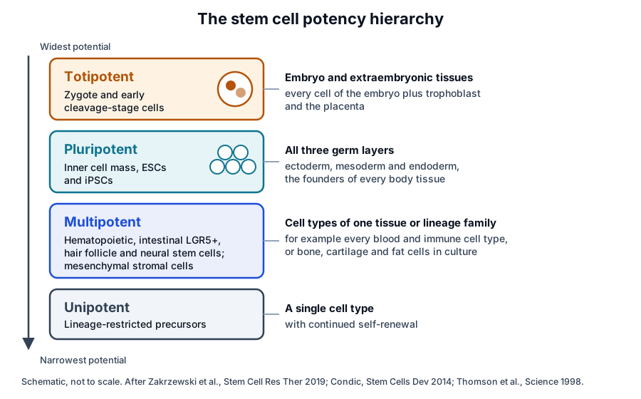
Potency describes the range of cell types a stem cell can produce, and it narrows at each step of development. Zakrzewski and colleagues classify stem cells as totipotent, pluripotent, multipotent, oligopotent and unipotent, from the widest potential to the narrowest (Fig. 1).[4]
- Totipotent cells, such as the zygote, form the embryo together with extraembryonic structures, including the placenta.[4]
- Pluripotent cells form cells of all three germ layers, ectoderm, mesoderm and endoderm; embryonic and induced pluripotent stem cells are the standard examples.[1][4]
- Multipotent cells form the cell types of one tissue or lineage family, as a hematopoietic stem cell forms the types of blood cells.[4]
- Unipotent cells form a single cell type and keep the capacity to divide repeatedly.[4]
Condic describes totipotency as the property of a cell that can form a whole organism, and links it to oocyte-derived cytoplasm. She proposed the term plenipotent to resolve confusion around totipotency.[6]
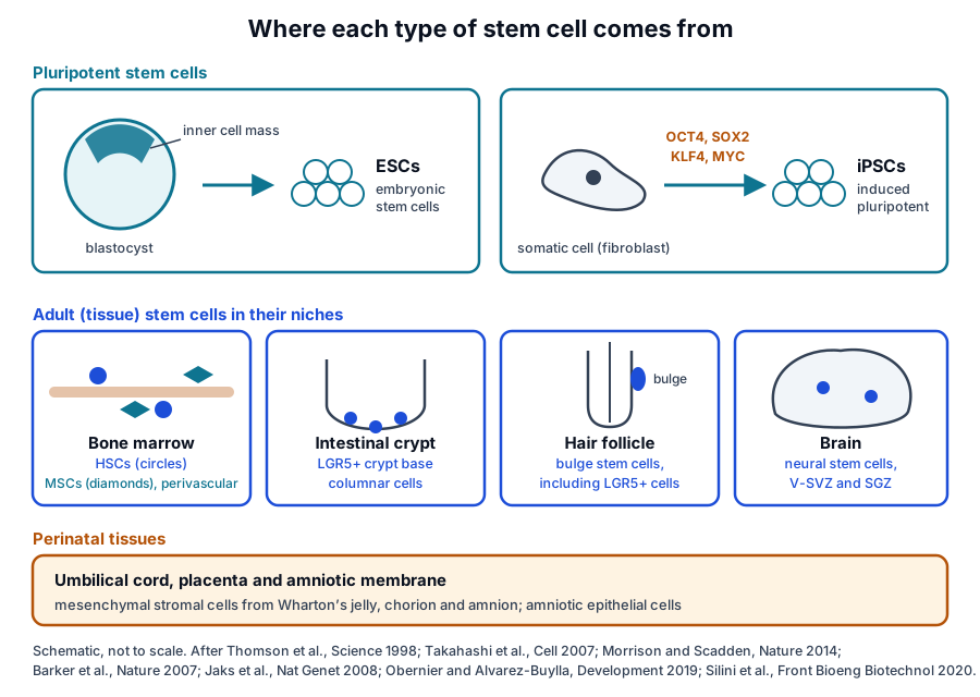
Embryonic stem cells come from the inner cell mass of the blastocyst
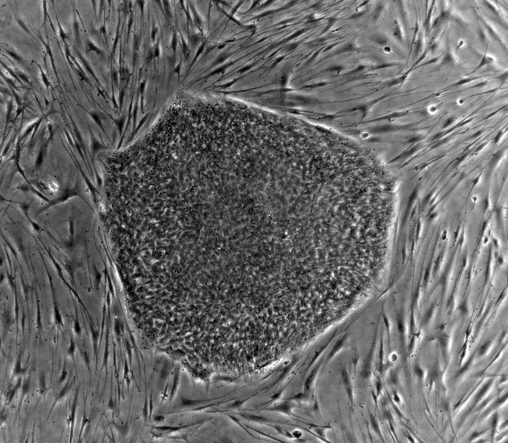
The early mammalian embryo at the blastocyst stage holds two cell types: the inner cell mass, which develops into every tissue of the body, and the trophectoderm, which contributes to the placenta.[1] Evans and Kaufman established pluripotential cell lines from cultured mouse embryos in 1981.[7]
Thomson and colleagues derived the first human embryonic stem cell lines from blastocysts in 1998. The lines had normal karyotypes, high telomerase activity and the surface markers of primate embryonic stem cells (Fig. 2).[8]
After four to five months of undifferentiated growth, the cells still formed trophoblast and derivatives of all three germ layers: gut epithelium, cartilage, bone, smooth and striated muscle, neural epithelium, embryonic ganglia and stratified squamous epithelium.[8]
Naive and primed states of pluripotency
Pluripotent cells can be held in culture in distinct configurations, termed naive and primed, that differ in their functional and molecular characteristics. Weinberger and colleagues review the signaling pathways that preserve each state and drive transitions between them in rodent and human cells.[9]
Induced pluripotent stem cells are reprogrammed from somatic cells
Takahashi and Yamanaka reported in 2006 that four transcription factors, Oct3/4, Sox2, c-Myc and Klf4, induced pluripotent stem cells from mouse embryonic and adult fibroblasts under embryonic stem cell culture conditions. Nanog was dispensable for this induction.[10]
The induced pluripotent stem (iPS) cells shared the morphology, growth properties and marker genes of embryonic stem cells, and after injection into blastocysts they contributed to mouse embryonic development.[10] Yamanaka later described iPSCs as the merging of three major scientific streams.[11]
In 2007, Takahashi and colleagues generated iPS cells from adult human dermal fibroblasts with the same four factors. The cells matched human embryonic stem cells in morphology, proliferation, surface antigens, gene expression, epigenetic status of pluripotency genes and telomerase activity.[12]
The same year, Yu and colleagues in the Thomson laboratory reprogrammed human somatic cells with a different set: OCT4, SOX2, NANOG and LIN28. Their lines had normal karyotypes and differentiated into advanced derivatives of all three primary germ layers.[13]
Takahashi and Yamanaka note that reprogramming needs only the host cell and a defined cocktail of transcription factors, and that its efficiency has risen considerably.[14] Whether iPSCs are functionally equivalent to embryonic stem cells remains an open research question.[11]
OCT4, SOX2 and NANOG form the core pluripotency network
OCT4 is a POU-family transcription factor encoded by POU5F1 on chromosome 6p21.33.[15] Nichols and colleagues found that Oct4-deficient mouse embryos reached the blastocyst stage with an inner cell mass restricted to the extraembryonic trophoblast lineage.[16]
NANOG, encoded on chromosome 12p13.31, is a homeodomain protein.[17] Chambers and colleagues found Nanog mRNA in pluripotent mouse and human cell lines and absent from differentiated cells, and raised Nanog expression sustained clonal expansion of mouse embryonic stem cells independently of Stat3.[18]
Mitsui and colleagues showed that Nanog maintained self-renewal independently of the LIF and Stat3 pathway. Nanog-deficient embryonic stem cells lost pluripotency and differentiated into the extraembryonic endoderm lineage.[19]
SOX2, an HMG-box transcription factor, is encoded on chromosome 3q26.33.[20] Boyer and colleagues mapped the binding sites of OCT4, SOX2 and NANOG across the genome of human embryonic stem cells and found that the three factors co-occupy a substantial portion of their target genes.[21]
Many of those targets encode transcription factors, among them developmentally important homeodomain proteins. The three factors form autoregulatory and feedforward loops, a circuitry that supports pluripotency and self-renewal.[21]
Crystal structures show OCT and SOX2 partners on DNA
Reményi and colleagues solved a ternary complex of the Oct1 POU domain and the Sox2 HMG domain bound to an enhancer DNA element (Fig. 3a).[22] Their models indicated that Oct4 and Sox2 dimerize on DNA in distinct arrangements, set by the layout of each enhancer.[22]
Esch and colleagues solved the Oct4 POU domain bound to DNA (Fig. 3b). The linker between its two DNA-binding domains forms an exposed alpha helix, where the Oct1 linker is unstructured, and point mutations in this helix altered or abolished reprogramming activity.[23]
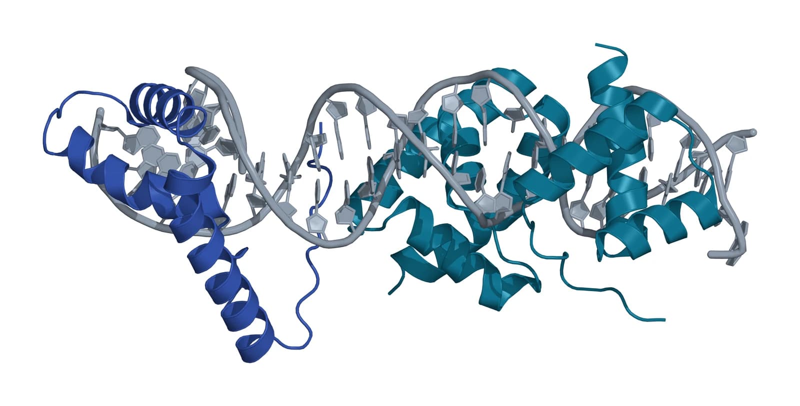
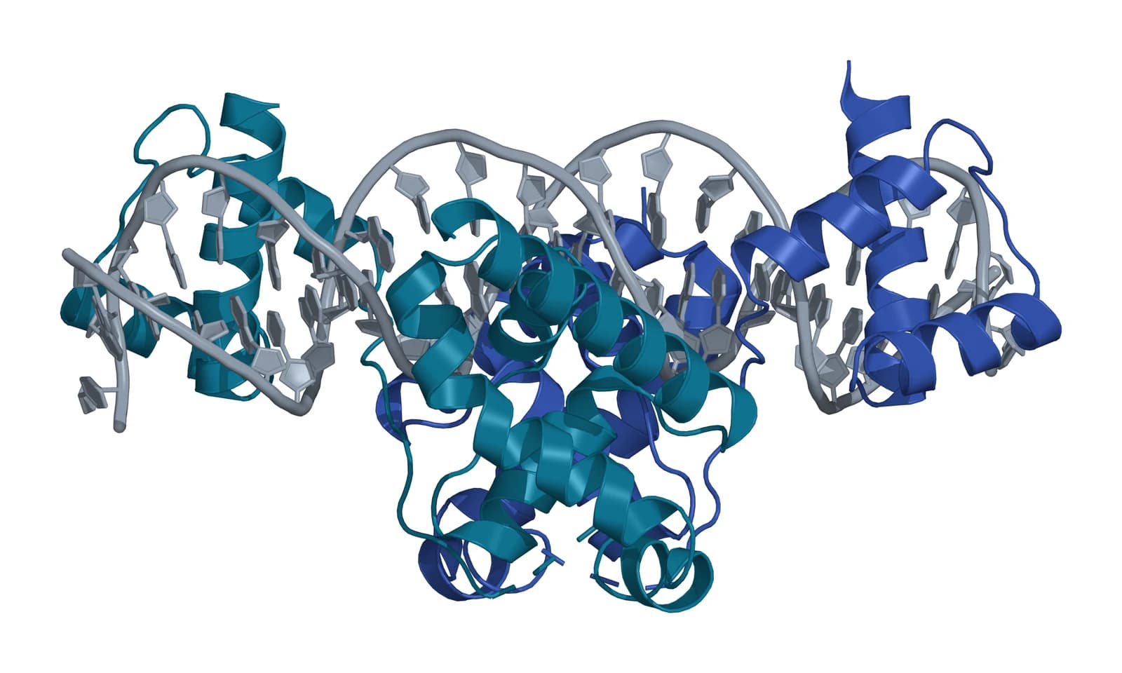
Fig. 3 | Oct1 POU and Sox2 HMG domains bound together on DNA and mouse Oct4 POU domain bound to DNA.
Adult stem cells maintain their tissues from defined niches
Adult stem cells are found in a tissue or organ and give rise to the specialized cell types of that tissue. They sit in specific anatomical locations and can remain quiescent for long periods until activated (Fig. 4).[1] Morrison and Scadden define niches as local microenvironments that maintain and regulate stem cells.[24]
Hematopoietic stem cells and the spleen colony assay
Till and McCulloch injected mouse bone marrow cells into irradiated recipient mice and counted the macroscopic colonies that formed in the spleen, a direct and quantitative measure of colony-forming marrow cells.[25] Becker, McCulloch and Till then used chromosome markers to show that each spleen colony arose from a single cell.[26]
Morrison and Scadden describe the bone marrow niche for hematopoietic stem cells (HSCs) as perivascular, created partly by mesenchymal stromal cells and endothelial cells and often located near trabecular bone.[24]
Pinho and Frenette review how the niche controls HSC proliferation, self-renewal, differentiation and migration. Candidate niche cells include perivascular mesenchymal and endothelial cells as well as HSC-derived megakaryocytes, macrophages and regulatory T cells.[27]
Intestinal stem cells express LGR5
Barker and colleagues found the Wnt target gene Lgr5 expressed exclusively in cycling columnar cells at the base of intestinal crypts. In lineage tracing in adult mice, these crypt base columnar cells generated all epithelial lineages over the following two months.[28] Human LGR5 maps to chromosome 12q21.1.[29]
Sato and colleagues established culture conditions in which single sorted Lgr5+ stem cells built crypt-villus organoids containing all differentiated cell types, with no non-epithelial cellular niche.[30] Gehart and Clevers describe progeny that differentiate into one of six mature cell types as they move up toward the villus.[31]
Skin and hair follicle stem cells occupy separate pools
Blanpain and Fuchs describe distinct resident stem cell pools that maintain the interfollicular epidermis, the hair follicles and the sebaceous glands.[32] In the hair follicle bulge, Blanpain and colleagues used clonal analysis to demonstrate self-renewal and multipotency, the two defining features of stem cells.[33]
Jaks and colleagues found Lgr5 on actively cycling cells in the bulge and secondary germ, and Lgr5+ cells maintained all hair follicle lineages over long periods.[34] Hsu and colleagues showed that early stem cell descendants home back to the bulge and become the primary stem cells for the next hair cycle.[35]
Hsu, Li and Fuchs review how genetic and imaging tools have clarified the lineage relationships between skin stem cells and their progeny, and the mutual influences between these cells and their niches.[36]
Neural stem cells persist in two zones of the adult brain
Reynolds and Weiss isolated cells from the adult mouse striatum that proliferated in culture with epidermal growth factor. The cells expressed nestin and then developed the morphology and antigenic properties of neurons and astrocytes.[37]
Obernier and Alvarez-Buylla describe neural stem cells in the ventricular-subventricular zone and the hippocampal subgranular zone of the adult rodent brain, which generate young neurons for the olfactory bulb and the hippocampus.[38] Bond, Ming and Song review five decades of this research.[39]
Mesenchymal stromal cells are multipotent adult cells defined in culture
Pittenger and colleagues isolated cells from the marrow aspirates of volunteer donors that differentiated into the adipocytic, chondrocytic or osteocytic lineages. Single cells expanded to colonies kept this multilineage potential.[2]
The International Society for Cellular Therapy (ISCT) set three minimal criteria for human MSCs: adherence to plastic in standard culture; expression of CD105, CD73 and CD90 without CD45, CD34, CD14 or CD11b, CD79alpha or CD19 and HLA-DR; and differentiation to osteoblasts, adipocytes and chondroblasts in vitro.[3]
The ISCT MSC committee keeps the acronym MSC, recommends that it be paired with the tissue of origin and reads it as mesenchymal stromal cells unless rigorous in vitro and in vivo evidence supports stemness.[5] In the marrow, MSCs also form part of the perivascular HSC niche.[24][27]
Perinatal tissues supply stromal and epithelial cells
Parolini and colleagues, reporting the first international workshop on placenta-derived stem cells, noted that most work on the term placenta concerns MSCs from its different parts and epithelial cells from the amniotic membrane. The workshop called for clear definitions of the region of origin and the isolation method for each cell type.[40]
Silini and colleagues followed with a roadmap of placental development and structure. It locates each perinatal cell population, describes its morphology and phenotype and proposes a shared nomenclature for perinatal cells and derivatives.[41]
Comparing the main types of stem cells side by side
The table brings the types of stem cells on this page together by origin, potency and the measurement that defines each one. Landmark derivations and assays are listed with their authors.[3][8][12][25][28]
| Type | Origin | Potency | Defining measurement | Landmark work |
|---|---|---|---|---|
| Embryonic stem cells | Inner cell mass of the blastocyst | Pluripotent | OCT4, SOX2 and NANOG; three germ layer derivatives | Evans and Kaufman 1981; Thomson 1998 |
| Induced pluripotent stem cells | Reprogrammed somatic cells | Pluripotent | ESC-like markers and three germ layer derivatives | Takahashi and Yamanaka 2006; Takahashi 2007; Yu 2007 |
| Hematopoietic stem cells | Perivascular bone marrow niche | Multipotent | Clonal spleen colonies | Till and McCulloch 1961; Becker 1963 |
| Intestinal stem cells | Crypt base | Multipotent | LGR5, lineage tracing, organoids | Barker 2007; Sato 2009 |
| Hair follicle stem cells | Bulge and secondary germ | Multipotent | Clonal analysis, LGR5 | Blanpain 2004; Jaks 2008 |
| Neural stem cells | V-SVZ and hippocampal SGZ | Multipotent | Nestin; neurons and astrocytes in culture | Reynolds and Weiss 1992 |
| Mesenchymal stromal cells | Bone marrow, adipose and perinatal tissues | Multipotent in culture | ISCT panel and trilineage differentiation | Pittenger 1999; Dominici 2006 |
Where to go from here
The ISCT minimal criteria reference explains the MSC marker panel in detail, and the stem versus stromal cells reference traces the naming. The source tissues reference compares bone marrow, adipose tissue, umbilical cord and placenta, and the iPSC-derived MSC reference follows MSCs made from pluripotent cells.
The secretome and immunobiology references describe what MSCs release and how they signal to immune cells, and the perinatal tissue donor eligibility reference covers the federal framework for cord and placental material. Verified practices can request lot documentation, and our team is glad to discuss the methods described on this page.
Referencias comentadas
Numeradas en el orden en que se citan en el texto. Cada fuente lleva una nota sobre lo que respalda en esta página; los artículos revisados por pares se listan con revista, volumen, número y páginas, y las fuentes primarias son regulaciones, documentos de agencias y bases de datos de referencia.
- 1.Official sourceNIH Stem Cell Information, Stem Cell Basics. Accessed 20 October 2026. https://stemcells.nih.gov/info/basics/stc-basics
- 2.Peer-reviewedPittenger MF, Mackay AM, Beck SC, et al. Multilineage potential of adult human mesenchymal stem cells. Science. 1999;284(5411):143-147. https://pubmed.ncbi.nlm.nih.gov/10102814/Annotation: Isolation and clonal differentiation of human marrow MSCs. Cited for adipocytic, chondrocytic and osteocytic differentiation and clonal multilineage potential.
- 3.Peer-reviewedDominici M, Le Blanc K, Mueller I, et al. Minimal criteria for defining multipotent mesenchymal stromal cells. The International Society for Cellular Therapy position statement. Cytotherapy. 2006;8(4):315-317. https://pubmed.ncbi.nlm.nih.gov/16923606/Annotation: ISCT position statement. Cited for the three minimal criteria: plastic adherence, the surface marker panel and trilineage differentiation.
- 4.Peer-reviewedZakrzewski W, Dobrzyński M, Szymonowicz M, et al. Stem cells: past, present, and future. Stem Cell Research & Therapy. 2019;10(1):68. https://pubmed.ncbi.nlm.nih.gov/30808416/Annotation: Review of stem cell types and culture. Cited for the potency classification from totipotent to unipotent and its examples, including the zygote and the hematopoietic stem cell.
- 5.Peer-reviewedViswanathan S, Shi Y, Galipeau J, et al. Mesenchymal stem versus stromal cells: International Society for Cell & Gene Therapy (ISCT®) Mesenchymal Stromal Cell committee position statement on nomenclature. Cytotherapy. 2019;21(10):1019-1024. https://pubmed.ncbi.nlm.nih.gov/31526643/Annotation: ISCT MSC committee nomenclature statement. Cited for pairing MSC with the tissue of origin and reserving "stem" for rigorous evidence of stemness.
- 6.Peer-reviewedCondic ML. Totipotency: what it is and what it is not. Stem Cells and Development. 2014;23(8):796-812. https://pubmed.ncbi.nlm.nih.gov/24368070/Annotation: Conceptual review of totipotency. Cited for the link to oocyte-derived cytoplasm and the term plenipotent, proposed to resolve confusion around totipotency.
- 7.Peer-reviewedEvans MJ, Kaufman MH. Establishment in culture of pluripotential cells from mouse embryos. Nature. 1981;292(5819):154-156. https://pubmed.ncbi.nlm.nih.gov/7242681/Annotation: Derivation of pluripotential cell lines from cultured mouse embryos. Cited for the 1981 mouse landmark.
- 8.Peer-reviewedThomson JA, Itskovitz-Eldor J, Shapiro SS, et al. Embryonic stem cell lines derived from human blastocysts. Science. 1998;282(5391):1145-1147. https://pubmed.ncbi.nlm.nih.gov/9804556/Annotation: First human embryonic stem cell lines. Cited for normal karyotypes, telomerase activity, surface markers, and trophoblast and three germ layer derivatives after months of culture.
- 9.Peer-reviewedWeinberger L, Ayyash M, Novershtern N, et al. Dynamic stem cell states: naive to primed pluripotency in rodents and humans. Nature Reviews Molecular Cell Biology. 2016;17(3):155-169. https://pubmed.ncbi.nlm.nih.gov/26860365/Annotation: Review of pluripotent cell states. Cited for naive and primed configurations and the pathways that hold and convert them.
- 10.Peer-reviewedTakahashi K, Yamanaka S. Induction of pluripotent stem cells from mouse embryonic and adult fibroblast cultures by defined factors. Cell. 2006;126(4):663-676. https://pubmed.ncbi.nlm.nih.gov/16904174/Annotation: First induced pluripotent stem cells, from mouse fibroblasts. Cited for the four factors, Nanog being dispensable, ES-like properties and contribution to embryonic development.
- 11.Peer-reviewedYamanaka S. Induced pluripotent stem cells: past, present, and future. Cell Stem Cell. 2012;10(6):678-684. https://pubmed.ncbi.nlm.nih.gov/22704507/Annotation: Perspective on iPSC development. Cited for the three scientific streams behind iPSCs and the open question of equivalence to ESCs.
- 12.Peer-reviewedTakahashi K, Tanabe K, Ohnuki M, et al. Induction of pluripotent stem cells from adult human fibroblasts by defined factors. Cell. 2007;131(5):861-872. https://pubmed.ncbi.nlm.nih.gov/18035408/Annotation: Human iPS cells from adult dermal fibroblasts. Cited for reprogramming with the same four factors and the close match to human embryonic stem cells.
- 13.Peer-reviewedYu J, Vodyanik MA, Smuga-Otto K, et al. Induced pluripotent stem cell lines derived from human somatic cells. Science. 2007;318(5858):1917-1920. https://pubmed.ncbi.nlm.nih.gov/18029452/Annotation: Human iPS cell lines from somatic cells. Cited for reprogramming with OCT4, SOX2, NANOG and LIN28 and three germ layer differentiation.
- 14.Peer-reviewedTakahashi K, Yamanaka S. A decade of transcription factor-mediated reprogramming to pluripotency. Nature Reviews Molecular Cell Biology. 2016;17(3):183-193. https://pubmed.ncbi.nlm.nih.gov/26883003/Annotation: Review of the first decade of iPSC reprogramming. Cited for the technical simplicity of the method and gains in efficiency.
- 15.DatabaseNCBI Gene 5460, POU5F1 POU class 5 homeobox 1 (Homo sapiens). Accessed 20 October 2026. https://www.ncbi.nlm.nih.gov/gene/5460
- 16.Peer-reviewedNichols J, Zevnik B, Anastassiadis K, et al. Formation of pluripotent stem cells in the mammalian embryo depends on the POU transcription factor Oct4. Cell. 1998;95(3):379-391. https://pubmed.ncbi.nlm.nih.gov/9814708/Annotation: Oct4-deficient mouse embryos. Cited for an inner cell mass restricted to the trophoblast lineage without Oct4.
- 17.DatabaseNCBI Gene 79923, NANOG Nanog homeobox (Homo sapiens). Accessed 20 October 2026. https://www.ncbi.nlm.nih.gov/gene/79923
- 18.Peer-reviewedChambers I, Colby D, Robertson M, et al. Functional expression cloning of Nanog, a pluripotency sustaining factor in embryonic stem cells. Cell. 2003;113(5):643-655. https://pubmed.ncbi.nlm.nih.gov/12787505/Annotation: Expression cloning of Nanog in mouse ES cells. Cited for Nanog mRNA in pluripotent lines and Stat3-independent clonal expansion.
- 19.Peer-reviewedMitsui K, Tokuzawa Y, Itoh H, et al. The homeoprotein Nanog is required for maintenance of pluripotency in mouse epiblast and ES cells. Cell. 2003;113(5):631-642. https://pubmed.ncbi.nlm.nih.gov/12787504/Annotation: Nanog in mouse ES cells and embryos. Cited for self-renewal independent of LIF and Stat3, and extraembryonic endoderm differentiation without Nanog.
- 20.DatabaseNCBI Gene 6657, SOX2 SRY-box transcription factor 2 (Homo sapiens). Accessed 20 October 2026. https://www.ncbi.nlm.nih.gov/gene/6657
- 21.Peer-reviewedBoyer LA, Lee TI, Cole MF, et al. Core transcriptional regulatory circuitry in human embryonic stem cells. Cell. 2005;122(6):947-956. https://pubmed.ncbi.nlm.nih.gov/16153702/Annotation: Genome-scale location analysis in human ES cells. Cited for co-occupied OCT4, SOX2 and NANOG targets and autoregulatory and feedforward loops.
- 22.Peer-reviewedReményi A, Lins K, Nissen LJ, et al. Crystal structure of a POU/HMG/DNA ternary complex suggests differential assembly of Oct4 and Sox2 on two enhancers. Genes & Development. 2003;17(16):2048-2059. https://pubmed.ncbi.nlm.nih.gov/12923055/Annotation: Crystal structure of Oct1 POU and Sox2 HMG domains on DNA (PDB 1GT0). Cited for enhancer-dependent arrangements of Oct4 and Sox2 on DNA.
- 23.Peer-reviewedEsch D, Vahokoski J, Groves MR, et al. A unique Oct4 interface is crucial for reprogramming to pluripotency. Nature Cell Biology. 2013;15(3):295-301. https://pubmed.ncbi.nlm.nih.gov/23376973/Annotation: Crystal structure of the Oct4 POU domain on DNA (PDB 3L1P). Cited for the helical linker and the effect of linker mutations on reprogramming.
- 24.Peer-reviewedMorrison SJ, Scadden DT. The bone marrow niche for haematopoietic stem cells. Nature. 2014;505(7483):327-334. https://pubmed.ncbi.nlm.nih.gov/24429631/Annotation: Review of the HSC niche. Cited for the definition of a niche and the perivascular niche built partly by MSCs and endothelial cells.
- 25.Peer-reviewedTill JE, McCulloch EA. A direct measurement of the radiation sensitivity of normal mouse bone marrow cells. Radiation Research. 1961;14:213-222. https://pubmed.ncbi.nlm.nih.gov/13776896/Annotation: The spleen colony assay in mice. Cited for counting spleen colonies as a direct measure of colony-forming marrow cells.
- 26.Peer-reviewedBecker AJ, McCulloch EA, Till JE. Cytological demonstration of the clonal nature of spleen colonies derived from transplanted mouse marrow cells. Nature. 1963;197:452-454. https://pubmed.ncbi.nlm.nih.gov/13970094/Annotation: Chromosome marker study of spleen colonies. Cited for the single-cell origin of each colony.
- 27.Peer-reviewedPinho S, Frenette PS. Haematopoietic stem cell activity and interactions with the niche. Nature Reviews Molecular Cell Biology. 2019;20(5):303-320. https://pubmed.ncbi.nlm.nih.gov/30745579/Annotation: Review of HSC regulation by the niche. Cited for niche control of HSC behavior and the candidate niche cell types.
- 28.Peer-reviewedBarker N, van Es JH, Kuipers J, et al. Identification of stem cells in small intestine and colon by marker gene Lgr5. Nature. 2007;449(7165):1003-1007. https://pubmed.ncbi.nlm.nih.gov/17934449/Annotation: Lgr5 knock-in and lineage-tracing study in adult mice. Cited for Lgr5 on cycling crypt base columnar cells that generate all epithelial lineages.
- 29.DatabaseNCBI Gene 8549, LGR5 leucine rich repeat containing G protein-coupled receptor 5 (Homo sapiens). Accessed 20 October 2026. https://www.ncbi.nlm.nih.gov/gene/8549
- 30.Peer-reviewedSato T, Vries RG, Snippert HJ, et al. Single Lgr5 stem cells build crypt-villus structures in vitro without a mesenchymal niche. Nature. 2009;459(7244):262-265. https://pubmed.ncbi.nlm.nih.gov/19329995/Annotation: Intestinal organoid culture. Cited for crypt-villus organoids built from single sorted Lgr5+ cells.
- 31.Peer-reviewedGehart H, Clevers H. Tales from the crypt: new insights into intestinal stem cells. Nature Reviews Gastroenterology & Hepatology. 2019;16(1):19-34. https://pubmed.ncbi.nlm.nih.gov/30429586/Annotation: Review of the intestinal stem cell niche. Cited for differentiation into six mature cell types along the crypt-villus axis.
- 32.Peer-reviewedBlanpain C, Fuchs E. Epidermal homeostasis: a balancing act of stem cells in the skin. Nature Reviews Molecular Cell Biology. 2009;10(3):207-217. https://pubmed.ncbi.nlm.nih.gov/19209183/Annotation: Review of skin stem cells. Cited for separate stem cell pools in interfollicular epidermis, hair follicles and sebaceous glands.
- 33.Peer-reviewedBlanpain C, Lowry WE, Geoghegan A, et al. Self-renewal, multipotency, and the existence of two cell populations within an epithelial stem cell niche. Cell. 2004;118(5):635-648. https://pubmed.ncbi.nlm.nih.gov/15339667/Annotation: Clonal analysis of hair follicle bulge cells in mice. Cited for the demonstration of self-renewal and multipotency.
- 34.Peer-reviewedJaks V, Barker N, Kasper M, et al. Lgr5 marks cycling, yet long-lived, hair follicle stem cells. Nature Genetics. 2008;40(11):1291-1299. https://pubmed.ncbi.nlm.nih.gov/18849992/Annotation: Lineage tracing of Lgr5+ hair follicle cells in mice. Cited for cycling bulge and secondary germ cells that maintain all follicle lineages.
- 35.Peer-reviewedHsu YC, Pasolli HA, Fuchs E. Dynamics between stem cells, niche, and progeny in the hair follicle. Cell. 2011;144(1):92-105. https://pubmed.ncbi.nlm.nih.gov/21215372/Annotation: Pulse-chase and lineage tracing in mouse hair follicles. Cited for early descendants that return to the bulge as the next cycle’s stem cells.
- 36.Peer-reviewedHsu YC, Li L, Fuchs E. Emerging interactions between skin stem cells and their niches. Nature Medicine. 2014;20(8):847-856. https://pubmed.ncbi.nlm.nih.gov/25100530/Annotation: Review of skin stem cell niches. Cited for lineage relationships and stem cell and niche interactions revealed by genetic and imaging tools.
- 37.Peer-reviewedReynolds BA, Weiss S. Generation of neurons and astrocytes from isolated cells of the adult mammalian central nervous system. Science. 1992;255(5052):1707-1710. https://pubmed.ncbi.nlm.nih.gov/1553558/Annotation: Culture of adult mouse striatal cells with EGF. Cited for nestin-positive cells that became neurons and astrocytes.
- 38.Peer-reviewedObernier K, Alvarez-Buylla A. Neural stem cells: origin, heterogeneity and regulation in the adult mammalian brain. Development. 2019;146(4):dev156059. https://pubmed.ncbi.nlm.nih.gov/30777863/Annotation: Review of adult neural stem cells. Cited for the V-SVZ and SGZ niches and the neurons they generate for the olfactory bulb and hippocampus.
- 39.Peer-reviewedBond AM, Ming GL, Song H. Adult Mammalian Neural Stem Cells and Neurogenesis: Five Decades Later. Cell Stem Cell. 2015;17(4):385-395. https://pubmed.ncbi.nlm.nih.gov/26431181/Annotation: Review of adult neurogenesis research. Cited for the five-decade history of the field.
- 40.Peer-reviewedParolini O, Alviano F, Bagnara GP, et al. Concise review: isolation and characterization of cells from human term placenta: outcome of the first international Workshop on Placenta Derived Stem Cells. Stem Cells. 2008;26(2):300-311. https://pubmed.ncbi.nlm.nih.gov/17975221/Annotation: Workshop consensus on placental cells. Cited for the focus on placental MSCs and amniotic epithelial cells and the call to define region of origin and isolation method.
- 41.Peer-reviewedSilini AR, Di Pietro R, Lang-Olip I, et al. Perinatal Derivatives: Where Do We Stand? A Roadmap of the Human Placenta and Consensus for Tissue and Cell Nomenclature. Frontiers in Bioengineering and Biotechnology. 2020;8:610544. https://pubmed.ncbi.nlm.nih.gov/33392174/Annotation: COST SPRINT consensus review of perinatal tissues. Cited for the placental roadmap, cell locations, morphology and phenotype, and proposed nomenclature.
Cite this page
ExaVeyra Sciences Editorial Team. Stem Cell Types: Embryonic, Induced Pluripotent, Adult and MSC. ExaVeyra Sciences; 20 October 2026. Available from: https://exaveyra.com/science/stem-cells/stem-cell-types. Accessed [date accessed].
Fuentes de las figuras
- 42.PDB 1GT0, Crystal structure of a POU/HMG/DNA ternary complex. ExaVeyra Sciences, rendered in PyMOL from PDB 1GT0. Structure data: RCSB Protein Data Bank (rcsb.org), CC0. Publicación de la estructura: Reményi A, Lins K, Nissen LJ, Reinbold R, Schöler HR, Wilmanns M. Crystal structure of a POU/HMG/DNA ternary complex suggests differential assembly of Oct4 and Sox2 on two enhancers. Genes Dev. 2003;17(16):2048-2059.
- 43.PDB 3L1P, POU protein:DNA complex (Oct4). ExaVeyra Sciences, rendered in PyMOL from PDB 3L1P. Structure data: RCSB Protein Data Bank (rcsb.org), CC0. Publicación de la estructura: Esch D, Vahokoski J, Groves MR, et al. A unique Oct4 interface is crucial for reprogramming to pluripotency. Nat Cell Biol. 2013;15(3):295-301.