Esta referencia está disponible actualmente en inglés. La versión en español está en preparación.
MSC Sources: Bone Marrow, Adipose, Umbilical Cord and Placenta
Por Benn Bluestein-Veyra, M.Sc. en Química Orgánica · Actualizado el 20 de octubre de 2026
ExaVeyra Sciences es un proveedor B2B con sede en Miami que suministra exosomas para uso estético tópico a clínicas con licencia, y remite a los prescriptores con licencia a una farmacia de preparados compuestos 503A que prepara formulaciones según prescripciones individuales.
| Pregunta | Respuesta |
|---|---|
| Qué cubre esta página | How MSCs from bone marrow, fat, umbilical cord, Wharton’s jelly and placenta compare in yield, growth, surface markers and secreted factors. |
| Quién puede hacer pedidos | Clínicas y profesionales con licencia profesional activa y NPI verificados por ExaVeyra. Los pedidos de exosomas requieren además un reconocimiento firmado de uso profesional. |
| Qué se envía y cómo | El material de exosomas para uso estético tópico, caracterizado por lote, se envía en cadena de frío por exprés nocturno. La entrega puede tardar 1 a 3 días hábiles. Se emite un certificado de análisis para cada lote y se entrega a solicitud. |
MSC source tissues include bone marrow, adipose tissue, the umbilical cord with its Wharton’s jelly, and the placenta, and comparative studies show that bone marrow vs adipose vs umbilical cord MSCs differ in colony frequency, growth and differentiation.[1] Cells from adult and neonatal tissues share core properties while differing in proliferative capacity, surface markers and secreted factors.[2]
This reference describes how each tissue is obtained, where the cells sit within it, and what side-by-side studies measured. Mattar and Bieback note that relatively few studies compare sources directly, so the comparisons below name the study behind each finding.[3]
Understanding the shared definition behind every MSC source
Friedenstein and colleagues described fibroblast colonies growing from single adherent cells in monolayer cultures of guinea pig bone marrow, the origin of the colony-forming unit fibroblast (CFU-F) assay.[4] The CFU-F assay remains the standard measure of progenitor frequency in a fresh tissue sample.[5]
The International Society for Cellular Therapy (ISCT) set minimal criteria in 2006: plastic adherence, expression of CD105, CD73 and CD90, absence of hematopoietic markers and HLA-DR, and differentiation to osteoblasts, adipocytes and chondroblasts in vitro.[6] Cells from every tissue on this page are compared against that common panel.[2]
CD73 is the ecto-5′-nucleotidase encoded by the NT5E gene, a plasma membrane enzyme that converts extracellular nucleotides to nucleosides.[7] Hass and colleagues describe MSCs as found in nearly all tissues and located mostly in perivascular niches.[2]
Bone marrow aspirates established the reference MSC population
Pittenger and colleagues isolated MSCs from marrow aspirates of volunteer donors in 1999. The cells kept a stable phenotype as a monolayer, and single cells expanded into colonies that retained adipocytic, chondrocytic and osteocytic potential.[8]
MSCs are present at low frequency in bone marrow, so they are isolated by expansion in culture. Baxter and colleagues measured telomere shortening during that expansion and reported that even minimal expansion used about half of the cells’ replicative lifespan.[9]
Ganguly and colleagues aspirated iliac crest marrow from 67 healthy donors aged 19 to 89 and measured MSC number directly by CFU-F assay and by CD45lowCD271+ cell counts. Both methods showed fewer MSCs per milliliter of aspirate with increasing age, with large variation between donors.[10]
Adipose tissue yields the highest colony frequency among adult sources
Zuk and colleagues isolated processed lipoaspirate cells from human liposuction tissue in 2002. The cells and their clones differentiated along osteogenic, adipogenic, myogenic and chondrogenic lineages and showed a CD marker profile similar to, though distinct from, marrow MSCs.[11]
Aust and colleagues measured a mean yield of 404,000 adipose-derived cells per milliliter of lipoaspirate across 18 donors. Among the female donors, yield fell as body mass index rose and showed no correlation with donor age.[12] Kern and colleagues found colony frequency highest in adipose tissue among the sources they compared.[1]
The stromal vascular fraction precedes the cultured cell
Enzymatic digestion of adipose tissue releases the stromal vascular fraction (SVF), a mixed population. The IFATS and ISCT joint statement identifies SVF stromal cells as CD45−CD235a−CD31−CD34+, with CD13, CD73, CD90 and CD105 adding further detail.[5]
After culture, adipose-derived stromal cells share CD90, CD73, CD105 and CD44 with other MSCs. The same statement notes that they can be told apart from marrow MSCs by CD36 expression and absent CD106.[5]
The umbilical cord holds MSCs in distinct anatomical zones
The term human umbilical cord is covered by a single layer of amniotic epithelium that encloses Wharton’s jelly, a mucoid connective tissue around two arteries and one vein (Fig. 1). Davies and colleagues proposed a histological nomenclature for these structures because studies had described the same cells under different anatomical names.[13]
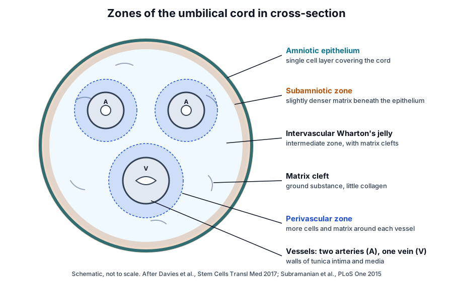
- Subamniotic zone: Wharton’s jelly just beneath the amniotic epithelium, with a slightly denser matrix and no clefts.[13]
- Intervascular, or intermediate, Wharton’s jelly: the central tissue between the vessels, crossed by clefts of ground substance.[13]
- Perivascular zone: a denser band of cells and matrix around each vessel, where CD146-positive cells accumulate during gestation.[13]
Karahuseyinoglu and colleagues found two cord stromal cell types in culture, distinguished by vimentin and cytokeratin filaments, and matched them to region-specific cells in cord sections. Both types differentiated to chondrogenic, adipogenic and osteogenic lineages.[14]
Perivascular cells give high colony frequency
Sarugaser and colleagues isolated human umbilical cord perivascular (HUCPV) cells and measured a CFU-F frequency of 1 in 333. Doubling time fell from 60 h at passage 0 to 20 h at passage 2, producing over 10¹⁰ cells within 30 days.[15]
Baksh and colleagues compared HUCPV cells with bone marrow stromal cells. The cord cells proliferated faster, differentiated to all three lineages, progressed through osteogenesis more rapidly and expressed more CD146.[16]
Wharton’s jelly gives the largest cell numbers
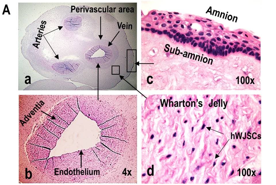
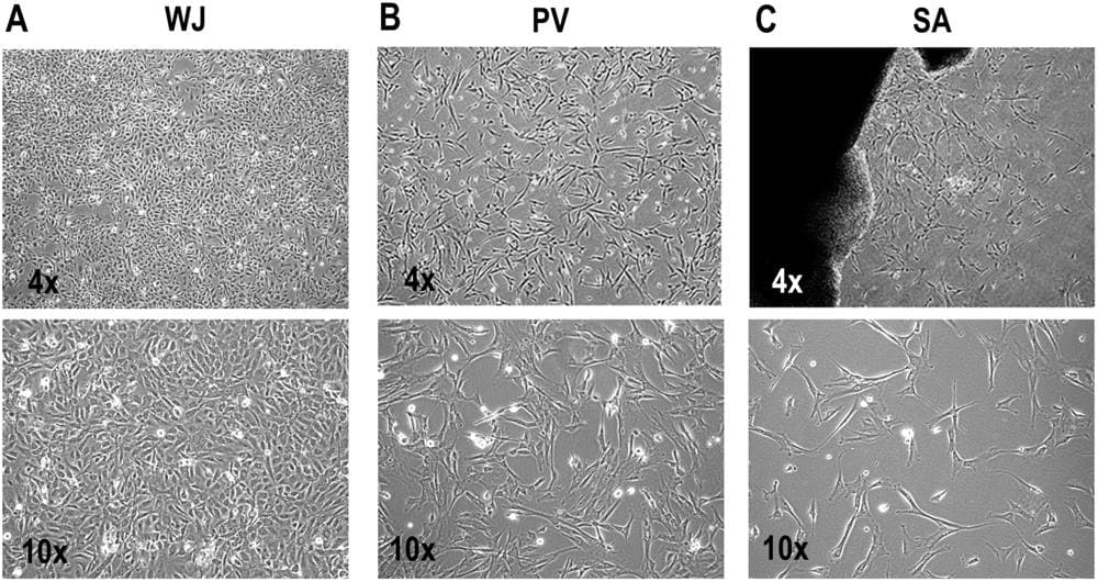
Subramanian and colleagues compared cells from the amnion, subamnion, perivascular zone, Wharton’s jelly and mixed cord of five cords (Fig. 2). Wharton’s jelly occupied the largest area and gave 4.61 × 10⁶ fresh cells per centimeter of cord without culture.[17]
Cells from all five compartments proliferated, expressed the typical MSC markers and formed adipocytes, chondrocytes and osteocytes. Cells from Wharton’s jelly showed a stellate epithelioid-like morphology in primary culture, while the other compartments gave short fibroblast-like cells (Fig. 3).[17]
Placental MSCs come from fetal and maternal layers
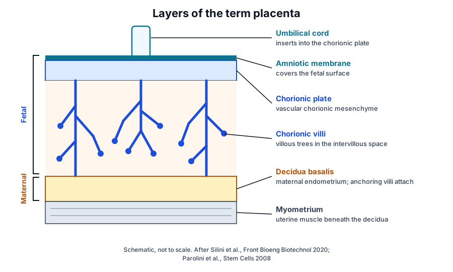
The term placenta has a fetal side, made of the amniotic membrane, the chorionic plate and the chorionic villi, and a maternal side, the decidua basalis (Fig. 4). Silini and colleagues describe the chorionic plate as vascularized chorionic mesenchyme from which stem villi branch into the intervillous space.[18]
The decidua basalis is a thin plate of maternal endometrial tissue that lies between the myometrium and the chorionic plate, and anchoring villi attach to it. Silini and colleagues proposed a shared nomenclature for each perinatal tissue and the cells isolated from it.[18]
The first international Workshop on Placenta Derived Stem Cells, summarized by Parolini and colleagues in 2008, called for each study to define the region of origin and the isolation method of its placental cells. Most published work had focused on stromal cells from various placental regions and on amniotic epithelial cells.[19]
Origin testing separates fetal from maternal cells
In ’t Anker and colleagues used HLA typing to show that amnion gives fetal MSCs and decidua basalis and parietalis give maternal MSCs. Phenotypes were comparable, and both placental MSC types expanded more than adult bone marrow MSCs.[20]
Wu and colleagues confirmed fetal origin for MSCs from the umbilical cord, amniotic membrane and chorionic plate, and maternal origin for decidua parietalis MSCs. Fetal MSCs expanded significantly more than maternal MSCs, and paracrine factor levels differed across sources.[21]
Sardesai and colleagues found maternal cells in cultures from every chorionic region tested, and maternal cells came to dominate standard-medium cultures started from fetal villi. Focal dissection of the cotyledon core with explant culture in endothelial growth medium selected fetal villous MSCs, which were smaller, more osteogenic and more proliferative.[22]
Region and donor both matter. Huang and colleagues reported that chorionic plate MSCs kept their fetal origin over serial passages and showed the strongest proliferation and migration among placental MSCs.[23] Tai and colleagues found stable surface markers across chorionic plate, amniotic and decidual MSCs from five placentas, alongside region- and donor-specific differences in growth and differentiation.[24]
Perinatal tissues are collected after delivery
The placenta and umbilical cord are delivered at birth, so collecting them requires no procedure on the donor. Parolini and colleagues describe placental tissue as readily available and easily procured without invasive procedures.[19]
Cord tissue also lends itself to a quick, standardized derivation. Subramanian and colleagues reported that Wharton’s jelly cells can be generated in large numbers with minimal culture.[17] Bone marrow and adipose tissue are obtained by aspiration and lipoaspiration.[8][11]
Dental pulp and synovium extend the range of MSC sources
Gronthos and colleagues isolated a clonogenic, rapidly proliferating population from adult human dental pulp in 2000. These dental pulp stem cells shared an immunophenotype with marrow stromal cells, formed sporadic densely calcified nodules in culture and generated a dentin-pulp-like complex after transplantation in mice.[26]
Mizuno and colleagues mapped synovium into surface (CD55+CD271−), stromal (CD55−CD271−) and perivascular (CD55−CD271+) regions. Synovial MSCs from the perivascular region showed the greatest proliferation and chondrogenic potential of the three.[27]
Comparing yield, growth, markers and secretome across MSC source tissues
Kern and colleagues isolated MSCs from every bone marrow and adipose sample and from 63% of umbilical cord blood samples. Cord blood MSCs could be cultured longest with the highest proliferation capacity, and bone marrow MSCs had the shortest culture period.[1]
| Source | How it is obtained | Colony frequency and yield | Growth in culture | Distinguishing markers | Differentiation tendencies |
|---|---|---|---|---|---|
| Bone marrow | Aspiration, usually iliac crest | Low; falls with donor age | Slowest of the main sources | Reference ISCT panel; CD106 positive | Reliable trilineage; strong osteogenic and adipogenic |
| Adipose tissue | Lipoaspiration, then SVF digestion | Highest colony frequency of the adult sources | Faster than bone marrow | CD36 positive, CD106 negative; SVF is CD34 positive | Trilineage, similar to bone marrow |
| Umbilical cord (Wharton’s jelly, perivascular) | Collected after delivery | High; perivascular CFU-F 1 in 333 | Fastest; doubling time near 20 h by passage 2 | Higher CD146; CD24 and CD108 in Wharton’s jelly | Rapid osteogenesis; adipocytes less mature |
| Placenta (chorionic plate, villi, decidua) | Collected after delivery | Varies by layer and method | Fetal layers expand more than maternal decidua | Similar core panel; origin confirmed by HLA or sex chromosome typing | Varies by region and donor |
| Dental pulp | Extracted teeth | Clonogenic population | Rapid | Immunophenotype similar to bone marrow | Calcified nodules; dentin-pulp-like tissue in vivo |
| Synovium | Synovial tissue | Perivascular region richest | Highest in the perivascular fraction | CD55 and CD271 mark synovial regions | Strong chondrogenic potential |
Proliferation and population doublings
Amable and colleagues found proliferation highest in Wharton’s jelly MSCs, followed by adipose MSCs, with bone marrow MSCs slowest.[28] Bourin and colleagues recommend the CFU-F assay for calculating the population doubling capacity of adipose-derived cells.[5]
Over ten passages, Kozlowska and colleagues found that bone marrow and adipose MSCs kept their differentiation capacity, while marker expression declined with passage in all sources.[29] Heo and colleagues, comparing bone marrow, adipose, placental and cord blood MSCs, found no significant differences in growth rate, colony-forming efficiency or immunophenotype.[30]
Surface markers beyond the ISCT panel
Amable and colleagues found surface marker differences between bone marrow, adipose and Wharton’s jelly MSCs only for CD54 and CD146.[28] Kozlowska and colleagues saw CD146 maintained in bone marrow MSCs during long-term culture and reduced in adipose MSCs.[29]
Subramanian and colleagues reported more CD24-positive and CD108-positive cells in Wharton’s jelly than in other cord compartments, markers they used to discriminate MSCs from other mesenchymal cells.[17]
Differentiation tendencies
Source affects lineage preference. Kern and colleagues saw no adipogenic differentiation in cord blood MSCs, and Karahuseyinoglu and colleagues found cord stromal adipocytes immature compared with marrow MSCs.[1][14] Heo and colleagues linked DLX5 expression to osteogenic potential across sources.[30]
Secretome differences
Amable and colleagues measured secreted proteins by Luminex and ELISA. Wharton’s jelly MSCs secreted more chemokines, inflammatory proteins and growth factors, and adipose MSCs showed a stronger pro-angiogenic profile with more extracellular matrix components and metalloproteinases.[28]
Hsieh and colleagues found that Wharton’s jelly MSCs expressed more genes for secreted factors linked to angiogenesis and neurogenesis than bone marrow MSCs.[31] In T cell proliferation co-culture, Heo and colleagues found the strongest inhibition with bone marrow MSCs, associated with high IL10 and TGFB1.[30]
Within the placenta, chorionic plate MSCs inhibited phytohemagglutinin-stimulated mononuclear cell proliferation in the study by Tai and colleagues.[24] Mattar and Bieback reviewed how bone marrow, adipose and birth-associated MSCs compare in their effects on T cell subsets, monocytes, macrophages and dendritic cells.[3]
Donor age effects
Choudhery and colleagues compared adipose MSCs from donors under 30, 35 to 50 and over 60 years. Cells from older donors showed senescent features, fewer population doublings and lower differentiation potential.[32]
In bone marrow, MSC number per milliliter of aspirate declined with age, and Ganguly and colleagues concluded that chronological age alone is an unreliable predictor of MSC number or potency.[10] Perinatal tissues come from a single developmental age, which Hass and colleagues discuss as one feature of neonatal MSC sources.[2]
Where to go from here
The ISCT minimal criteria reference explains the surface marker panel that every source on this page is measured against, and the stem versus stromal cells reference traces the naming. The MSC secretome reference describes conditioned medium and vesicle fractions, and the immunobiology reference covers HLA, IDO and PGE2.
The perinatal tissue donor eligibility reference explains how cord and placenta donors qualify, and the surface receptor chemistry and exosome references continue from cells to vesicles. Verified practices can request lot documentation, and our team is glad to discuss the methods described on this page.
Referencias comentadas
Numeradas en el orden en que se citan en el texto. Cada fuente lleva una nota sobre lo que respalda en esta página; los artículos revisados por pares se listan con revista, volumen, número y páginas, y las fuentes primarias son regulaciones, documentos de agencias y bases de datos de referencia.
- 1.Peer-reviewedKern S, Eichler H, Stoeve J, et al. Comparative analysis of mesenchymal stem cells from bone marrow, umbilical cord blood, or adipose tissue. Stem Cells. 2006;24(5):1294-1301. https://pubmed.ncbi.nlm.nih.gov/16410387/Annotation: Side-by-side comparison of bone marrow, cord blood and adipose MSCs. Cited for isolation success, colony frequency highest in adipose tissue, culture longevity and the absent adipogenesis of cord blood MSCs.
- 2.Peer-reviewedHass R, Kasper C, Böhm S, et al. Different populations and sources of human mesenchymal stem cells (MSC): A comparison of adult and neonatal tissue-derived MSC. Cell Communication and Signaling. 2011;9:12. https://pubmed.ncbi.nlm.nih.gov/21569606/Annotation: Review comparing adult and neonatal MSC sources. Cited for shared and differing properties, perivascular niches and neonatal sources.
- 3.Peer-reviewedMattar P, Bieback K. Comparing the Immunomodulatory Properties of Bone Marrow, Adipose Tissue, and Birth-Associated Tissue Mesenchymal Stromal Cells. Frontiers in Immunology. 2015;6:560. https://pubmed.ncbi.nlm.nih.gov/26579133/Annotation: Review of direct source comparisons. Cited for the small number of head-to-head studies and the immune cell types compared.
- 4.Peer-reviewedFriedenstein AJ, Chailakhjan RK, Lalykina KS. The development of fibroblast colonies in monolayer cultures of guinea-pig bone marrow and spleen cells. Cell and Tissue Kinetics. 1970;3(4):393-403. https://pubmed.ncbi.nlm.nih.gov/5523063/Annotation: Founding description of fibroblast colonies from guinea pig marrow monolayer cultures. Cited as the origin of the CFU-F assay.
- 5.Peer-reviewedBourin P, Bunnell BA, Casteilla L, et al. Stromal cells from the adipose tissue-derived stromal vascular fraction and culture expanded adipose tissue-derived stromal/stem cells: a joint statement of the International Federation for Adipose Therapeutics and Science (IFATS) and the International Society for Cellular Therapy (ISCT). Cytotherapy. 2013;15(6):641-648. https://pubmed.ncbi.nlm.nih.gov/23570660/Annotation: IFATS and ISCT definitions for adipose SVF and cultured cells. Cited for SVF markers, CD36 and CD106 as distinguishing markers, and the CFU-F assay.
- 6.Peer-reviewedDominici M, Le Blanc K, Mueller I, et al. Minimal criteria for defining multipotent mesenchymal stromal cells. The International Society for Cellular Therapy position statement. Cytotherapy. 2006;8(4):315-317. https://pubmed.ncbi.nlm.nih.gov/16923606/Annotation: ISCT minimal criteria. Cited for plastic adherence, the CD105, CD73 and CD90 panel, the negative markers and trilineage differentiation.
- 7.DatabaseNCBI Gene 4907, NT5E 5′-nucleotidase ecto (CD73) (Homo sapiens). Accessed 20 October 2026. https://www.ncbi.nlm.nih.gov/gene/4907
- 8.Peer-reviewedPittenger MF, Mackay AM, Beck SC, et al. Multilineage potential of adult human mesenchymal stem cells. Science. 1999;284(5411):143-147. https://pubmed.ncbi.nlm.nih.gov/10102814/Annotation: Isolation of human MSCs from marrow aspirates. Cited for the stable monolayer phenotype and clonal trilineage potential.
- 9.Peer-reviewedBaxter MA, Wynn RF, Jowitt SN, et al. Study of telomere length reveals rapid aging of human marrow stromal cells following in vitro expansion. Stem Cells. 2004;22(5):675-682. https://pubmed.ncbi.nlm.nih.gov/15342932/Annotation: Telomere measurement during marrow stromal cell expansion. Cited for the low marrow frequency of MSCs and the share of replicative lifespan used by expansion.
- 10.Peer-reviewedGanguly P, El-Jawhari JJ, Burska AN, et al. The Analysis of In Vivo Aging in Human Bone Marrow Mesenchymal Stromal Cells Using Colony-Forming Unit-Fibroblast Assay and the CD45(low)CD271(+) Phenotype. Stem Cells International. 2019;2019:5197983. https://pubmed.ncbi.nlm.nih.gov/31467563/Annotation: Marrow aspirates from 67 healthy donors aged 19 to 89. Cited for the age-related fall in MSCs per milliliter and for wide donor-to-donor variation.
- 11.Peer-reviewedZuk PA, Zhu M, Ashjian P, et al. Human adipose tissue is a source of multipotent stem cells. Molecular Biology of the Cell. 2002;13(12):4279-4295. https://pubmed.ncbi.nlm.nih.gov/12475952/Annotation: Processed lipoaspirate cells and clones from human liposuction tissue. Cited for multilineage differentiation and a CD marker profile similar to, yet distinct from, marrow MSCs.
- 12.Peer-reviewedAust L, Devlin B, Foster SJ, et al. Yield of human adipose-derived adult stem cells from liposuction aspirates. Cytotherapy. 2004;6(1):7-14. https://pubmed.ncbi.nlm.nih.gov/14985162/Annotation: Cell yield from lipoaspirate across 18 donors. Cited for the mean of 404,000 cells per milliliter and its relation to body mass index and donor age.
- 13.Peer-reviewedDavies JE, Walker JT, Keating A. Concise Review: Wharton’s Jelly: The Rich, but Enigmatic, Source of Mesenchymal Stromal Cells. Stem Cells Translational Medicine. 2017;6(7):1620-1630. https://pubmed.ncbi.nlm.nih.gov/28488282/Annotation: Histological nomenclature for umbilical cord structures. Cited for the amniotic epithelium, vessels, subamniotic, intermediate and perivascular zones, clefts and CD146-positive cells.
- 14.Peer-reviewedKarahuseyinoglu S, Cinar O, Kilic E, et al. Biology of stem cells in human umbilical cord stroma: in situ and in vitro surveys. Stem Cells. 2007;25(2):319-331. https://pubmed.ncbi.nlm.nih.gov/17053211/Annotation: In situ and culture study of cord stromal cells. Cited for two filament-defined cell types with region-specific localization and for adipocytes less mature than those of marrow MSCs.
- 15.Peer-reviewedSarugaser R, Lickorish D, Baksh D, et al. Human umbilical cord perivascular (HUCPV) cells: a source of mesenchymal progenitors. Stem Cells. 2005;23(2):220-229. https://pubmed.ncbi.nlm.nih.gov/15671145/Annotation: Isolation of cord perivascular cells. Cited for the 1 in 333 CFU-F frequency, the fall in doubling time from 60 to 20 h and expansion beyond 10^10 cells.
- 16.Peer-reviewedBaksh D, Yao R, Tuan RS. Comparison of proliferative and multilineage differentiation potential of human mesenchymal stem cells derived from umbilical cord and bone marrow. Stem Cells. 2007;25(6):1384-1392. https://pubmed.ncbi.nlm.nih.gov/17332507/Annotation: Comparison of cord perivascular cells with marrow stromal cells. Cited for faster proliferation, more rapid osteogenesis and higher CD146 in the cord cells.
- 17.Peer-reviewedSubramanian A, Fong CY, Biswas A, et al. Comparative Characterization of Cells from the Various Compartments of the Human Umbilical Cord Shows that the Wharton’s Jelly Compartment Provides the Best Source of Clinically Utilizable Mesenchymal Stem Cells. PLoS One. 2015;10(6):e0127992. https://pubmed.ncbi.nlm.nih.gov/26061052/Annotation: Comparison of cells from five umbilical cord compartments. Cited for fresh cell yield per centimeter, morphology, CD24 and CD108, and quick derivation from Wharton’s jelly; source of Figs. 2 and 3.
- 18.Peer-reviewedSilini AR, Di Pietro R, Lang-Olip I, et al. Perinatal Derivatives: Where Do We Stand? A Roadmap of the Human Placenta and Consensus for Tissue and Cell Nomenclature. Frontiers in Bioengineering and Biotechnology. 2020;8:610544. https://pubmed.ncbi.nlm.nih.gov/33392174/Annotation: COST SPRINT consensus on perinatal tissue structure and nomenclature. Cited for the fetal and maternal layers, the chorionic plate and the decidua basalis.
- 19.Peer-reviewedParolini O, Alviano F, Bagnara GP, et al. Concise review: isolation and characterization of cells from human term placenta: outcome of the first international Workshop on Placenta Derived Stem Cells. Stem Cells. 2008;26(2):300-311. https://pubmed.ncbi.nlm.nih.gov/17975221/Annotation: Workshop consensus review on placenta-derived cells. Cited for defining region of origin and isolation method, and for placenta as readily available without invasive procedures.
- 20.Peer-reviewedIn ’t Anker PS, Scherjon SA, Kleijburg-van der Keur C, et al. Isolation of mesenchymal stem cells of fetal or maternal origin from human placenta. Stem Cells. 2004;22(7):1338-1345. https://pubmed.ncbi.nlm.nih.gov/15579651/Annotation: HLA typing of MSCs from amnion and decidua. Cited for fetal amnion and maternal decidual MSCs and their greater expansion than adult marrow MSCs.
- 21.Peer-reviewedWu M, Zhang R, Zou Q, et al. Comparison of the Biological Characteristics of Mesenchymal Stem Cells Derived from the Human Placenta and Umbilical Cord. Scientific Reports. 2018;8(1):5014. https://pubmed.ncbi.nlm.nih.gov/29568084/Annotation: Comparison of cord, amnion, chorionic plate and decidua parietalis MSCs. Cited for fetal and maternal origin by HLA typing, greater fetal expansion and differing paracrine factors.
- 22.Peer-reviewedSardesai VS, Shafiee A, Fisk NM, et al. Avoidance of Maternal Cell Contamination and Overgrowth in Isolating Fetal Chorionic Villi Mesenchymal Stem Cells from Human Term Placenta. Stem Cells Translational Medicine. 2017;6(4):1070-1084. https://pubmed.ncbi.nlm.nih.gov/28205414/Annotation: Fetal and maternal cell tracking in chorionic villus cultures. Cited for maternal cell predominance in standard media and the isolation method that selects fetal villous MSCs.
- 23.Peer-reviewedHuang Q, Yang Y, Luo C, et al. An efficient protocol to generate placental chorionic plate-derived mesenchymal stem cells with superior proliferative and immunomodulatory properties. Stem Cell Research & Therapy. 2019;10(1):301. https://pubmed.ncbi.nlm.nih.gov/31623677/Annotation: Isolation of MSCs from several placental regions. Cited for chorionic plate MSCs keeping fetal origin and showing the strongest proliferation and migration.
- 24.Peer-reviewedTai C, Wang L, Xie Y, et al. Analysis of Key Distinct Biological Characteristics of Human Placenta-Derived Mesenchymal Stromal Cells and Individual Heterogeneity Attributing to Donors. Cells, Tissues, Organs. 2021;210(1):45-57. https://pubmed.ncbi.nlm.nih.gov/33780947/Annotation: Chorionic plate, amniotic and decidual MSCs from five placentas. Cited for stable surface markers, region and donor differences, and mononuclear cell proliferation in co-culture.
- 25.Regulatory21 CFR Part 1271, Human Cells, Tissues, and Cellular and Tissue-Based Products (eCFR). Accessed 20 October 2026. https://www.ecfr.gov/current/title-21/chapter-I/subchapter-L/part-1271
- 26.Peer-reviewedGronthos S, Mankani M, Brahim J, et al. Postnatal human dental pulp stem cells (DPSCs) in vitro and in vivo. Proceedings of the National Academy of Sciences of the United States of America. 2000;97(25):13625-13630. https://pubmed.ncbi.nlm.nih.gov/11087820/Annotation: Isolation of dental pulp stem cells compared with marrow stromal cells. Cited for clonogenicity, shared immunophenotype, calcified nodules and the dentin-pulp-like complex formed in mice.
- 27.Peer-reviewedMizuno M, Katano H, Mabuchi Y, et al. Specific markers and properties of synovial mesenchymal stem cells in the surface, stromal, and perivascular regions. Stem Cell Research & Therapy. 2018;9(1):123. https://pubmed.ncbi.nlm.nih.gov/29720268/Annotation: Regional markers of synovial MSCs. Cited for CD55 and CD271 region markers and the proliferative and chondrogenic potential of perivascular cells.
- 28.Peer-reviewedAmable PR, Teixeira MV, Carias RB, et al. Protein synthesis and secretion in human mesenchymal cells derived from bone marrow, adipose tissue and Wharton’s jelly. Stem Cell Research & Therapy. 2014;5(2):53. https://pubmed.ncbi.nlm.nih.gov/24739658/Annotation: Comparison of bone marrow, adipose and Wharton’s jelly MSCs. Cited for proliferation ranking, CD54 and CD146 differences and the secreted protein profiles.
- 29.Peer-reviewedKozlowska U, Krawczenko A, Futoma K, et al. Similarities and differences between mesenchymal stem/progenitor cells derived from various human tissues. World Journal of Stem Cells. 2019;11(6):347-374. https://pubmed.ncbi.nlm.nih.gov/31293717/Annotation: Ten-passage culture of MSCs from four adult tissues. Cited for marker expression over passage, CD146 retention in bone marrow MSCs and maintained differentiation.
- 30.Peer-reviewedHeo JS, Choi Y, Kim HS, et al. Comparison of molecular profiles of human mesenchymal stem cells derived from bone marrow, umbilical cord blood, placenta and adipose tissue. International Journal of Molecular Medicine. 2016;37(1):115-125. https://pubmed.ncbi.nlm.nih.gov/26719857/Annotation: Four-source comparison of MSC gene expression and function. Cited for similar growth and immunophenotype, DLX5 and osteogenic potential, and T cell co-culture with IL10 and TGFB1.
- 31.Peer-reviewedHsieh JY, Wang HW, Chang SJ, et al. Mesenchymal stem cells from human umbilical cord express preferentially secreted factors related to neuroprotection, neurogenesis, and angiogenesis. PLoS One. 2013;8(8):e72604. https://pubmed.ncbi.nlm.nih.gov/23991127/Annotation: Secretome comparison of Wharton’s jelly and bone marrow MSCs. Cited for more secreted factor genes linked to angiogenesis and neurogenesis in Wharton’s jelly MSCs.
- 32.Peer-reviewedChoudhery MS, Badowski M, Muise A, et al. Donor age negatively impacts adipose tissue-derived mesenchymal stem cell expansion and differentiation. Journal of Translational Medicine. 2014;12:8. https://pubmed.ncbi.nlm.nih.gov/24397850/Annotation: Adipose MSCs from three donor age groups. Cited for senescent features, population doublings and differentiation potential in cells from older donors.
Cite this page
ExaVeyra Sciences Editorial Team. MSC Sources: Bone Marrow, Adipose, Umbilical Cord and Placenta. ExaVeyra Sciences; 20 October 2026. Available from: https://exaveyra.com/science/stem-cells/msc-source-tissues. Accessed [date accessed].