Esta referencia está disponible actualmente en inglés. La versión en español está en preparación.
Mesenchymal Stem Cells vs Mesenchymal Stromal Cells
Por Benn Bluestein-Veyra, M.Sc. en Química Orgánica · Actualizado el 20 de octubre de 2026
ExaVeyra Sciences es un proveedor B2B con sede en Miami que suministra exosomas para uso estético tópico a clínicas con licencia, y remite a los prescriptores con licencia a una farmacia de preparados compuestos 503A que prepara formulaciones según prescripciones individuales.
| Pregunta | Respuesta |
|---|---|
| Qué cubre esta página | Why the ISCT distinguishes mesenchymal stem cells from mesenchymal stromal cells, what each term requires, and how to read MSC in the literature. |
| Quién puede hacer pedidos | Clínicas y profesionales con licencia profesional activa y NPI verificados por ExaVeyra. Los pedidos de exosomas requieren además un reconocimiento firmado de uso profesional. |
| Qué se envía y cómo | El material de exosomas para uso estético tópico, caracterizado por lote, se envía en cadena de frío por exprés nocturno. La entrega puede tardar 1 a 3 días hábiles. Se emite un certificado de análisis para cada lote y se entrega a solicitud. |
Mesenchymal stem cells vs mesenchymal stromal cells is a question of evidence. The ISCT uses "mesenchymal stromal cells" for plastic-adherent populations from bone marrow and other tissues, and reserves "mesenchymal stem cells" for cells that meet specified stem cell criteria.[1] The acronym MSC covers both, so each paper has to say which one it means.[2]
This reference traces the term from Friedenstein's colony-forming cells to the ISCT statements of 2005 and 2019, sets out what a stem cell claim requires, and explains how to read "MSC" in a methods section. Bianco, Robey and Simmons framed the same questions around the experimental origin of the concept and the assays needed to test stemness.[3]
Tracing the term from colony-forming cells to mesenchymal stem cells
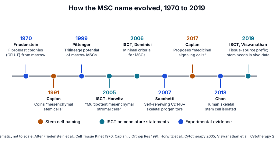
The story begins in bone marrow transplantation experiments. Friedenstein and colleagues analyzed the marrow precursor cells for osteogenic and hematopoietic tissues in heterotopic grafts, placed away from their native site.[4] In 1970 they described the development of fibroblast colonies in monolayer cultures of guinea pig bone marrow and spleen cells (Fig. 1).[5]
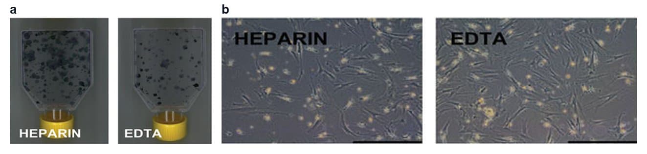
Each colony grows from a single precursor called the colony-forming unit-fibroblast, or CFU-F (Fig. 2).[6] Kuznetsov and colleagues expanded single-colony strains of human marrow stromal fibroblasts and transplanted them into mice: 20 of 34 strains from four donors formed bone, and every multicolony strain did.[6]
Arnold Caplan gave these cells their best-known name in 1991. He described bone and cartilage formation in the embryo, and turnover in the adult, as the work of the progeny of a small number of cells he called mesenchymal stem cells.[7] Their progeny commit to lineage pathways with discrete steps that end in cells making a specific tissue, such as cartilage or bone.[7]
Pittenger and colleagues then isolated human mesenchymal stem cells from marrow aspirates of volunteer donors. The cells kept a stable phenotype in monolayer culture, could be induced into the adipocytic, chondrocytic or osteocytic lineages, and individual cells expanded into colonies retained this multilineage potential.[8]
Understanding the 2005 ISCT clarification of MSC nomenclature
By 2005 the plastic-adherent cells from marrow and other sources were widely known as mesenchymal stem cells. Horwitz and colleagues judged that the biologic properties of the unfractionated population fell short of the accepted criteria for stem cell activity, while recognizing that a bona fide MSC exists.[1]
Their ISCT position statement proposed two names. The fibroblast-like, plastic-adherent cells, from any tissue, became multipotent mesenchymal stromal cells; mesenchymal stem cells became the name for cells that meet specified stem cell criteria.[1] The shared acronym MSC was kept, which places the duty to state the correct designation on each author.[1]
The 2006 minimal criteria describe the stromal population
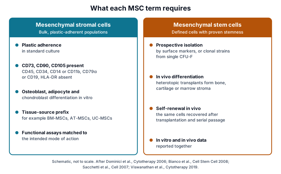
A year later, Dominici and colleagues set out three minimal criteria for human MSCs. The cells adhere to plastic in standard culture; express CD105, CD73 and CD90 while lacking CD45, CD34, CD14 or CD11b, CD79α or CD19 and HLA-DR; and differentiate to osteoblasts, adipocytes and chondroblasts in vitro.[9]
Every one of these criteria is measured on a cultured population in vitro. The authors expected the criteria to change as knowledge grew and presented them as a shared standard to make data comparable between laboratories.[9] Nombela-Arrieta and colleagues later described MSCs as operationally defined by in vitro differentiation after expansion.[10]
The 2017 proposal to rename MSCs
In 2017 Caplan, who had coined the name, asked the field to change it. He recalled that MSCs were named for cells from marrow and periosteum that could be expanded in culture while keeping the capacity to form mesodermal phenotypes, and that this in vitro capacity became the identifying assay.[11]
Caplan proposed the name "medicinal signaling cells" to place the emphasis on the factors the cells secrete, keeping the acronym MSC.[11] He presented the new name as a more accurate reflection of what the cells do.[11]
Applying the 2019 ISCT position on stem and stromal claims
The ISCT Mesenchymal Stromal Cell committee, writing with Viswanathan as first author, kept the acronym MSCs and attached three recommendations to it (Fig. 3).[2] Each recommendation tells a reader what a given use of the term is expected to carry.
- Supplement MSCs with the tissue-source origin of the cells, which highlights tissue-specific properties.[2]
- Read MSCs as mesenchymal stromal cells unless rigorous evidence for stemness exists, supported by both in vitro and in vivo data.[2]
- Pair MSCs with a robust matrix of functional assays, chosen for the intended mode of action and defined case by case.[2]
In practice, a tissue prefix, for example bone marrow, adipose tissue or umbilical cord, travels with every MSC preparation under this scheme.[2] The word "stem" becomes a claim with a defined burden of proof, met by data from living tissue as well as from the culture dish.[2][3]
Defining stem cell evidence through skeletal stem cell studies
Skeletal biology supplies the clearest examples of what stemness evidence looks like. Bianco and Robey describe skeletal stem cells in postnatal marrow that give rise to cartilage, bone, hematopoiesis-supportive stroma and marrow adipocytes in defined in vivo assays, together forming one bone and bone marrow organ.[12]
CD146-positive progenitors rebuild their own niche
Sacchetti and colleagues showed in 2007 that CD146-expressing subendothelial cells of human marrow stroma transfer the hematopoietic microenvironment to heterotopic sites upon transplantation. The grafts established identical subendothelial cells within a miniature bone organ.[13]
The re-establishment of identical cells after transplantation is the in vivo evidence of self-renewal named in the study's title. These subendothelial cells on the sinusoidal wall were also major producers of angiopoietin-1, a molecule of the hematopoietic stem cell niche.[13]
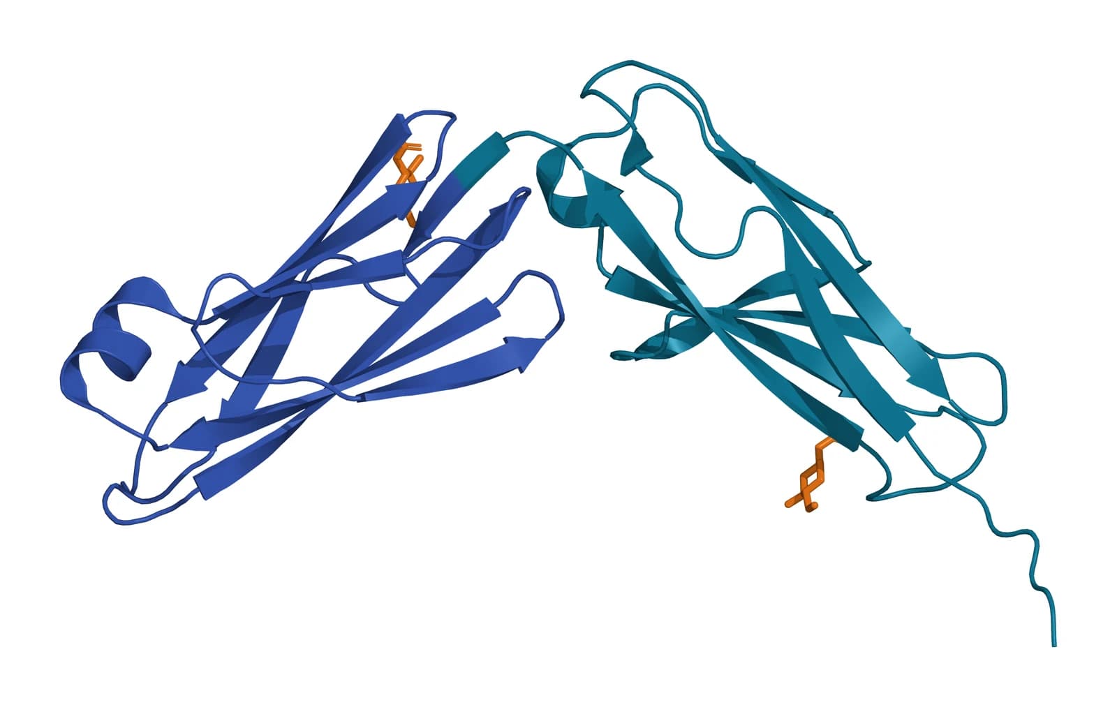
Crisan and colleagues also used CD146, with NG2 and PDGF-Rβ, to identify perivascular cells in multiple human organs (Fig. 4). Cultured perivascular cells showed osteogenic, chondrogenic and adipogenic potential at the clonal level and expressed MSC markers.[14]
Clonal strains and serial transplantation test potency
Sworder and colleagues transplanted 24 human single colony-derived strains: 20.8% formed fibrous tissue, 66.7% formed bone, and 12.5% formed a complete bone and marrow organ.[15] The multipotent strains showed over-representation of genes for skeletogenesis, hematopoiesis and extracellular matrix.[15]
Méndez-Ferrer and colleagues found that nestin-positive cells in mouse marrow contain all the marrow CFU-F activity. Grown as non-adherent "mesenspheres," these cells self-renewed and expanded in serial transplantations.[16]
Mabuchi and colleagues found that selecting human MSCs on three surface markers, LNGFR, THY-1 and VCAM-1, enriched clonogenic cells to about one in three isolated cells.[17] Clonal analysis of LNGFR+THY-1+ cells revealed heterogeneity among the clones, and rapidly expanding clones showed robust multilineage differentiation and self-renewal potency.[17]
Lineage tracing identifies skeletal stem cells in vivo
Zhou and colleagues found that leptin receptor marks about 0.3% of mouse marrow cells, a population accounting for 94% of marrow CFU-F. Fate mapping showed these cells arise after birth and give rise to most of the bone and adipocytes formed in adult marrow.[18]
Worthley and colleagues identified gremlin 1 as a marker of osteochondroreticular stem cells, concentrated in the metaphysis of long bones. These cells self-renew and generate osteoblasts, chondrocytes and reticular marrow stromal cells.[19]
Chan and colleagues mapped bone, cartilage and stromal development from highly pure mouse skeletal stem cells to their downstream progenitors.[20] In 2018 the same group isolated a self-renewing, multipotent human skeletal stem cell in fetal and adult bone, whose stroma maintained human hematopoietic stem cells in serum-free culture.[21]
Weighing the critiques of Bianco and colleagues
Paolo Bianco wrote the most detailed analysis of the two meanings. He identified one view of MSCs as the self-renewing, multipotent stem cells of the skeleton, coinciding with a specific marrow perivascular cell.[22] The second view sees MSCs as ubiquitous in connective tissues, defined by in vitro characteristics and by their capacity to modulate host tissue function.[22]
With an international group of authors, Bianco noted that more than three decades had passed since the concept that several connective tissues could arise from one progenitor in postnatal marrow. They listed open questions about the nature, identity, function, isolation and experimental handling of MSCs.[23]
Sacchetti and colleagues tested the ubiquity view directly in 2016. Human cell populations from different anatomical sources, all regarded as MSCs by in vitro criteria, differed widely in their transcriptomes and in vivo differentiation potential.[24]
The same populations shared the capacity to guide the assembly of functional microvessels in vivo, whatever their source. Cord blood-derived stromal cells formed cartilage spontaneously in vivo, and the authors proposed that mesoderm derivatives include distinct classes of tissue-specific committed progenitors.[24]
Reading MSC in a research paper
Library indexing groups the two names together. The National Library of Medicine descriptor for mesenchymal stem cells notes that they are also called multipotent stromal cells or mesenchymal stromal cells, and lists bone marrow, adipose tissue and Wharton's jelly among their tissues.[25]
A literature search therefore returns both meanings together, and the methods section decides which one a paper uses. Three questions resolve it: which meaning the authors intend, which tissue the cells came from, and which assays support the claims.[1][2]
| Question | What to look for in the methods | What it tells you |
|---|---|---|
| Which meaning? | The expanded term at first use, or a statement of ISCT nomenclature | Whether the paper claims stromal or stem cell identity |
| Which tissue? | A tissue prefix such as bone marrow, adipose tissue or umbilical cord, and the donor age | Which tissue-specific properties to expect |
| Which population? | Plastic adherence alone, prospective sorting, or clonal strains | Whether results describe a bulk population or defined cells |
| Which assays? | Marker panel, trilineage staining, CFU-F counts, transplantation, serial passage | Whether stemness rests on in vitro data, in vivo data or both |
Two checks follow from the history above. Trilineage staining in culture is evidence for the ISCT stromal criteria, and in vivo differentiation with self-renewal is evidence for stemness.[3][9] The tissue of origin shapes the result, since populations from different sources differ in transcriptome and in vivo potential.[24]
Clonal studies add a third check. Kuznetsov and colleagues found that individual human CFU-F differ in osteogenic capacity, and later clonal work confirmed a range of potencies within one marrow.[6][15] A result from a bulk culture therefore reflects a mixed population of clones.[17]
Where to go from here
The ISCT minimal criteria reference covers the marker panel and trilineage assays in detail, and the source tissues reference compares bone marrow, adipose and perinatal MSCs. The secretome reference and the immunobiology reference follow the signaling activity Caplan placed at the center of the medicinal signaling cell proposal.
The perinatal tissue donor eligibility reference explains how birth tissues are screened under federal rules. The surface receptor chemistry reference and the exosome library describe the membrane proteins and vesicles studied alongside stromal cells, and the quality references explain how vesicle preparations are tested.
Verified practices can request lot documentation, and our team is glad to discuss the methods described on this page.
Referencias comentadas
Numeradas en el orden en que se citan en el texto. Cada fuente lleva una nota sobre lo que respalda en esta página; los artículos revisados por pares se listan con revista, volumen, número y páginas, y las fuentes primarias son regulaciones, documentos de agencias y bases de datos de referencia.
- 1.Peer-reviewedHorwitz EM, Le Blanc K, Dominici M, et al. Clarification of the nomenclature for MSC: The International Society for Cellular Therapy position statement. Cytotherapy. 2005;7(5):393-395. https://pubmed.ncbi.nlm.nih.gov/16236628/Annotation: ISCT position statement on MSC nomenclature. Cited for the terms multipotent mesenchymal stromal cells and mesenchymal stem cells, and for the shared acronym MSC.
- 2.Peer-reviewedViswanathan S, Shi Y, Galipeau J, et al. Mesenchymal stem versus stromal cells: International Society for Cell & Gene Therapy (ISCT®) Mesenchymal Stromal Cell committee position statement on nomenclature. Cytotherapy. 2019;21(10):1019-1024. https://pubmed.ncbi.nlm.nih.gov/31526643/Annotation: ISCT MSC committee position on nomenclature. Cited for the tissue-source prefix, the in vitro and in vivo evidence needed for a stemness claim, and the matrix of functional assays.
- 3.Peer-reviewedBianco P, Robey PG, Simmons PJ. Mesenchymal stem cells: revisiting history, concepts, and assays. Cell Stem Cell. 2008;2(4):313-319. https://pubmed.ncbi.nlm.nih.gov/18397751/Annotation: Commentary on the experimental origin of the MSC concept. Cited for the framework of potency and self-renewal assays needed to assess stemness.
- 4.Peer-reviewedFriedenstein AJ, Petrakova KV, Kurolesova AI, et al. Heterotopic of bone marrow. Analysis of precursor cells for osteogenic and hematopoietic tissues. Transplantation. 1968;6(2):230-247. https://pubmed.ncbi.nlm.nih.gov/5654088/Annotation: Early heterotopic marrow transplantation study, indexed without an abstract. Cited only for its stated subject: the analysis of marrow precursor cells for osteogenic and hematopoietic tissues.
- 5.Peer-reviewedFriedenstein AJ, Chailakhjan RK, Lalykina KS. The development of fibroblast colonies in monolayer cultures of guinea-pig bone marrow and spleen cells. Cell and Tissue Kinetics. 1970;3(4):393-403. https://pubmed.ncbi.nlm.nih.gov/5523063/Annotation: The founding description of fibroblast colonies from marrow, indexed without an abstract. Cited for its stated subject, fibroblast colony development in guinea pig marrow and spleen cultures.
- 6.Peer-reviewedKuznetsov SA, Krebsbach PH, Satomura K, et al. Single-colony derived strains of human marrow stromal fibroblasts form bone after transplantation in vivo. Journal of Bone and Mineral Research. 1997;12(9):1335-1347. https://pubmed.ncbi.nlm.nih.gov/9286749/Annotation: Transplantation of single-colony human marrow stromal strains into mice. Cited for the CFU-F as the colony precursor and for bone formation by 20 of 34 single-colony strains.
- 7.Peer-reviewedCaplan AI. Mesenchymal stem cells. Journal of Orthopaedic Research. 1991;9(5):641-650. https://pubmed.ncbi.nlm.nih.gov/1870029/Annotation: The paper that named mesenchymal stem cells. Cited for the lineage model of bone and cartilage formation from the progeny of a small number of such cells.
- 8.Peer-reviewedPittenger MF, Mackay AM, Beck SC, et al. Multilineage potential of adult human mesenchymal stem cells. Science. 1999;284(5411):143-147. https://pubmed.ncbi.nlm.nih.gov/10102814/Annotation: Isolation of human marrow cells from volunteer donor aspirates. Cited for adipocytic, chondrocytic and osteocytic differentiation and for clonal retention of multilineage potential.
- 9.Peer-reviewedDominici M, Le Blanc K, Mueller I, et al. Minimal criteria for defining multipotent mesenchymal stromal cells. The International Society for Cellular Therapy position statement. Cytotherapy. 2006;8(4):315-317. https://pubmed.ncbi.nlm.nih.gov/16923606/Annotation: ISCT minimal criteria for human MSCs. Cited for plastic adherence, the CD105, CD73 and CD90 positive and hematopoietic marker negative panel, and trilineage differentiation in vitro.
- 10.Peer-reviewedNombela-Arrieta C, Ritz J, Silberstein LE. The elusive nature and function of mesenchymal stem cells. Nature Reviews Molecular Cell Biology. 2011;12(2):126-131. https://pubmed.ncbi.nlm.nih.gov/21253000/Annotation: Review of MSC identity in vivo. Cited for the operational definition of MSCs by in vitro differentiation into osteoblasts, adipocytes and chondrocytes after expansion.
- 11.Peer-reviewedCaplan AI. Mesenchymal Stem Cells: Time to Change the Name! Stem Cells Translational Medicine. 2017;6(6):1445-1451. https://pubmed.ncbi.nlm.nih.gov/28452204/Annotation: Perspective by the author of the MSC name. Cited for the history of the in vitro identifying assay and the proposed name medicinal signaling cells.
- 12.Peer-reviewedBianco P, Robey PG. Skeletal stem cells. Development. 2015;142(6):1023-1027. https://pubmed.ncbi.nlm.nih.gov/25758217/Annotation: Concise review of skeletal stem cell biology. Cited for the lineages these cells form in defined in vivo assays and the bone and bone marrow organ they compose.
- 13.Peer-reviewedSacchetti B, Funari A, Michienzi S, et al. Self-renewing osteoprogenitors in bone marrow sinusoids can organize a hematopoietic microenvironment. Cell. 2007;131(2):324-336. https://pubmed.ncbi.nlm.nih.gov/17956733/Annotation: Heterotopic transplantation of human CD146-expressing marrow stromal cells. Cited for transfer of the hematopoietic microenvironment, re-establishment of identical subendothelial cells and angiopoietin-1 production.
- 14.Peer-reviewedCrisan M, Yap S, Casteilla L, et al. A perivascular origin for mesenchymal stem cells in multiple human organs. Cell Stem Cell. 2008;3(3):301-313. https://pubmed.ncbi.nlm.nih.gov/18786417/Annotation: Prospective identification of human perivascular cells. Cited for the CD146, NG2 and PDGF-Rbeta markers and for clonal osteogenic, chondrogenic and adipogenic potential with MSC marker expression.
- 15.Peer-reviewedSworder BJ, Yoshizawa S, Mishra PJ, et al. Molecular profile of clonal strains of human skeletal stem/progenitor cells with different potencies. Stem Cell Research. 2015;14(3):297-306. https://pubmed.ncbi.nlm.nih.gov/25814434/Annotation: In vivo transplantation and microarray profiling of 24 human single colony-derived strains. Cited for the share forming fibrous tissue, bone or a bone and marrow organ, and the genes enriched in multipotent strains.
- 16.Peer-reviewedMéndez-Ferrer S, Michurina TV, Ferraro F, et al. Mesenchymal and haematopoietic stem cells form a unique bone marrow niche. Nature. 2010;466(7308):829-834. https://pubmed.ncbi.nlm.nih.gov/20703299/Annotation: Mouse study of nestin-positive marrow cells. Cited for their containing all marrow CFU-F activity and for mesenspheres that self-renew and expand in serial transplantations.
- 17.Peer-reviewedMabuchi Y, Morikawa S, Harada S, et al. LNGFR(+)THY-1(+)VCAM-1(hi+) cells reveal functionally distinct subpopulations in mesenchymal stem cells. Stem Cell Reports. 2013;1(2):152-165. https://pubmed.ncbi.nlm.nih.gov/24052950/Annotation: Prospective clonal isolation of human MSCs. Cited for the LNGFR, THY-1 and VCAM-1 sort, its clonal efficiency, and functional heterogeneity among clones.
- 18.Peer-reviewedZhou BO, Yue R, Murphy MM, et al. Leptin-receptor-expressing mesenchymal stromal cells represent the main source of bone formed by adult bone marrow. Cell Stem Cell. 2014;15(2):154-168. https://pubmed.ncbi.nlm.nih.gov/24953181/Annotation: Mouse fate-mapping study of leptin receptor-positive marrow cells. Cited for their frequency, their share of marrow CFU-F, and their postnatal origin of most adult marrow bone and adipocytes.
- 19.Peer-reviewedWorthley DL, Churchill M, Compton JT, et al. Gremlin 1 identifies a skeletal stem cell with bone, cartilage, and reticular stromal potential. Cell. 2015;160(1-2):269-284. https://pubmed.ncbi.nlm.nih.gov/25594183/Annotation: Mouse lineage-tracing study. Cited for gremlin 1 marking self-renewing osteochondroreticular stem cells in the metaphysis that generate osteoblasts, chondrocytes and reticular stromal cells.
- 20.Peer-reviewedChan CK, Seo EY, Chen JY, et al. Identification and specification of the mouse skeletal stem cell. Cell. 2015;160(1-2):285-298. https://pubmed.ncbi.nlm.nih.gov/25594184/Annotation: Isolation of highly pure mouse skeletal stem cells. Cited for mapping bone, cartilage and stromal development from these cells to their downstream progenitors.
- 21.Peer-reviewedChan CKF, Gulati GS, Sinha R, et al. Identification of the Human Skeletal Stem Cell. Cell. 2018;175(1):43-56.e21. https://pubmed.ncbi.nlm.nih.gov/30241615/Annotation: Isolation of a self-renewing, multipotent human skeletal stem cell. Cited for its presence in fetal and adult bone and for its stroma maintaining human hematopoietic stem cells in serum-free culture.
- 22.Peer-reviewedBianco P. "Mesenchymal" stem cells. Annual Review of Cell and Developmental Biology. 2014;30:677-704. https://pubmed.ncbi.nlm.nih.gov/25150008/Annotation: Review of the two descriptions of MSCs. Cited for the skeletal stem cell view and the ubiquitous connective tissue view and how each developed.
- 23.Peer-reviewedBianco P, Cao X, Frenette PS, et al. The meaning, the sense and the significance: translating the science of mesenchymal stem cells into medicine. Nature Medicine. 2013;19(1):35-42. https://pubmed.ncbi.nlm.nih.gov/23296015/Annotation: Perspective by an international author group. Cited for the history of the common-progenitor concept and the open questions on MSC nature, identity, isolation and handling.
- 24.Peer-reviewedSacchetti B, Funari A, Remoli C, et al. No Identical "Mesenchymal Stem Cells" at Different Times and Sites: Human Committed Progenitors of Distinct Origin and Differentiation Potential Are Incorporated as Adventitial Cells in Microvessels. Stem Cell Reports. 2016;6(6):897-913. https://pubmed.ncbi.nlm.nih.gov/27304917/Annotation: Comparison of human stromal populations from several anatomical sources by in vivo assays and transcriptomics. Cited for their distinct profiles and potentials, shared microvessel assembly, and in vivo cartilage formation by cord blood-derived cells.
- 25.DatabaseNational Library of Medicine, MeSH descriptor D059630, Mesenchymal Stem Cells. Accessed 20 October 2026. https://www.ncbi.nlm.nih.gov/mesh/68059630
Cite this page
ExaVeyra Sciences Editorial Team. Mesenchymal Stem Cells vs Mesenchymal Stromal Cells. ExaVeyra Sciences; 20 October 2026. Available from: https://exaveyra.com/science/stem-cells/mesenchymal-stem-vs-stromal-cells. Accessed [date accessed].
Fuentes de las figuras
- 26.PDB 6LYN, CD146 immunoglobulin-like domains 4 and 5. ExaVeyra Sciences, rendered in PyMOL from PDB 6LYN. Structure data: RCSB Protein Data Bank (rcsb.org), CC0. Publicación de la estructura: Chen X, Yan H, Liu D, et al. Structure basis for AA98 inhibition on the activation of endothelial cells mediated by CD146. iScience. 2021;24(5):102417.