Esta referencia está disponible actualmente en inglés. La versión en español está en preparación.
ISCT Minimal Criteria for Mesenchymal Stromal Cells (MSCs)
Por Benn Bluestein-Veyra, M.Sc. en Química Orgánica · Actualizado el 20 de octubre de 2026
ExaVeyra Sciences es un proveedor B2B con sede en Miami que suministra exosomas para uso estético tópico a clínicas con licencia, y remite a los prescriptores con licencia a una farmacia de preparados compuestos 503A que prepara formulaciones según prescripciones individuales.
| Pregunta | Respuesta |
|---|---|
| Qué cubre esta página | The three ISCT criteria that define MSCs: plastic adherence, the CD73, CD90 and CD105 surface panel, and trilineage differentiation, with the 2019 update. |
| Quién puede hacer pedidos | Clínicas y profesionales con licencia profesional activa y NPI verificados por ExaVeyra. Los pedidos de exosomas requieren además un reconocimiento firmado de uso profesional. |
| Qué se envía y cómo | El material de exosomas para uso estético tópico, caracterizado por lote, se envía en cadena de frío por exprés nocturno. La entrega puede tardar 1 a 3 días hábiles. Se emite un certificado de análisis para cada lote y se entrega a solicitud. |
The ISCT criteria for MSCs are three laboratory tests that define human multipotent mesenchymal stromal cells: adherence to plastic, a surface marker panel and differentiation to bone, fat and cartilage cells in vitro.[1] The International Society for Cellular Therapy (ISCT) published these minimal criteria in 2006.[1]
This reference explains each criterion, the molecules behind the marker panel and how a flow cytometry report is read. It then follows the later ISCT statements and the studies that refined the definition, which matters to readers because MSC papers report their cells in very different ways.[2]
The 2005 and 2006 ISCT statements gave MSC research a shared definition
The field began with Friedenstein and colleagues, who in 1970 described fibroblast colonies growing in monolayer cultures of guinea pig bone marrow and spleen cells.[3] In 1999 Pittenger and colleagues expanded cells from human marrow that could be induced along adipocytic, chondrocytic or osteocytic lineages, and single expanded cells kept this multilineage potential.[4]
In 2005 the ISCT addressed the name. Horwitz and colleagues proposed that fibroblast-like plastic-adherent cells from any tissue be called multipotent mesenchymal stromal cells, with "mesenchymal stem cells" reserved for cells that meet specified stem cell criteria.[5] The acronym MSC could serve both, provided authors state which they mean.[5]
A year later the Mesenchymal and Tissue Stem Cell Committee, writing with Dominici as first author, set out the minimal criteria. Laboratories were isolating, expanding and characterizing MSCs by different methods, and a shared standard would let investigators compare results and exchange data.[1]
The committee expected the criteria to require modification as new knowledge unfolded, and presented them as a minimal set.[1] Bianco and colleagues later described the ambiguities that remained about the nature, identity, function, isolation and experimental handling of MSCs, which keeps the criteria in use as a common floor for comparison.[7]
Plastic adherence under standard culture conditions is the first criterion
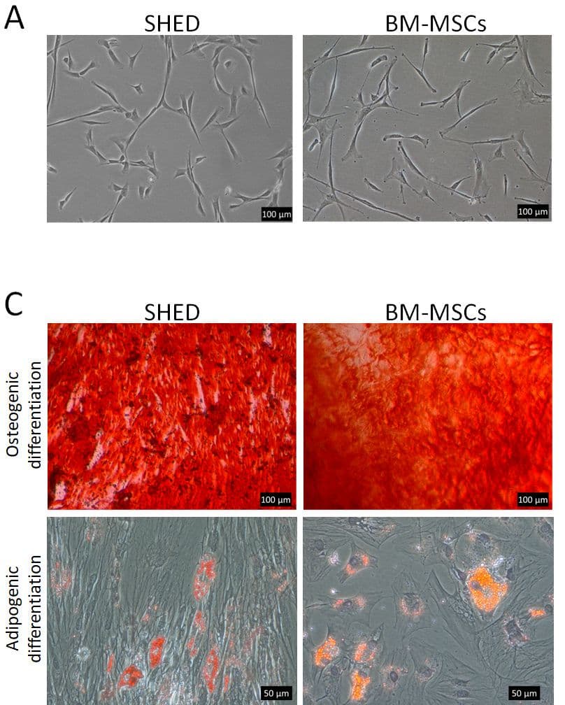
The first criterion asks that MSCs adhere to tissue culture plastic when kept in standard culture conditions.[1] Adherence is also how the cells are isolated: the ISCT nomenclature statement describes MSCs as the fibroblast-like, plastic-adherent cells recovered from bone marrow and other sources, which grow as spindle-shaped cells (Fig. 1).[5]
Friedenstein's fibroblast colonies are counted today in the colony-forming unit fibroblast (CFU-F) assay.[3] The joint IFATS and ISCT statement on adipose-derived cells recommends the CFU-F assay to estimate progenitor frequency and to calculate the population doubling capacity of expanded cells.[8]
Adherence selects a mixed population. Lv and colleagues note that plastic-adherent MSC cultures contain subpopulations with different levels of stemness, and that the search continues for one surface marker able to isolate cells meeting the minimal criteria from every tissue.[9]
Understanding the ISCT surface marker panel
The second criterion is a phenotype measured by flow cytometry. At least 95% of the population must express CD105, CD73 and CD90, and at most 2% may express CD45, CD34, CD14 or CD11b, CD79α or CD19, and HLA-DR (Fig. 2).[1][6]
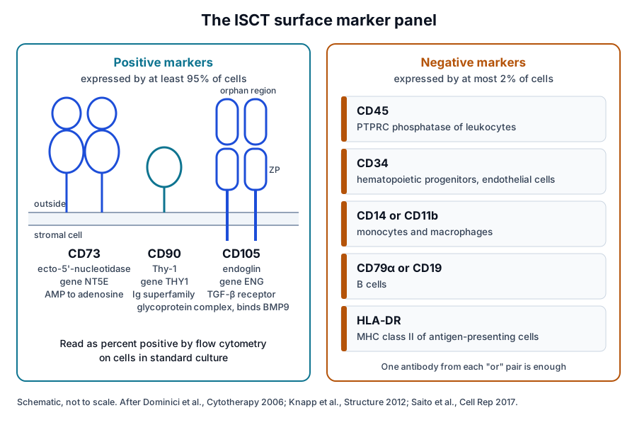
The negative markers detect the blood and immune cells most likely to remain in a stromal culture. Dominici and colleagues allowed one antibody from each of two pairs, CD14 or CD11b and CD79α or CD19, and noted that MSCs can express HLA-DR when stimulated, for example by interferon gamma.[1]
| Marker | Gene | ISCT requirement | Cells it usually marks |
|---|---|---|---|
| CD73 | NT5E | At least 95% positive | Stromal cells; a lymphocyte differentiation marker |
| CD90 | THY1 | At least 95% positive | Stromal, immune and neural cells |
| CD105 | ENG | At least 95% positive | Stromal and vascular endothelial cells |
| CD45 | PTPRC | At most 2% positive | Leukocytes |
| CD34 | CD34 | At most 2% positive | Hematopoietic progenitors, endothelial cells |
| CD14 or CD11b | CD14, ITGAM | At most 2% positive | Monocytes and macrophages |
| CD79α or CD19 | CD79A, CD19 | At most 2% positive | B cells |
| HLA-DR | HLA-DRA | At most 2% positive | Antigen-presenting cells |
The positive markers are stromal surface proteins
- CD73 is the ecto-5'-nucleotidase encoded by NT5E, a plasma membrane enzyme that converts extracellular nucleotides to membrane-permeable nucleosides.[10]
- CD90 is Thy-1, encoded by THY1, a cell surface glycoprotein of the immunoglobulin superfamily that takes part in cell adhesion and cell communication.[11]
- CD105 is endoglin, encoded by ENG, a homodimeric transmembrane glycoprotein that is a component of the transforming growth factor beta receptor complex.[12]
The negative markers identify blood and immune lineages
- CD45, encoded by PTPRC, is a receptor-type protein tyrosine phosphatase that regulates T and B cell antigen receptor signaling.[13]
- CD34 is a highly glycosylated single-pass membrane protein that may attach stem cells to the bone marrow matrix or to stromal cells.[14]
- CD14 is preferentially expressed on monocytes and macrophages, and CD11b, encoded by ITGAM, is the alpha M chain of the leukocyte integrin Mac-1.[15][16]
- CD79α is the Ig-alpha chain of the B cell antigen receptor, and CD19 expression is restricted to B lymphocytes.[17][18]
- HLA-DR is a class II major histocompatibility molecule expressed on antigen-presenting cells such as B lymphocytes, dendritic cells and monocytes.[19]
Crystal structures show CD73 and CD105 as an enzyme and a co-receptor
Knapp and colleagues solved the crystal structure of dimeric human CD73, the enzyme that hydrolyzes extracellular AMP to adenosine and sets extracellular adenosine levels (Fig. 3a).[20] They found a 114 degree switch between open and closed forms, and the C-terminal domains formed the dimer interface.[20]
Complexes with adenosine and the AMP analog AMPCP showed that control of this domain movement determines the enzyme's selectivity for monophosphate nucleotides.[20] CD73 is therefore an active enzyme at the cell surface as well as a marker.[10]
Saito and colleagues determined crystal structures of the human endoglin ectodomain and its complex with bone morphogenetic protein 9 (BMP9). The ligand binds a hydrophobic surface of the N-terminal orphan domain, which adopts a duplicated fold generated by circular permutation (Fig. 3b).[21]
The structure of the C-terminal zona pellucida module suggested how two endoglin copies embrace the BMP9 dimer, in a way compatible with ligand recognition by type I receptors.[21] Endoglin therefore sits in the bone morphogenetic protein branch of transforming growth factor beta signaling.[21]
CD90 also has a reported role within MSCs. Saalbach and Anderegg reviewed evidence that Thy-1 promotes osteogenic differentiation and inhibits adipogenic differentiation, which links a panel marker to the balance between two of the three ISCT lineages.[22]
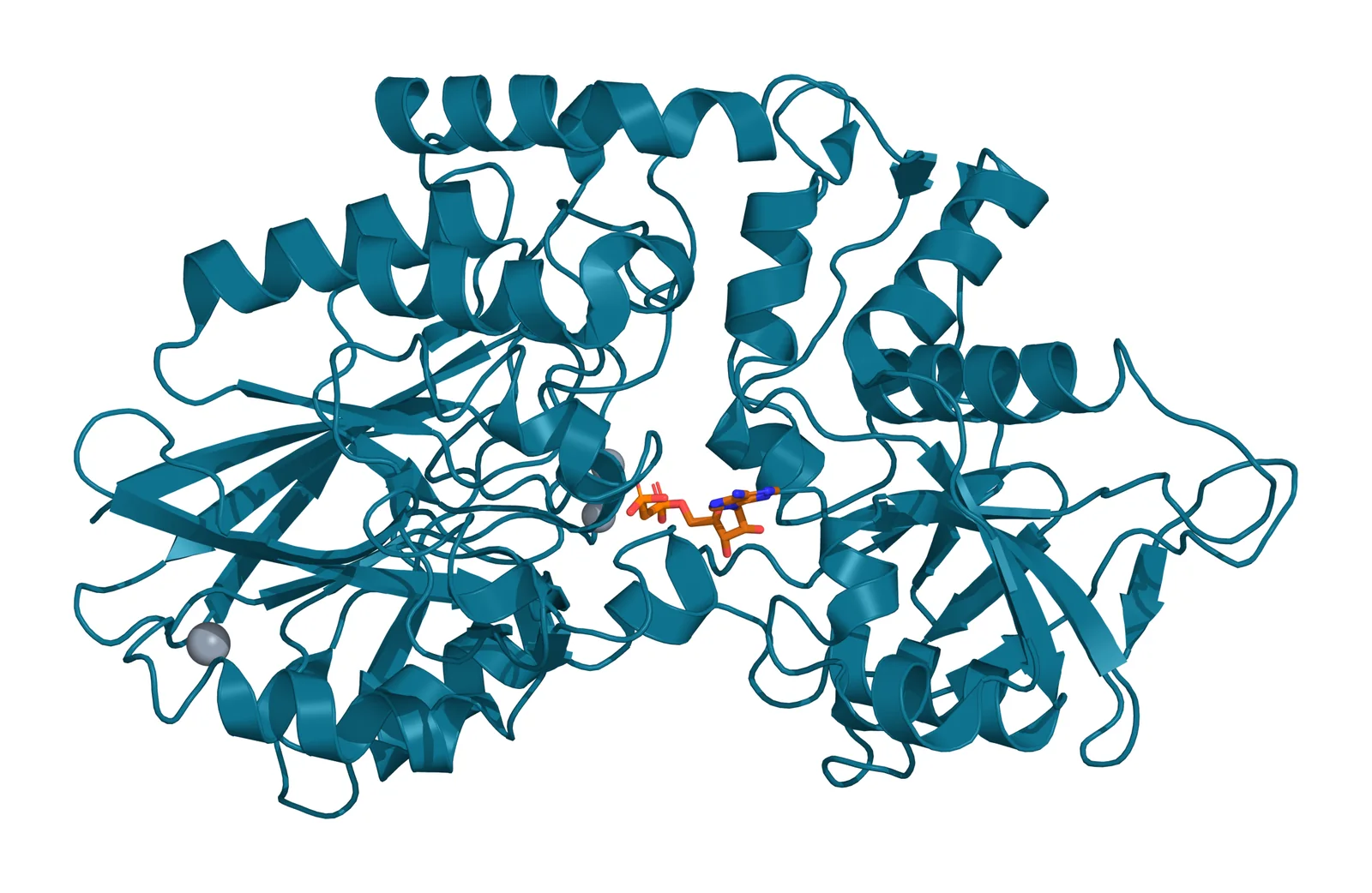
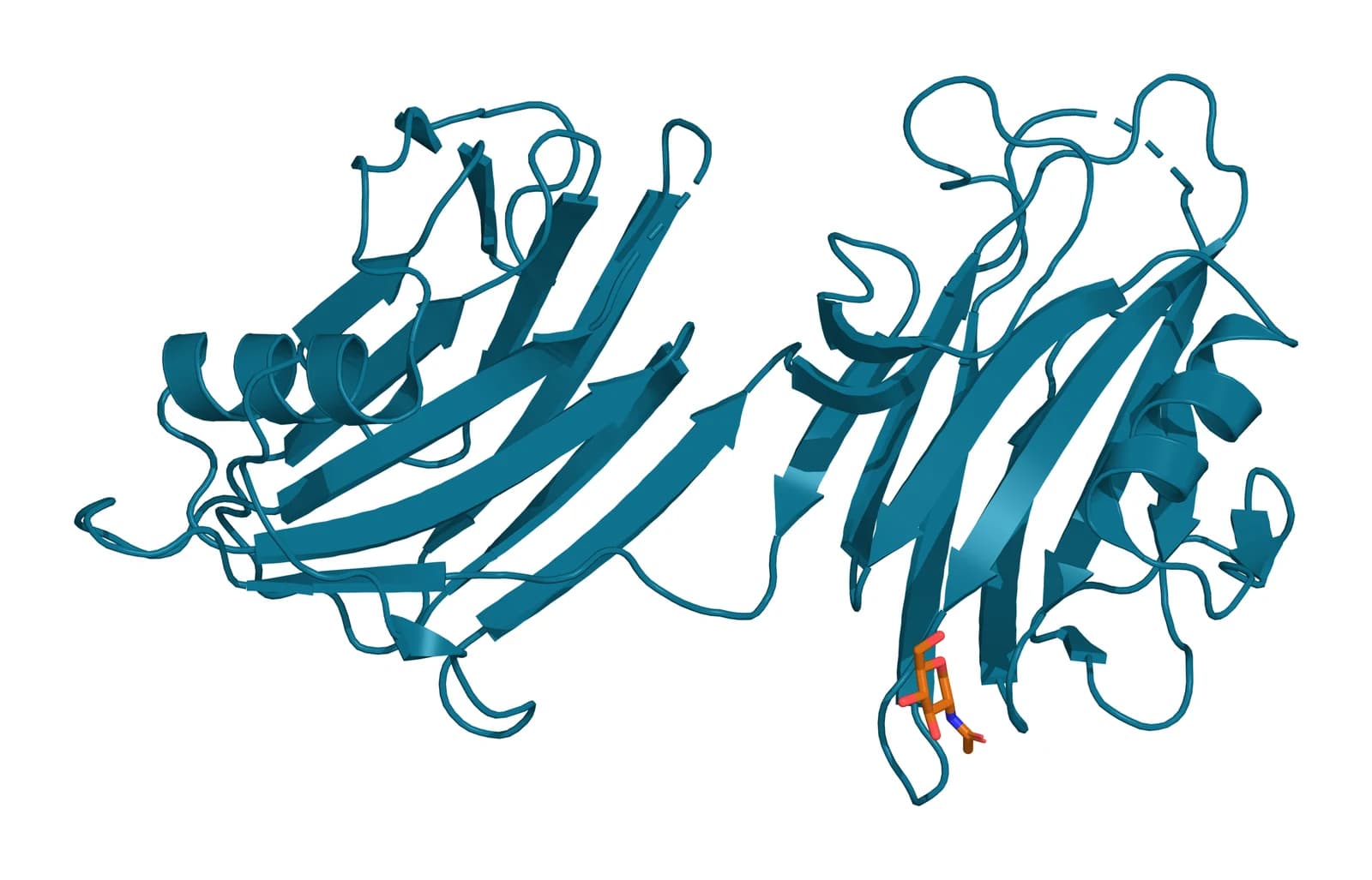
Fig. 3 | Human CD73 (ecto-5'-nucleotidase), closed form and Human endoglin (CD105) orphan region.
Flow cytometry reports show the panel as gated histograms
A flow cytometry report shows each marker as a histogram of cell count against fluorescence, with the stained sample overlaid on a control and a gate marking the positive region (Fig. 4). The percentage of cells inside the gate is the value laboratories compare with the ISCT thresholds.[1]
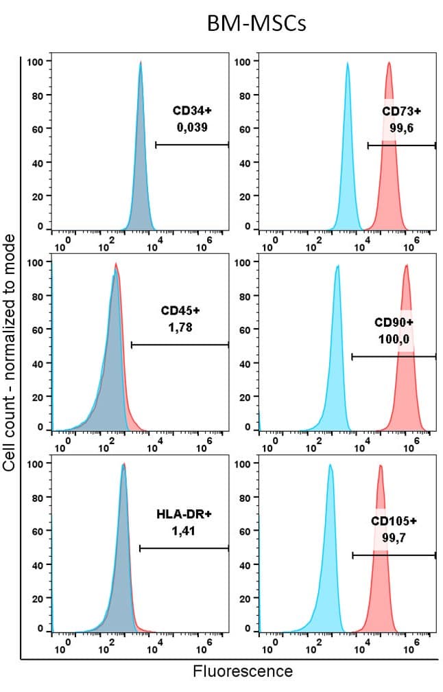
In the bone marrow MSC example shown, CD73, CD90 and CD105 read 99.6%, 100.0% and 99.7% positive, and CD34, CD45 and HLA-DR read 0.039%, 1.78% and 1.41% (Fig. 4). Each value falls inside the ISCT limits of at least 95% for positive markers and at most 2% for negative ones.[1]
What a complete phenotype report states
- The percentage of cells positive for each marker, set against the ISCT thresholds.[1]
- Values for each cell lot. Wilson and colleagues found individual lot values in 11 of 84 clinical trial reports, and averages across lots in 45.[23]
- Cell viability, which 57% of the trial reports in the same analysis included.[23]
- The tissue source and the culture medium, because both shape the surface phenotype.[24][25]
Fitzgerald and colleagues screened bone marrow MSCs grown in five media against a panel of 243 surface markers. Every preparation strongly expressed CD73, CD90 and CD105, and one third of the tested markers varied with the medium.[25]
Trilineage differentiation in vitro is the third criterion
The third criterion asks that MSCs differentiate to osteoblasts, adipocytes and chondroblasts in vitro (Fig. 5).[1] Dominici and colleagues named the readouts: Alizarin red or von Kossa staining for osteoblasts, Oil Red O for adipocytes, and Alcian blue or collagen type II immunostaining for chondroblasts.[1] Later reviews list the same histochemical stains: Oil Red O for adipogenesis, Alizarin red S and von Kossa for osteogenesis, and Alcian blue for chondrogenesis.[26]
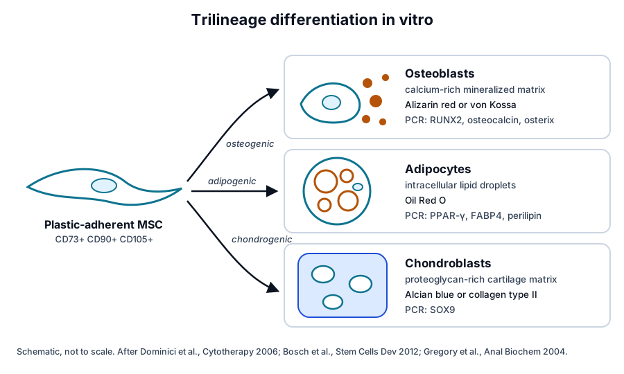
Each stain is paired with lineage genes
- Alizarin red S stains the calcium-rich deposits that cells lay down in culture, and the dye can be extracted from the monolayer and measured at 405 nm.[27]
- Osteogenic differentiation is confirmed by PCR for RUNX2, bone sialoprotein, osteocalcin and osterix.[28]
- Adipogenic differentiation is read with Oil Red O and PCR for PPAR-gamma, fatty acid-binding protein 4 and perilipin.[28]
- Chondrogenic differentiation is read with Alcian blue periodic acid-Schiff and Safranin O staining, with PCR for SOX9.[28]
The IFATS and ISCT statement asks that differentiation assays be completed with a quantitative evaluation, either biochemical or by RT-PCR of lineage genes.[8] Yuan and colleagues found manual scoring of adipogenesis prone to error, and built an image-based algorithm that reports the percentage of differentiating cells.[29]
In stained cultures, osteogenic wells turn red with Alizarin red and adipogenic wells show red lipid droplets with Oil Red O (Fig. 1). Bourin and colleagues present the three differentiation assays as part of both cell identification and potency assessment.[8]
Later ISCT statements added immune assays, source naming and functional testing
In 2013 Krampera and colleagues, for the ISCT MSC Committee, put forward a working proposal for standardized immunological assays of MSCs, aimed at shared, reproducible and consistent data across laboratories.[30] The same year, the IFATS and ISCT joint statement defined adipose-derived cells both before and after culture.[8]
For the uncultured stromal vascular fraction, Bourin and colleagues defined the phenotype CD45-negative, CD235a-negative, CD31-negative and CD34-positive.[8] Cultured adipose stromal cells keep CD90, CD73, CD105 and CD44, and differ from bone marrow MSCs by expressing CD36 and lacking CD106.[8]
In 2016 Galipeau and colleagues reported an ISCT workshop consensus on immune functional assays. They proposed a matrix of quantitative RNA analysis, flow cytometry of functionally relevant surface markers and protein assays of the secretome, measured after in vitro licensing.[31]
The 2019 ISCT position statement, led by Viswanathan, kept the acronym MSCs and recommended three practices. Authors should name the tissue source, use "stem" only with in vitro and in vivo evidence of stemness, and support each preparation with functional assays chosen for its intended mode of action.[24]
A 2025 Delphi study led by Renesme gathered 87 international participants in its first round and reached consensus on nine items for an MSC definition. These covered terminology, five cell marker items, tissue origin, stemness and critical quality attributes, alongside 33 items for clinical study reports.[32]
Research has refined what the minimal criteria can distinguish
The panel is shared with fibroblasts. Haniffa and colleagues reviewed evidence that MSCs and fibroblasts share anti-proliferative effects and differentiation potential.[33] Denu and colleagues found that fibroblast strains from three tissues expressed the same surface markers as MSCs and also formed adipocytes, chondrocytes and osteoblasts.[34]
Halfon and colleagues found CD105, CD73, CD90 and other MSC markers on skin and lung fibroblasts. CD146 appeared only on MSCs, and CD106, integrin alpha 11 and insulin-like growth factor 2 were at least tenfold higher in MSCs. CD106 and CD146 declined by passage 6.[35]
CD146 also marks the stromal cells in their native position. Sacchetti and colleagues showed that CD146-expressing subendothelial cells in human marrow stroma could transfer the hematopoietic microenvironment to heterotopic sites.[36] Lv and colleagues list Stro-1, SSEA-4, CD271 and CD146 among candidate markers whose expression differs widely between MSC sources.[9]
Source and species shape the phenotype
Kern and colleagues compared MSCs from bone marrow, umbilical cord blood and adipose tissue and found similar morphology and immunophenotype. Cord blood cells grew longest, adipose tissue gave the highest colony frequency, and adipogenic differentiation appeared in bone marrow and adipose cells only.[37]
Perinatal tissues show the same variety. Beeravolu and colleagues isolated adherent cells from cord lining, Wharton's jelly, the cord-placenta junction and fetal placenta that expressed CD73, CD90 and CD105 and differentiated along all three lineages.[38] Bosch and colleagues found that differentiation potential varied between cord blood and umbilical cord cell preparations, which is why each preparation is tested along all three lineages.[28]
Camilleri and colleagues validated nine additional markers on adipose-derived MSCs from 15 donors, including CD36, CD200, CD146 and CD271, as candidates for release testing.[39] In mouse MSCs, Anderson and colleagues described stable CD105-positive and CD105-negative subpopulations with different differentiation and immunoregulatory properties.[40]
Reporting practice is still catching up
Renesme and colleagues analyzed 318 MSC articles and found that 18% explicitly referred to the ISCT minimal criteria and 20% reported at least one functional assay.[2] In an FDA perspective, Mendicino and colleagues described growing variability in donor and tissue sources, manufacturing and characterization methods among MSC-based product proposals.[41]
Where to go from here
The mesenchymal stem versus stromal cells reference follows the naming question from 2005 to today, and the source tissues reference compares bone marrow, adipose and perinatal MSCs. The immunobiology reference covers HLA, IDO and PGE2, and the secretome reference describes what MSCs release into conditioned medium.
The perinatal tissue donor eligibility reference explains how birth tissues are screened under federal rules, and the surface receptor chemistry reference explains how markers like CD73 sit in the membrane. Verified practices can request lot documentation, and our team is glad to discuss the methods described on this page.
Referencias comentadas
Numeradas en el orden en que se citan en el texto. Cada fuente lleva una nota sobre lo que respalda en esta página; los artículos revisados por pares se listan con revista, volumen, número y páginas, y las fuentes primarias son regulaciones, documentos de agencias y bases de datos de referencia.
- 1.Peer-reviewedDominici M, Le Blanc K, Mueller I, et al. Minimal criteria for defining multipotent mesenchymal stromal cells. The International Society for Cellular Therapy position statement. Cytotherapy. 2006;8(4):315-317. https://pubmed.ncbi.nlm.nih.gov/16923606/Annotation: ISCT committee position statement. Cited for the three minimal criteria, the 95% and 2% marker thresholds, the reasons for the negative markers, the HLA-DR note on stimulated cells and the named differentiation stains.
- 2.Peer-reviewedRenesme L, Pierro M, Cobey KD, et al. Definition and Characteristics of Mesenchymal Stromal Cells in Preclinical and Clinical Studies: A Scoping Review. Stem Cells Translational Medicine. 2022;11(1):44-54. https://pubmed.ncbi.nlm.nih.gov/35641170/Annotation: Scoping review of 318 analyzed MSC articles. Cited for 18% explicitly referring to the ISCT criteria, 20% reporting a functional assay and inconsistent characterization reporting.
- 3.Peer-reviewedFriedenstein AJ, Chailakhjan RK, Lalykina KS. The development of fibroblast colonies in monolayer cultures of guinea-pig bone marrow and spleen cells. Cell and Tissue Kinetics. 1970;3(4):393-403. https://pubmed.ncbi.nlm.nih.gov/5523063/Annotation: Founding culture study of marrow stromal cells. Cited for fibroblast colonies forming in monolayer cultures of guinea pig marrow and spleen cells.
- 4.Peer-reviewedPittenger MF, Mackay AM, Beck SC, et al. Multilineage potential of adult human mesenchymal stem cells. Science. 1999;284(5411):143-147. https://pubmed.ncbi.nlm.nih.gov/10102814/Annotation: Culture and differentiation of human marrow cells. Cited for adipocytic, chondrocytic and osteocytic differentiation and retained multilineage potential of single expanded cells.
- 5.Peer-reviewedHorwitz EM, Le Blanc K, Dominici M, et al. Clarification of the nomenclature for MSC: The International Society for Cellular Therapy position statement. Cytotherapy. 2005;7(5):393-395. https://pubmed.ncbi.nlm.nih.gov/16236628/Annotation: ISCT nomenclature statement. Cited for "multipotent mesenchymal stromal cells" as the name for plastic-adherent cells, "mesenchymal stem cells" reserved for cells meeting stem cell criteria, and the shared acronym MSC.
- 6.Peer-reviewedGarcía-Muñoz E, Vives J. Towards the standardization of methods of tissue processing for the isolation of mesenchymal stromal cells for clinical use. Cytotechnology. 2021;73(3):513-522. https://pubmed.ncbi.nlm.nih.gov/33994662/Annotation: Open-access review of MSC isolation from bone marrow, adipose tissue and Wharton’s jelly. Its full text restates the minimal criteria: at least 95% CD105, CD73 and CD90, at most 2% of the negative panel, and trilineage differentiation.
- 7.Peer-reviewedBianco P, Cao X, Frenette PS, et al. The meaning, the sense and the significance: translating the science of mesenchymal stem cells into medicine. Nature Medicine. 2013;19(1):35-42. https://pubmed.ncbi.nlm.nih.gov/23296015/Annotation: Perspective on MSC biology. Cited for the open questions on the nature, identity, function, isolation and experimental handling of MSCs.
- 8.Peer-reviewedBourin P, Bunnell BA, Casteilla L, et al. Stromal cells from the adipose tissue-derived stromal vascular fraction and culture expanded adipose tissue-derived stromal/stem cells: a joint statement of the International Federation for Adipose Therapeutics and Science (IFATS) and the International Society for Cellular Therapy (ISCT). Cytotherapy. 2013;15(6):641-648. https://pubmed.ncbi.nlm.nih.gov/23570660/Annotation: IFATS and ISCT joint statement. Cited for the stromal vascular fraction phenotype, CD36 and CD106 as adipose versus marrow markers, the CFU-F assay and quantitative evaluation of differentiation.
- 9.Peer-reviewedLv FJ, Tuan RS, Cheung KM, et al. Concise review: the surface markers and identity of human mesenchymal stem cells. Stem Cells. 2014;32(6):1408-1419. https://pubmed.ncbi.nlm.nih.gov/24578244/Annotation: Review of MSC surface markers. Cited for heterogeneity of plastic-adherent cultures and candidate markers Stro-1, SSEA-4, CD271 and CD146 varying by source.
- 10.DatabaseNCBI Gene 4907, NT5E 5'-nucleotidase ecto (Homo sapiens). Accessed 20 October 2026. https://www.ncbi.nlm.nih.gov/gene/4907
- 11.DatabaseNCBI Gene 7070, THY1 Thy-1 cell surface antigen (Homo sapiens). Accessed 20 October 2026. https://www.ncbi.nlm.nih.gov/gene/7070
- 12.DatabaseNCBI Gene 2022, ENG endoglin (Homo sapiens). Accessed 20 October 2026. https://www.ncbi.nlm.nih.gov/gene/2022
- 13.DatabaseNCBI Gene 5788, PTPRC protein tyrosine phosphatase receptor type C (Homo sapiens). Accessed 20 October 2026. https://www.ncbi.nlm.nih.gov/gene/5788
- 14.DatabaseNCBI Gene 947, CD34 molecule (Homo sapiens). Accessed 20 October 2026. https://www.ncbi.nlm.nih.gov/gene/947
- 15.DatabaseNCBI Gene 929, CD14 molecule (Homo sapiens). Accessed 20 October 2026. https://www.ncbi.nlm.nih.gov/gene/929
- 16.DatabaseNCBI Gene 3684, ITGAM integrin subunit alpha M (Homo sapiens). Accessed 20 October 2026. https://www.ncbi.nlm.nih.gov/gene/3684
- 17.DatabaseNCBI Gene 973, CD79A CD79a molecule (Homo sapiens). Accessed 20 October 2026. https://www.ncbi.nlm.nih.gov/gene/973
- 18.DatabaseNCBI Gene 930, CD19 molecule (Homo sapiens). Accessed 20 October 2026. https://www.ncbi.nlm.nih.gov/gene/930
- 19.DatabaseNCBI Gene 3122, HLA-DRA major histocompatibility complex, class II, DR alpha (Homo sapiens). Accessed 20 October 2026. https://www.ncbi.nlm.nih.gov/gene/3122
- 20.Peer-reviewedKnapp K, Zebisch M, Pippel J, et al. Crystal structure of the human ecto-5'-nucleotidase (CD73): insights into the regulation of purinergic signaling. Structure. 2012;20(12):2161-2173. https://pubmed.ncbi.nlm.nih.gov/23142347/Annotation: Crystal structures of human CD73. Cited for AMP hydrolysis to adenosine, the 114 degree open-closed switch, the C-terminal dimer interface and selectivity for monophosphate nucleotides.
- 21.Peer-reviewedSaito T, Bokhove M, Croci R, et al. Structural Basis of the Human Endoglin-BMP9 Interaction: Insights into BMP Signaling and HHT1. Cell Reports. 2017;19(9):1917-1928. https://pubmed.ncbi.nlm.nih.gov/28564608/Annotation: Crystal structures of the endoglin ectodomain and its BMP9 complex. Cited for the orphan domain fold, the BMP9 binding surface and the zona pellucida module.
- 22.Peer-reviewedSaalbach A, Anderegg U. Thy-1: more than a marker for mesenchymal stromal cells. FASEB Journal. 2019;33(6):6689-6696. https://pubmed.ncbi.nlm.nih.gov/30811954/Annotation: Review of Thy-1 in MSC fate. Cited for Thy-1 promoting osteogenic and inhibiting adipogenic differentiation.
- 23.Peer-reviewedWilson AJ, Rand E, Webster AJ, et al. Characterisation of mesenchymal stromal cells in clinical trial reports: analysis of published descriptors. Stem Cell Research & Therapy. 2021;12(1):360. https://pubmed.ncbi.nlm.nih.gov/34158116/Annotation: Structured analysis of 84 published MSC trial reports. Cited for per-lot versus averaged marker values and the share reporting viability.
- 24.Peer-reviewedViswanathan S, Shi Y, Galipeau J, et al. Mesenchymal stem versus stromal cells: International Society for Cell & Gene Therapy (ISCT®) Mesenchymal Stromal Cell committee position statement on nomenclature. Cytotherapy. 2019;21(10):1019-1024. https://pubmed.ncbi.nlm.nih.gov/31526643/Annotation: ISCT MSC committee position statement. Cited for keeping the acronym MSCs, naming the tissue source, reserving "stem" for cells with in vitro and in vivo evidence, and functional assays matched to the mode of action.
- 25.Peer-reviewedFitzgerald JC, Shaw G, Murphy JM, et al. Media matters: culture medium-dependent hypervariable phenotype of mesenchymal stromal cells. Stem Cell Research & Therapy. 2023;14(1):363. https://pubmed.ncbi.nlm.nih.gov/38087388/Annotation: Comparison of five culture media on marrow MSCs with a 243-marker flow screen. Cited for uniform CD73, CD90 and CD105 expression and medium-dependent variation in one third of markers.
- 26.Peer-reviewedDessels C, Potgieter M, Pepper MS. Making the Switch: Alternatives to Fetal Bovine Serum for Adipose-Derived Stromal Cell Expansion. Frontiers in Cell and Developmental Biology. 2016;4:115. https://pubmed.ncbi.nlm.nih.gov/27800478/Annotation: Open-access review of adipose-derived stromal cell culture. Its full text lists the histochemical stains used to read differentiation: Oil Red O, Alizarin red S, von Kossa and Alcian blue.
- 27.Peer-reviewedGregory CA, Gunn WG, Peister A, et al. An Alizarin red-based assay of mineralization by adherent cells in culture: comparison with cetylpyridinium chloride extraction. Analytical Biochemistry. 2004;329(1):77-84. https://pubmed.ncbi.nlm.nih.gov/15136169/Annotation: Methods study of Alizarin red S. Cited for staining calcium-rich deposits in culture and extraction of the dye for measurement at 405 nm.
- 28.Peer-reviewedBosch J, Houben AP, Radke TF, et al. Distinct differentiation potential of "MSC" derived from cord blood and umbilical cord: are cord-derived cells true mesenchymal stromal cells? Stem Cells and Development. 2012;21(11):1977-1988. https://pubmed.ncbi.nlm.nih.gov/22087798/Annotation: Differentiation study of cord blood, umbilical cord and marrow cells. Cited for the stains and lineage genes used to read each lineage and for differentiation potential varying between cord-derived preparations.
- 29.Peer-reviewedYuan C, Chakraborty S, Chitta KK, et al. Fast Adipogenesis Tracking System (FATS)-a robust, high-throughput, automation-ready adipogenesis quantification technique. Stem Cell Research & Therapy. 2019;10(1):38. https://pubmed.ncbi.nlm.nih.gov/30670100/Annotation: Image-analysis method for adipogenesis. Cited for observer error in manual scoring and automated measurement of the percentage of differentiating cells.
- 30.Peer-reviewedKrampera M, Galipeau J, Shi Y, et al. Immunological characterization of multipotent mesenchymal stromal cells--The International Society for Cellular Therapy (ISCT) working proposal. Cytotherapy. 2013;15(9):1054-1061. https://pubmed.ncbi.nlm.nih.gov/23602578/Annotation: ISCT MSC Committee working proposal. Cited for a standardized approach to immunological assays of MSCs aimed at shared, reproducible data.
- 31.Peer-reviewedGalipeau J, Krampera M, Barrett J, et al. International Society for Cellular Therapy perspective on immune functional assays for mesenchymal stromal cells as potency release criterion for advanced phase clinical trials. Cytotherapy. 2016;18(2):151-159. https://pubmed.ncbi.nlm.nih.gov/26724220/Annotation: ISCT workshop consensus. Cited for a matrix of RNA analysis, flow cytometry of functional surface markers and secretome protein assays after in vitro licensing.
- 32.Peer-reviewedRenesme L, Cobey KD, Lalu MM, et al. Delphi-driven consensus definition for mesenchymal stromal cells and clinical reporting guidelines for mesenchymal stromal cell-based therapeutics. Cytotherapy. 2025;27(2):146-168. https://pubmed.ncbi.nlm.nih.gov/39580717/Annotation: Modified Delphi consensus. Cited for 87 first-round participants, nine consensus items for an MSC definition and 33 reporting items.
- 33.Peer-reviewedHaniffa MA, Collin MP, Buckley CD, et al. Mesenchymal stem cells: the fibroblasts' new clothes? Haematologica. 2009;94(2):258-263. https://pubmed.ncbi.nlm.nih.gov/19109217/Annotation: Review comparing MSCs and fibroblasts. Cited for shared anti-proliferative effects and differentiation potential.
- 34.Peer-reviewedDenu RA, Nemcek S, Bloom DD, et al. Fibroblasts and Mesenchymal Stromal/Stem Cells Are Phenotypically Indistinguishable. Acta Haematologica. 2016;136(2):85-97. https://pubmed.ncbi.nlm.nih.gov/27188909/Annotation: Comparison of fibroblast strains and MSC strains. Cited for fibroblasts sharing MSC morphology, surface markers and trilineage differentiation.
- 35.Peer-reviewedHalfon S, Abramov N, Grinblat B, et al. Markers distinguishing mesenchymal stem cells from fibroblasts are downregulated with passaging. Stem Cells and Development. 2011;20(1):53-66. https://pubmed.ncbi.nlm.nih.gov/20528146/Annotation: Flow cytometry and PCR comparison of marrow MSCs and fibroblasts. Cited for shared panel markers, CD146 on MSCs only, higher CD106, integrin alpha 11 and IGF2 in MSCs, and loss of CD106 and CD146 by passage 6.
- 36.Peer-reviewedSacchetti B, Funari A, Michienzi S, et al. Self-renewing osteoprogenitors in bone marrow sinusoids can organize a hematopoietic microenvironment. Cell. 2007;131(2):324-336. https://pubmed.ncbi.nlm.nih.gov/17956733/Annotation: Transplantation study of human marrow stromal cells. Cited for CD146-expressing subendothelial stromal cells transferring the hematopoietic microenvironment to heterotopic sites.
- 37.Peer-reviewedKern S, Eichler H, Stoeve J, et al. Comparative analysis of mesenchymal stem cells from bone marrow, umbilical cord blood, or adipose tissue. Stem Cells. 2006;24(5):1294-1301. https://pubmed.ncbi.nlm.nih.gov/16410387/Annotation: Side-by-side comparison of three MSC sources. Cited for similar morphology and immunophenotype, source differences in colony frequency and culture span, and adipogenic differentiation by source.
- 38.Peer-reviewedBeeravolu N, McKee C, Alamri A, et al. Isolation and Characterization of Mesenchymal Stromal Cells from Human Umbilical Cord and Fetal Placenta. Journal of Visualized Experiments. 2017. https://pubmed.ncbi.nlm.nih.gov/28447991/Annotation: Explant isolation of MSCs from four perinatal regions. Cited for plastic adherence, CD73, CD90 and CD105 expression and trilineage differentiation of cells from all four sources.
- 39.Peer-reviewedCamilleri ET, Gustafson MP, Dudakovic A, et al. Identification and validation of multiple cell surface markers of clinical-grade adipose-derived mesenchymal stromal cells as novel release criteria for good manufacturing practice-compliant production. Stem Cell Research & Therapy. 2016;7(1):107. https://pubmed.ncbi.nlm.nih.gov/27515308/Annotation: Surface marker transcriptome and flow validation in adipose-derived MSCs from 15 donors. Cited for nine non-classical candidate markers including CD36, CD200, CD146 and CD271.
- 40.Peer-reviewedAnderson P, Carrillo-Gálvez AB, García-Pérez A, et al. CD105 (endoglin)-negative murine mesenchymal stromal cells define a new multipotent subpopulation with distinct differentiation and immunoregulatory capacities. PLoS One. 2013;8(10):e76979. https://pubmed.ncbi.nlm.nih.gov/24124603/Annotation: Characterization of mouse MSC subpopulations. Cited for stable CD105-positive and CD105-negative subpopulations with different differentiation and immunoregulatory properties.
- 41.Peer-reviewedMendicino M, Bailey AM, Wonnacott K, et al. MSC-based product characterization for clinical trials: an FDA perspective. Cell Stem Cell. 2014;14(2):141-145. https://pubmed.ncbi.nlm.nih.gov/24506881/Annotation: FDA staff perspective. Cited for variability in donor and tissue sources, manufacturing and characterization methods in MSC-based product proposals.
Cite this page
ExaVeyra Sciences Editorial Team. ISCT Minimal Criteria for Mesenchymal Stromal Cells (MSCs). ExaVeyra Sciences; 20 October 2026. Available from: https://exaveyra.com/science/stem-cells/isct-msc-minimal-criteria. Accessed [date accessed].
Fuentes de las figuras
- 42.PDB 4H2I, Human ecto-5'-nucleotidase (CD73), closed form with AMPCP. ExaVeyra Sciences, rendered in PyMOL from PDB 4H2I. Structure data: RCSB Protein Data Bank (rcsb.org), CC0. Publicación de la estructura: Knapp K, Zebisch M, Pippel J, et al. Crystal structure of the human ecto-5'-nucleotidase (CD73): insights into the regulation of purinergic signaling. Structure. 2012;20(12):2161-2173.
- 43.PDB 5I04, Orphan region of human endoglin/CD105. ExaVeyra Sciences, rendered in PyMOL from PDB 5I04. Structure data: RCSB Protein Data Bank (rcsb.org), CC0. Publicación de la estructura: Saito T, Bokhove M, Croci R, et al. Structural Basis of the Human Endoglin-BMP9 Interaction: Insights into BMP Signaling and HHT1. Cell Rep. 2017;19(9):1917-1928.