Esta referencia está disponible actualmente en inglés. La versión en español está en preparación.
iPSC-Derived MSCs (iMSCs): Derivation, Identity and Consistency
Por Benn Bluestein-Veyra, M.Sc. en Química Orgánica · Actualizado el 20 de octubre de 2026
ExaVeyra Sciences es un proveedor B2B con sede en Miami que suministra exosomas para uso estético tópico a clínicas con licencia, y remite a los prescriptores con licencia a una farmacia de preparados compuestos 503A que prepara formulaciones según prescripciones individuales.
| Pregunta | Respuesta |
|---|---|
| Qué cubre esta página | How MSCs are derived from induced pluripotent stem cells, how iMSCs compare with tissue-derived MSCs, and why consistency at scale draws research interest. |
| Quién puede hacer pedidos | Clínicas y profesionales con licencia profesional activa y NPI verificados por ExaVeyra. Los pedidos de exosomas requieren además un reconocimiento firmado de uso profesional. |
| Qué se envía y cómo | El material de exosomas para uso estético tópico, caracterizado por lote, se envía en cadena de frío por exprés nocturno. La entrega puede tardar 1 a 3 días hábiles. Se emite un certificado de análisis para cada lote y se entrega a solicitud. |
iPSC-derived mesenchymal stromal cells (iMSCs), also called iPSC-derived mesenchymal stem cells, are made in culture by differentiating induced pluripotent stem cells, and they meet the marker and differentiation criteria applied to MSCs isolated from tissue.[1][2] Each lot can start from one banked iPSC line, which makes iMSCs a renewable MSC source suited to lot-to-lot consistency.[3]
This reference covers how iMSCs are derived and characterized, and what side-by-side studies measured against bone marrow MSCs. It also describes master cell banking, iMSC-derived extracellular vesicles and the assays that measure residual pluripotent cells in a differentiated culture.
Understanding iPSC-derived mesenchymal stromal cells as a renewable cell source
Takahashi and colleagues reprogrammed adult human dermal fibroblasts into induced pluripotent stem cells in 2007 with four transcription factors: OCT3/4, SOX2, KLF4 and c-MYC. The cells resembled embryonic stem cells in morphology, proliferation, surface antigens, gene expression and telomerase activity.[4]
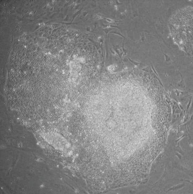
Human iPSCs grow as compact colonies that resemble embryonic stem cell colonies (Fig. 1). They differentiate in vitro and in mice into cell types of all three germ layers, which makes a single iPSC line a starting point for mesenchymal derivation.[4]
Chen and colleagues pointed to the rarity of MSCs in adult organs, their heterogeneity and the harvest procedures they require as reasons to derive MSCs from iPSCs.[5] Wruck and colleagues describe the iMSC concept as a way to reduce heterogeneity tied to tissue origin and donor age.[6]
Derivation routes guide pluripotent cells toward a mesenchymal fate
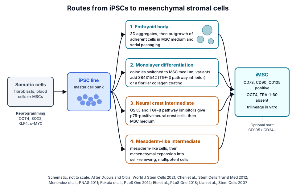
Dupuis and Oltra grouped published iMSC protocols into five categories: MSC switch, embryoid body formation, specific differentiation, pathway inhibitor and platelet lysate methods (Fig. 2). They also compiled the positive and negative markers used to characterize the resulting cells.[3]
The routes build on embryonic stem cell work. In 2005, Barberi and colleagues described culture conditions that gave pure mesenchymal precursors from human embryonic stem cells, which differentiated into fat, cartilage, bone and skeletal muscle cells.[7]
Embryoid body formation
In embryoid body protocols, iPSCs are first aggregated into three-dimensional bodies, and the adherent cells that grow out are expanded in MSC medium.[3] Ababneh and colleagues compared an embryoid body protocol using retinoic acid with four monolayer protocols, and every protocol gave cells expressing MSC markers and lacking pluripotency markers.[8]
Monolayer differentiation
Hynes and colleagues cultured iPSC colonies directly in MSC medium for 2 weeks and then used serial passaging to select fast-growing fibroblastic cells. They obtained MSC-like cells from iPSC lines reprogrammed from gingiva, periodontal ligament and lung.[1]
Chen and colleagues cultured iPSCs in serum-free medium with SB431542, an inhibitor of the transforming growth factor-β pathway, which gave epithelial-like monolayers. Transfer to conventional MSC medium then drove an epithelial-to-mesenchymal transition into MSC-like cells.[5]
Liu and colleagues plated single iPSCs on thin fibrillar type I collagen coatings in medium with ascorbate and dexamethasone. Within 10 d, homogeneous colonies of spindle-shaped cells formed that expressed CD73, CD90, CD105, CD146 and CD166.[9]
Neural crest and mesoderm intermediates
Neural crest cells contribute to the craniofacial skeleton and can give rise to mesenchymal cells.[10] Lee and colleagues derived neural crest stem cells from human embryonic stem cells that differentiated toward smooth muscle, adipogenic, osteogenic and chondrogenic cells.[11]
Menendez and colleagues activated canonical Wnt signaling while blocking the Activin A and Nodal pathway, converting pluripotent cells into p75, HNK1 and AP2 positive neural crest-like cells over 12 to 14 d. These cells gave mesenchymal precursors that formed osteocytes, chondrocytes and adipocytes.[12]
Fukuta and colleagues combined a GSK3β inhibitor and a TGFβ inhibitor in chemically defined medium and induced neural crest cells at 70 to 80% efficiency. Culture in a defined MSC medium then produced cells with MSC-like differentiation characteristics.[10]
Eto and colleagues derived iMSCs through mesoderm-like and neuroepithelium-like intermediates, and both populations self-renewed and were multipotent.[13]
Characterizing iMSCs against the ISCT minimal criteria
The ISCT minimal criteria define MSCs by plastic adherence, expression of CD105, CD73 and CD90, absence of CD45, CD34, CD14 or CD11b, CD79α or CD19 and HLA-DR, and differentiation to osteoblasts, adipocytes and chondroblasts in vitro.[14] iMSC studies test their cells against this same panel.[3]
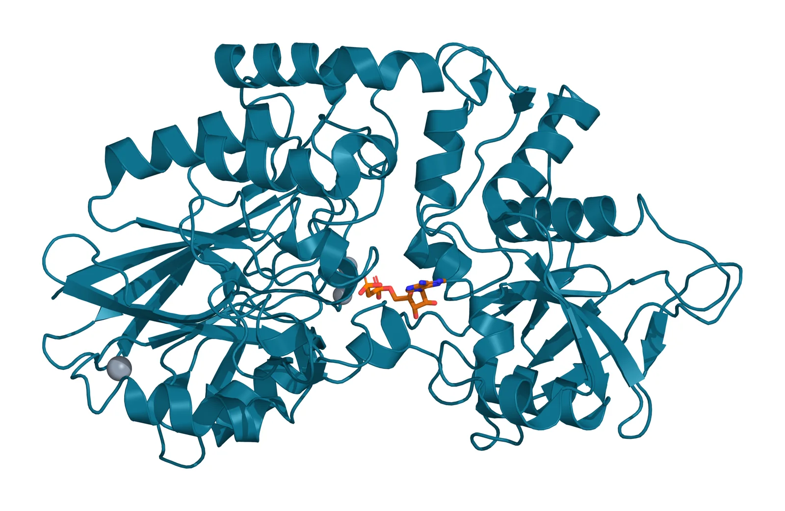
Kang and colleagues found two iMSC lines positive for CD29, CD44, CD73, CD90 and CD105 and negative for CD11b, CD14, CD31, CD34, CD45 and HLA-DR, matching bone marrow MSCs.[2] CD73 is the ecto-5′-nucleotidase encoded by NT5E, a membrane enzyme that converts extracellular nucleotides to nucleosides (Fig. 3).[15]
Pluripotency markers form a second, negative panel. Hynes and colleagues confirmed that iMSCs lacked TRA-1-60, TRA-1-81 and alkaline phosphatase, and Kang and colleagues found OCT4, SOX2, GDF3 and LIN28A expression absent in iMSCs and their differentiated progeny.[1][2]
Surface marker sorting
Lian and colleagues found CD105 highly expressed on embryonic stem cell-derived MSCs and CD24 on the parent stem cells. CD105-positive, CD24-negative isolates had typical MSC gene expression and were highly correlated with each other, with r² above 0.90.[16]
Diederichs and Tuan derived MSC-like progenitors by four methods from an iPSC line reprogrammed from bone marrow stromal cells and compared them with the parental cells. The derived cells met the morphology, marker and trilineage criteria and showed a distinct pattern of mesenchymal and pluripotency gene expression.[17]
How iMSCs compare with tissue-derived MSCs in growth, lineage and secretome
Side-by-side studies compare iMSCs with bone marrow MSCs and, in some designs, with parental cells from the same donor (Fig. 4).[2][17] The table summarizes the attributes measured most often, and the sections below give the study behind each finding.
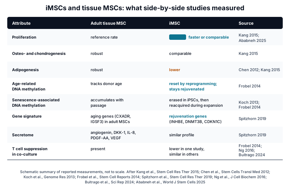
| Attribute | Tissue-derived MSC | iMSC | Reported by |
|---|---|---|---|
| Proliferation | Reference rate | Faster, or comparable, in most protocols | Kang 2015; Ababneh 2025 |
| Osteogenic and chondrogenic differentiation | Robust | Comparable | Kang 2015; Chen 2012 |
| Adipogenic differentiation | Robust | Lower in several protocols | Chen 2012; Kang 2015; Ababneh 2025 |
| Senescence measures | Accumulate with passage | Lower in four of five protocols | Ababneh 2025 |
| Age-related DNA methylation | Reflects donor age | Reset by reprogramming | Frobel 2014 |
| Secretome | Angiogenin, DKK-1, IL-8, PDGF-AA, VEGF | Similar profile | Spitzhorn 2019 |
| T cell proliferation in co-culture | Suppressed | Lower suppression in one study, similar in others | Frobel 2014; Ng 2016; Buitrago 2024 |
Proliferation and senescence
Kang and colleagues saw faster proliferation in both iMSC lines than in bone marrow MSCs.[2] Ababneh and colleagues found proliferation comparable to or above that of bone marrow MSCs in four of five protocols, with lower reactive oxygen species in all iMSCs and lower senescence in four.[8]
Lineage preference
Kang and colleagues reported adequate osteogenic and chondrogenic capacity in iMSCs and less adipogenic potential.[2] Chen and colleagues likewise saw robust osteogenesis and chondrogenesis with limited adipogenesis, a pattern also reported for fetal MSCs and embryonic stem cell-derived MSCs.[5]
Ababneh and colleagues found enhanced RUNX2 and reduced adipsin in embryoid body-derived iMSCs, so the protocol shapes lineage balance.[8] Hynes and colleagues saw differentiation capacity vary between iPSC lines, and only cells from the periodontal ligament line formed mineralized structures after implantation in mice.[1]
Secretome and co-culture assays
Spitzhorn and colleagues measured an iMSC secretome that included angiogenin, DKK-1, IL-8, PDGF-AA, osteopontin, SERPINE1 and VEGF. The profile was similar to that of fetal and adult MSCs, which points to paracrine signaling as a shared feature.[18]
T cell co-culture results vary between studies. Frobel and colleagues found iMSCs less able than primary MSCs to suppress T cell proliferation, while Ng and colleagues found suppression of mitogen-stimulated mononuclear cells similar to the parental fibroblasts, with raised IDO1 and IL6 transcripts.[19][20]
Buitrago and colleagues reported that iMSCs regulated lymphocyte proliferation and shifted monocytes toward an anti-inflammatory phenotype to a degree comparable with umbilical cord MSCs. They also measured lower TNFα and higher IL-10 in these co-cultures.[21]
Reprogramming resets the epigenetic age of iPSC-derived MSCs
Long-term culture of MSCs brings reproducible senescence-associated DNA methylation changes, enriched in nonpromoter regions of developmental genes. Koch and colleagues showed that reprogramming into iPSCs prevented almost the entire set of these changes.[22]
Lapasset and colleagues reprogrammed senescent and centenarian fibroblasts into iPSCs with reset telomere size, gene expression, oxidative stress and mitochondrial metabolism. Cells redifferentiated from these iPSCs were described as fully rejuvenated.[23]
Frobel and colleagues reprogrammed MSCs into iPSCs and differentiated them back to MSCs. Tissue-specific, senescence-associated and age-related methylation patterns were erased, and the iMSCs reacquired senescence-associated methylation during expansion while staying rejuvenated in age-related methylation.[19]
Spitzhorn and colleagues made iMSCs from iPSCs of fetal and aged adult MSCs. Irrespective of donor age, the iMSCs acquired a rejuvenation signature including INHBE, DNMT3B, CDKN1C and GCNT2, and shared more genes with fetal MSCs than with adult MSCs.[18]
Frobel and colleagues also found that iPSC methylation profiles kept donor-specific features.[19] In a meta-analysis of MSC transcriptomes, Wruck and colleagues linked heterogeneity mainly to tissue origin and proposed iMSCs as a way to bypass tissue- and age-related variation.[6]
A master iPSC bank supports consistency and scale
Dupuis and Oltra describe iPSCs as a reliable, unlimited source for generating MSC lines.[3] Sullivan and colleagues set out consensus recommendations on the minimum dataset of critical quality attributes and assays for a clinical-grade iPSC line, so lines from different donors and facilities can be compared.[24]
Umekage and colleagues describe the iPSC stock at the Center for iPS Cell Research and Application in Kyoto, made from peripheral blood or cord blood of consenting HLA-homozygous donors. In 2019 the stock offered 21 clones from 5 donors.[25]
Lian and colleagues generated karyotypically stable, identical batches of embryonic stem cell-derived MSCs without virus, mouse cells or serum.[16] Diederichs and Tuan noted that the proliferative capacity of pluripotent cells opens the possibility of large, uniform MSC batches.[17]
Rogers and colleagues expanded iMSCs on degradable gelatin methacryloyl microcarriers in vertical wheel bioreactors, reaching 8.8 to 16.3-fold expansion over 8 d. Microcarrier digestion released cells at over 95% viability with immunomodulatory potential in vitro equal to or above monolayer-expanded iMSCs.[26]
iMSC-derived extracellular vesicles are an active research direction
Kim and colleagues established iPSCs from Wharton’s jelly MSCs, differentiated them into iMSCs, and collected exosomes from the culture supernatant of both cell types. Both preparations showed the basic characteristics of exosomes and were taken up by keratinocytes and dermal fibroblasts in culture, and the authors highlighted iMSCs as producer cells for exosomes.[27]
Buitrago and colleagues reported that iMSC vesicles inhibited T cell proliferation and influenced macrophage polarization in vitro, similar to their parent cells.[21]
Residual pluripotent cell testing is a defined quality attribute
Tano and colleagues describe the fraction of undifferentiated cells as a quality attribute of any cell preparation derived from pluripotent stem cells. Assays for it are designed to find a few iPSCs among a large number of differentiated cells.[28]
Their culture assay on laminin-521 in Essential 8 medium amplifies trace iPSCs into countable colonies. It detected iPSCs spiked into primary MSCs at 0.001 to 0.01%, and it detected undifferentiated cells during iMSC differentiation from iPSCs.[28]
Kuroda and colleagues built a droplet digital PCR assay for LIN28 transcripts that detected 0.001% iPSCs in cardiomyocytes, one cell in 10⁵. LIN28 expression was very low in the adult human tissues they tested.[29] LIN28A encodes an RNA-binding regulator of self-renewal in embryonic stem cells.[30]
Yasuda and colleagues ran an international multisite evaluation of a one-step RT-ddPCR assay for ESRG, LINC00678 and LIN28A, with iPSCs spiked into iPSC-derived cardiomyocytes. The assay was sensitive and robust, and most variation between laboratories came from the spiking step.[31]
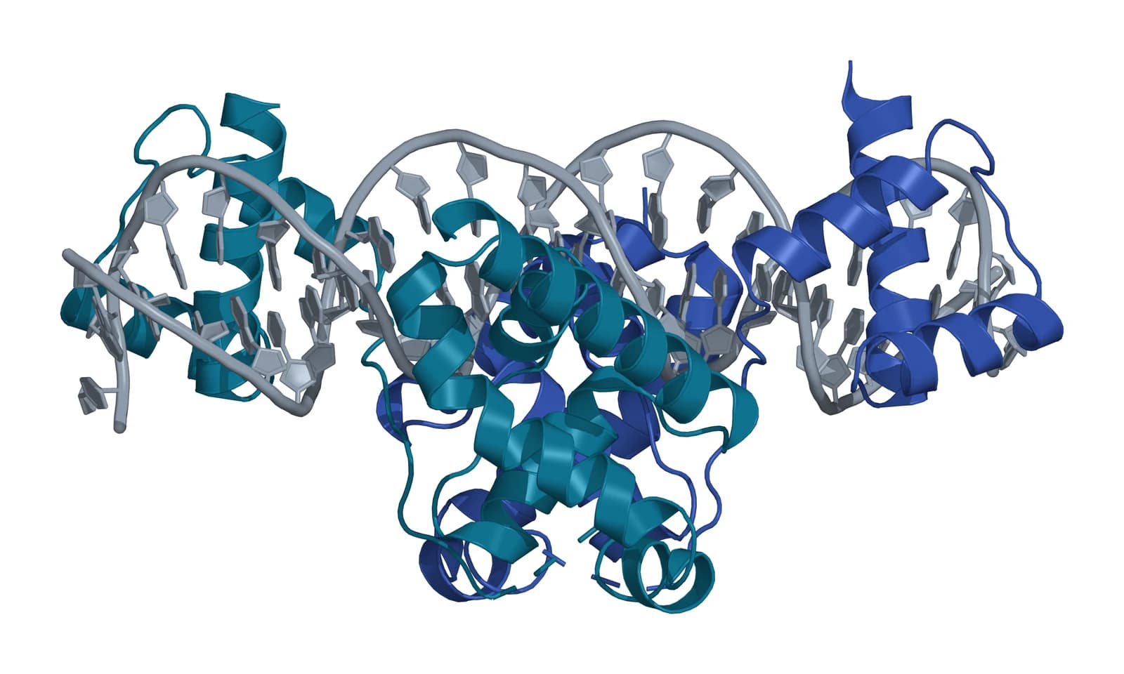
OCT4, encoded by POU5F1, is a POU homeodomain transcription factor with a key role in embryonic development and stem cell pluripotency (Fig. 5).[32] Kang and colleagues reported OCT4 transcripts absent from iMSCs, which makes OCT4 one of the negative markers in iMSC characterization.[2]
Where to go from here
The ISCT minimal criteria reference explains the marker panel every iMSC study measures against, and the MSC source tissues reference compares bone marrow, adipose, cord and placental cells. The stem cell types reference places iPSCs among the other stem cell classes, and the single-cell heterogeneity reference shows how varied a tissue MSC culture can be.
The MSC secretome reference covers conditioned medium and vesicle fractions, and the exosome biochemistry and surface receptor chemistry references continue from cells to vesicles. Verified practices can request lot documentation, and our team is glad to discuss the methods described on this page.
Referencias comentadas
Numeradas en el orden en que se citan en el texto. Cada fuente lleva una nota sobre lo que respalda en esta página; los artículos revisados por pares se listan con revista, volumen, número y páginas, y las fuentes primarias son regulaciones, documentos de agencias y bases de datos de referencia.
- 1.Peer-reviewedHynes K, Menicanin D, Mrozik K, et al. Generation of functional mesenchymal stem cells from different induced pluripotent stem cell lines. Stem Cells and Development. 2014;23(10):1084-1096. https://pubmed.ncbi.nlm.nih.gov/24367908/Annotation: MSC switch route from three iPSC lines. Cited for serial passaging selection, the MSC and pluripotency marker panels and line-to-line variation in differentiation.
- 2.Peer-reviewedKang R, Zhou Y, Tan S, et al. Mesenchymal stem cells derived from human induced pluripotent stem cells retain adequate osteogenicity and chondrogenicity but less adipogenicity. Stem Cell Research & Therapy. 2015;6(1):144. https://pubmed.ncbi.nlm.nih.gov/26282538/Annotation: Two iMSC lines compared with bone marrow MSCs. Cited for the surface marker profile, faster proliferation, lower adipogenesis and absent stemness gene expression.
- 3.Peer-reviewedDupuis V, Oltra E. Methods to produce induced pluripotent stem cell-derived mesenchymal stem cells: Mesenchymal stem cells from induced pluripotent stem cells. World Journal of Stem Cells. 2021;13(8):1094-1111. https://pubmed.ncbi.nlm.nih.gov/34567428/Annotation: Review of iMSC derivation protocols. Cited for the five protocol categories, the marker lists used for characterization and iPSCs as an unlimited source of MSC lines.
- 4.Peer-reviewedTakahashi K, Tanabe K, Ohnuki M, et al. Induction of pluripotent stem cells from adult human fibroblasts by defined factors. Cell. 2007;131(5):861-872. https://pubmed.ncbi.nlm.nih.gov/18035408/Annotation: First human iPSCs, from adult dermal fibroblasts with OCT3/4, SOX2, KLF4 and c-MYC. Cited for their similarity to embryonic stem cells and differentiation into all three germ layers.
- 5.Peer-reviewedChen YS, Pelekanos RA, Ellis RL, et al. Small molecule mesengenic induction of human induced pluripotent stem cells to generate mesenchymal stem/stromal cells. Stem Cells Translational Medicine. 2012;1(2):83-95. https://pubmed.ncbi.nlm.nih.gov/23197756/Annotation: SB431542 monolayer route from iPSCs to MSCs. Cited for the rationale for iMSCs, the epithelial-to-mesenchymal transition step and limited adipogenesis with robust osteogenesis and chondrogenesis.
- 6.Peer-reviewedWruck W, Graffmann N, Spitzhorn LS, et al. Human Induced Pluripotent Stem Cell-Derived Mesenchymal Stem Cells Acquire Rejuvenation and Reduced Heterogeneity. Frontiers in Cell and Developmental Biology. 2021;9:717772. https://pubmed.ncbi.nlm.nih.gov/34604216/Annotation: Review and transcriptome meta-analysis of MSC heterogeneity. Cited for tissue origin as the main driver of heterogeneity and the iMSC concept for reducing it.
- 7.Peer-reviewedBarberi T, Willis LM, Socci ND, et al. Derivation of multipotent mesenchymal precursors from human embryonic stem cells. PLoS Medicine. 2005;2(6):e161. https://pubmed.ncbi.nlm.nih.gov/15971941/Annotation: Early derivation of mesenchymal precursors from human embryonic stem cells. Cited for multilineage differentiation into fat, cartilage, bone and skeletal muscle cells.
- 8.Peer-reviewedAbabneh NA, Alwohoush E, AlDiqs R, et al. Impact of differentiation protocols on the functionality of mesenchymal stem cells derived from induced pluripotent stem cells. World Journal of Stem Cells. 2025;17(12):110564. https://pubmed.ncbi.nlm.nih.gov/41480400/Annotation: Five iMSC protocols compared with bone marrow MSCs. Cited for the embryoid body protocol, marker results, proliferation, lineage balance, reactive oxygen species and senescence.
- 9.Peer-reviewedLiu Y, Goldberg AJ, Dennis JE, et al. One-step derivation of mesenchymal stem cell (MSC)-like cells from human pluripotent stem cells on a fibrillar collagen coating. PLoS One. 2012;7(3):e33225. https://pubmed.ncbi.nlm.nih.gov/22457746/Annotation: Fibrillar collagen route from embryonic and induced pluripotent cells. Cited for spindle-shaped colonies within 10 d expressing CD73, CD90, CD105, CD146 and CD166.
- 10.Peer-reviewedFukuta M, Nakai Y, Kirino K, et al. Derivation of mesenchymal stromal cells from pluripotent stem cells through a neural crest lineage using small molecule compounds with defined media. PLoS One. 2014;9(12):e112291. https://pubmed.ncbi.nlm.nih.gov/25464501/Annotation: Neural crest route to MSCs in chemically defined media. Cited for GSK3β and TGFβ inhibition, 70 to 80% induction efficiency and the craniofacial contribution of neural crest cells.
- 11.Peer-reviewedLee G, Kim H, Elkabetz Y, et al. Isolation and directed differentiation of neural crest stem cells derived from human embryonic stem cells. Nature Biotechnology. 2007;25(12):1468-1475. https://pubmed.ncbi.nlm.nih.gov/18037878/Annotation: Derivation of human neural crest stem cells from embryonic stem cells. Cited for their differentiation toward smooth muscle, adipogenic, osteogenic and chondrogenic cells.
- 12.Peer-reviewedMenendez L, Yatskievych TA, Antin PB, et al. Wnt signaling and a Smad pathway blockade direct the differentiation of human pluripotent stem cells to multipotent neural crest cells. Proceedings of the National Academy of Sciences of the United States of America. 2011;108(48):19240-19245. https://pubmed.ncbi.nlm.nih.gov/22084120/Annotation: Small-molecule neural crest induction from pluripotent cells. Cited for Wnt activation with Activin A and Nodal blockade, the p75, HNK1 and AP2 markers and trilineage mesenchymal precursors.
- 13.Peer-reviewedEto S, Goto M, Soga M, et al. Mesenchymal stem cells derived from human iPS cells via mesoderm and neuroepithelium have different features and therapeutic potentials. PLoS One. 2018;13(7):e0200790. https://pubmed.ncbi.nlm.nih.gov/30044827/Annotation: Two developmental routes to iMSCs. Cited only for derivation through mesoderm-like and neuroepithelium-like cells, self-renewal and multipotency.
- 14.Peer-reviewedDominici M, Le Blanc K, Mueller I, et al. Minimal criteria for defining multipotent mesenchymal stromal cells. The International Society for Cellular Therapy position statement. Cytotherapy. 2006;8(4):315-317. https://pubmed.ncbi.nlm.nih.gov/16923606/Annotation: ISCT minimal criteria. Cited for plastic adherence, the CD105, CD73 and CD90 panel, the negative markers and trilineage differentiation.
- 15.DatabaseNCBI Gene 4907, NT5E 5′-nucleotidase ecto (CD73) (Homo sapiens). Accessed 20 October 2026. https://www.ncbi.nlm.nih.gov/gene/4907
- 16.Peer-reviewedLian Q, Lye E, Suan Yeo K, et al. Derivation of clinically compliant MSCs from CD105+, CD24- differentiated human ESCs. Stem Cells. 2007;25(2):425-436. https://pubmed.ncbi.nlm.nih.gov/17053208/Annotation: Embryonic stem cell-derived MSCs made without virus, mouse cells or serum. Cited for CD105 and CD24 as sorting markers, correlated clonal isolates and identical, karyotypically stable batches.
- 17.Peer-reviewedDiederichs S, Tuan RS. Functional comparison of human-induced pluripotent stem cell-derived mesenchymal cells and bone marrow-derived mesenchymal stromal cells from the same donor. Stem Cells and Development. 2014;23(14):1594-1610. https://pubmed.ncbi.nlm.nih.gov/24625206/Annotation: Same-donor comparison of iPSC-derived MSC-like progenitors with parental marrow stromal cells. Cited for four derivation methods, a distinct gene expression pattern and the prospect of large uniform batches.
- 18.Peer-reviewedSpitzhorn LS, Megges M, Wruck W, et al. Human iPSC-derived MSCs (iMSCs) from aged individuals acquire a rejuvenation signature. Stem Cell Research & Therapy. 2019;10(1):100. https://pubmed.ncbi.nlm.nih.gov/30885246/Annotation: iMSCs from fetal and aged adult MSC-derived iPSCs. Cited for the rejuvenation gene signature, shared genes with fetal MSCs and the secretome profile.
- 19.Peer-reviewedFrobel J, Hemeda H, Lenz M, et al. Epigenetic rejuvenation of mesenchymal stromal cells derived from induced pluripotent stem cells. Stem Cell Reports. 2014;3(3):414-422. https://pubmed.ncbi.nlm.nih.gov/25241740/Annotation: MSCs reprogrammed to iPSCs and redifferentiated to MSCs. Cited for erased and reacquired methylation patterns, retained donor-specific features and reduced T cell suppression in co-culture.
- 20.Peer-reviewedNg J, Hynes K, White G, et al. Immunomodulatory Properties of Induced Pluripotent Stem Cell-Derived Mesenchymal Cells. Journal of Cellular Biochemistry. 2016;117(12):2844-2853. https://pubmed.ncbi.nlm.nih.gov/27167148/Annotation: Co-culture of iMSCs with stimulated mononuclear cells. Cited for suppression similar to parental fibroblasts and raised IDO1 and IL6 transcripts.
- 21.Peer-reviewedBuitrago JC, Morris SL, Backhaus A, et al. Unveiling the Immunomodulatory and regenerative potential of iPSC-derived mesenchymal stromal cells and their extracellular vesicles. Scientific Reports. 2024;14(1):24098. https://pubmed.ncbi.nlm.nih.gov/39407038/Annotation: iMSCs and their vesicles compared with umbilical cord MSCs in vitro. Cited for lymphocyte and monocyte co-culture results, TNFα and IL-10, and vesicle effects on T cells and macrophages.
- 22.Peer-reviewedKoch CM, Reck K, Shao K, et al. Pluripotent stem cells escape from senescence-associated DNA methylation changes. Genome Research. 2013;23(2):248-259. https://pubmed.ncbi.nlm.nih.gov/23080539/Annotation: Methylation profiling of MSCs in long-term culture and after reprogramming. Cited for reproducible senescence-associated methylation and its near-complete prevention by reprogramming.
- 23.Peer-reviewedLapasset L, Milhavet O, Prieur A, et al. Rejuvenating senescent and centenarian human cells by reprogramming through the pluripotent state. Genes & Development. 2011;25(21):2248-2253. https://pubmed.ncbi.nlm.nih.gov/22056670/Annotation: Reprogramming of senescent and centenarian cells. Cited for reset telomere size, gene expression, oxidative stress and mitochondrial metabolism, and rejuvenated redifferentiated cells.
- 24.Peer-reviewedSullivan S, Stacey GN, Akazawa C, et al. Quality control guidelines for clinical-grade human induced pluripotent stem cell lines. Regenerative Medicine. 2018;13(7):859-866. https://pubmed.ncbi.nlm.nih.gov/30205750/Annotation: Consensus workshop recommendations for iPSC lines. Cited for the minimum dataset of critical quality attributes and assays that allows lines to be compared.
- 25.Peer-reviewedUmekage M, Sato Y, Takasu N. Overview: an iPS cell stock at CiRA. Inflammation and Regeneration. 2019;39:17. https://pubmed.ncbi.nlm.nih.gov/31497180/Annotation: Description of the CiRA iPSC stock. Cited for HLA-homozygous donors, blood and cord blood starting cells, and 21 clones from 5 donors.
- 26.Peer-reviewedRogers RE, Haskell A, White BP, et al. A scalable system for generation of mesenchymal stem cells derived from induced pluripotent cells employing bioreactors and degradable microcarriers. Stem Cells Translational Medicine. 2021;10(12):1650-1665. https://pubmed.ncbi.nlm.nih.gov/34505405/Annotation: Bioreactor expansion of iMSCs on GelMA microcarriers. Cited for 8.8 to 16.3-fold expansion over 8 d, over 95% viability at harvest and in vitro immunomodulatory potential.
- 27.Peer-reviewedKim S, Lee SK, Kim H, et al. Exosomes Secreted from Induced Pluripotent Stem Cell-Derived Mesenchymal Stem Cells Accelerate Skin Cell Proliferation. International Journal of Molecular Sciences. 2018;19(10):3119. https://pubmed.ncbi.nlm.nih.gov/30314356/Annotation: Exosomes from Wharton’s jelly MSCs and the iMSCs derived from them. Cited for exosome characteristics, uptake by skin cells in culture and iMSCs as producer cells for exosomes.
- 28.Peer-reviewedTano K, Yasuda S, Kuroda T, et al. A novel in vitro method for detecting undifferentiated human pluripotent stem cells as impurities in cell therapy products using a highly efficient culture system. PLoS One. 2014;9(10):e110496. https://pubmed.ncbi.nlm.nih.gov/25347300/Annotation: Laminin-521 and Essential 8 amplification assay for residual iPSCs. Cited for detection at 0.001 to 0.01% in primary MSCs and during iMSC differentiation.
- 29.Peer-reviewedKuroda T, Yasuda S, Matsuyama S, et al. Highly sensitive droplet digital PCR method for detection of residual undifferentiated cells in cardiomyocytes derived from human pluripotent stem cells. Regenerative Therapy. 2015;2:17-23. https://pubmed.ncbi.nlm.nih.gov/31245455/Annotation: LIN28 droplet digital PCR for residual iPSCs. Cited for detection of 0.001%, one cell in 10^5, and low LIN28 expression in adult tissues.
- 30.DatabaseNCBI Gene 79727, LIN28A lin-28 RNA binding posttranscriptional regulator A (Homo sapiens). Accessed 20 October 2026. https://www.ncbi.nlm.nih.gov/gene/79727
- 31.Peer-reviewedYasuda S, Bando K, Henry MP, et al. Detection of residual pluripotent stem cells in cell therapy products utilizing droplet digital PCR: an international multisite evaluation study. Stem Cells Translational Medicine. 2024;13(10):1001-1014. https://pubmed.ncbi.nlm.nih.gov/39120125/Annotation: Multisite evaluation of RT-ddPCR for residual iPSCs. Cited for the ESRG, LINC00678 and LIN28A targets, assay sensitivity and the spiking step as the main source of variation.
- 32.DatabaseNCBI Gene 5460, POU5F1 POU class 5 homeobox 1 (OCT4) (Homo sapiens). Accessed 20 October 2026. https://www.ncbi.nlm.nih.gov/gene/5460
Cite this page
ExaVeyra Sciences Editorial Team. iPSC-Derived MSCs (iMSCs): Derivation, Identity and Consistency. ExaVeyra Sciences; 20 October 2026. Available from: https://exaveyra.com/science/stem-cells/ipsc-derived-mscs. Accessed [date accessed].
Fuentes de las figuras
- 33.PDB 4H2I, Human ecto-5'-nucleotidase (CD73), closed form with AMPCP. ExaVeyra Sciences, rendered in PyMOL from PDB 4H2I. Structure data: RCSB Protein Data Bank (rcsb.org), CC0. Publicación de la estructura: Knapp K, Zebisch M, Pippel J, et al. Crystal structure of the human ecto-5'-nucleotidase (CD73): insights into the regulation of purinergic signaling. Structure. 2012;20(12):2161-2173.
- 34.PDB 3L1P, POU protein:DNA complex (Oct4). ExaVeyra Sciences, rendered in PyMOL from PDB 3L1P. Structure data: RCSB Protein Data Bank (rcsb.org), CC0. Publicación de la estructura: Esch D, Vahokoski J, Groves MR, et al. A unique Oct4 interface is crucial for reprogramming to pluripotency. Nat Cell Biol. 2013;15(3):295-301.