Esta referencia está disponible actualmente en inglés. La versión en español está en preparación.
The MSC Secretome: Conditioned Medium, Growth Factors and EVs
Por Benn Bluestein-Veyra, M.Sc. en Química Orgánica · Actualizado el 20 de octubre de 2026
ExaVeyra Sciences es un proveedor B2B con sede en Miami que suministra exosomas para uso estético tópico a clínicas con licencia, y remite a los prescriptores con licencia a una farmacia de preparados compuestos 503A que prepara formulaciones según prescripciones individuales.
| Pregunta | Respuesta |
|---|---|
| Qué cubre esta página | What MSCs secrete into culture medium, how conditioned medium differs from purified EV fractions, and how the secretome is characterized. |
| Quién puede hacer pedidos | Clínicas y profesionales con licencia profesional activa y NPI verificados por ExaVeyra. Los pedidos de exosomas requieren además un reconocimiento firmado de uso profesional. |
| Qué se envía y cómo | El material de exosomas para uso estético tópico, caracterizado por lote, se envía en cadena de frío por exprés nocturno. La entrega puede tardar 1 a 3 días hábiles. Se emite un certificado de análisis para cada lote y se entrega a solicitud. |
The MSC secretome is the full set of molecules that mesenchymal stromal cells release into their surroundings, and MSC conditioned medium is the culture medium that collects them.[1] It contains soluble growth factors, cytokines and chemokines together with extracellular vesicles that enclose proteins and RNA.[1][2]
This reference describes what the secretome contains, how conditioned medium is produced, how culture conditions change it, how vesicle fractions are separated from it and how each fraction is measured. Witwer and colleagues, writing for four scientific societies, framed these measurements as quantifiable metrics that let one laboratory compare its preparations with another.[3]
Understanding the paracrine model of MSC biology
In 2006 Caplan and Dennis described marrow-derived mesenchymal stem cells as trophic mediators. Beyond their capacity to differentiate into osteoblasts, chondrocytes, adipocytes and other mesenchymal phenotypes, the cells secrete a variety of cytokines and growth factors with paracrine and autocrine activity.[4]
Caplan and Dennis grouped the reported activities of these secreted factors, including modulation of local immune responses, support of angiogenesis and stimulation of mitosis and differentiation in neighboring cells, under the term trophic effects. They described these effects as distinct from the direct differentiation of the cells themselves.[4]
Experimental support for soluble mediators came from co-culture work. Di Nicola and colleagues separated bone marrow stromal cells from T cells with a Transwell membrane and still observed suppressed T cell proliferation, and neutralizing antibodies identified TGF-beta1 and hepatocyte growth factor (HGF) as the mediators.[5]
Konala and colleagues describe the secretome as the combined repertoire of soluble factors and factors released in extracellular vesicles such as exosomes and microvesicles.[1] Research on MSC conditioned medium therefore studies both classes of material, together and apart.[6]
The MSC secretome combines soluble proteins, matrix components, metabolites and vesicles
MSC conditioned medium holds soluble growth factors, cytokines, chemokines, matrix proteins and metabolites alongside a population of extracellular vesicles (Fig. 1).[1][7] The vesicles enclose their own proteins, RNA and metabolites within a lipid bilayer.[8]
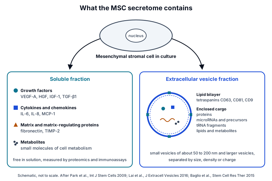
Sze and colleagues produced one of the first systematic catalogs. They conditioned a chemically defined, serum-free medium with MSCs derived from human embryonic stem cells, analyzed it by multidimensional protein identification technology and cytokine antibody arrays, and identified 201 unique gene products, 86 to 88 percent of which had detectable transcripts in the cells.[9]
Computational analysis of that protein set predicted activation of signaling pathways including Jak-STAT, MAPK, Toll-like receptor, TGF-beta and mTOR.[9] Later proteomic studies extended this catalog to MSCs from bone marrow, adipose tissue, placenta and Wharton’s jelly.[10]
Growth factors: VEGF-A, HGF, IGF-1 and TGF-beta1
Hsiao and colleagues compared paracrine factor expression in MSCs from bone marrow, adipose and dermal tissue. VEGF-A, angiogenin, basic fibroblast growth factor and nerve growth factor were expressed at comparable levels across the populations, while IGF-1, VEGF-D and IL-8 were higher in adipose-derived cells.[11]
Protein measurements in their conditioned media confirmed comparable VEGF-A and angiogenin secretion across all the populations. In endothelial tube formation assays, neutralizing antibodies identified VEGF-A and VEGF-D as two of the main secreted factors supporting tubulogenesis by adipose-derived cell media.[11]
VEGF-A belongs to the PDGF and VEGF growth factor family and is a heparin-binding protein that exists as a disulfide-linked homodimer (Fig. 2a). It induces proliferation and migration of vascular endothelial cells.[12]
HGF binds its receptor to regulate cell growth, cell motility and morphogenesis in many cell and tissue types, and it is made as a preproprotein that is proteolytically processed.[13] In the co-culture experiments of Di Nicola and colleagues, HGF acted together with TGF-beta1 as a soluble stromal cell signal.[5]
TGF-beta1 is secreted as a preproprotein that is processed into a latency-associated peptide and a mature peptide. The mature homodimer binds TGF-beta receptors to recruit and activate SMAD transcription factors (Fig. 2b).[14]
Cytokines and chemokines
Park and colleagues profiled the media of human bone marrow MSCs on an antibody array recognizing 120 cytokines and chemokines. IL-6 gave the predominant signal, with moderately elevated signals for IL-8, TIMP-2, MCP-1, VEGF and osteoprotegerin.[15]
The same profile appeared in cells from three different donors and was similar, with some differences, to that of umbilical cord blood-derived cells. The authors suggested that the profile might depend slightly on cell origin, with little effect of the individual donor.[15]
Matrix proteins and metabolites
The secretome also carries matrix and matrix-regulating proteins. Park and colleagues detected the metalloproteinase inhibitor TIMP-2 among the abundant secreted factors, and Lai and colleagues found fibronectin containing the EDA domain on one class of MSC vesicles.[8][15]
Showalter and colleagues reported that MSCs exposed to culture conditions designed to mimic the in vivo microenvironmental niche underwent glycolytic reprogramming. Their exosomes were packaged with metabolites previously associated with immunomodulation, including M2 macrophage polarization and regulatory T cell induction.[7]
Extracellular vesicles and their RNA cargo
Chen and colleagues studied conditioned medium from MSCs derived from human embryonic stem cells. It held small RNAs enclosed in cholesterol-rich phospholipid vesicles, which protected them from RNase until the membranes were disrupted. MicroRNAs such as let-7b and let-7g were present with a high ratio of precursor to mature forms.[2]
Baglio and colleagues sequenced the small RNAs of exosomes from bone marrow and adipose MSCs. MicroRNAs made up only 2 to 5 percent of the exosomal small RNA, a defined subset was enriched relative to the cells, and tRNA species, in particular tRNA halves, were preferentially released.[16]
Vesicles in the secretome are themselves heterogeneous. Lai and colleagues separated at least three MSC vesicle types by their affinity for lipid-binding ligands, and each carried a different combination of membrane lipid, protein and RNA.[8]
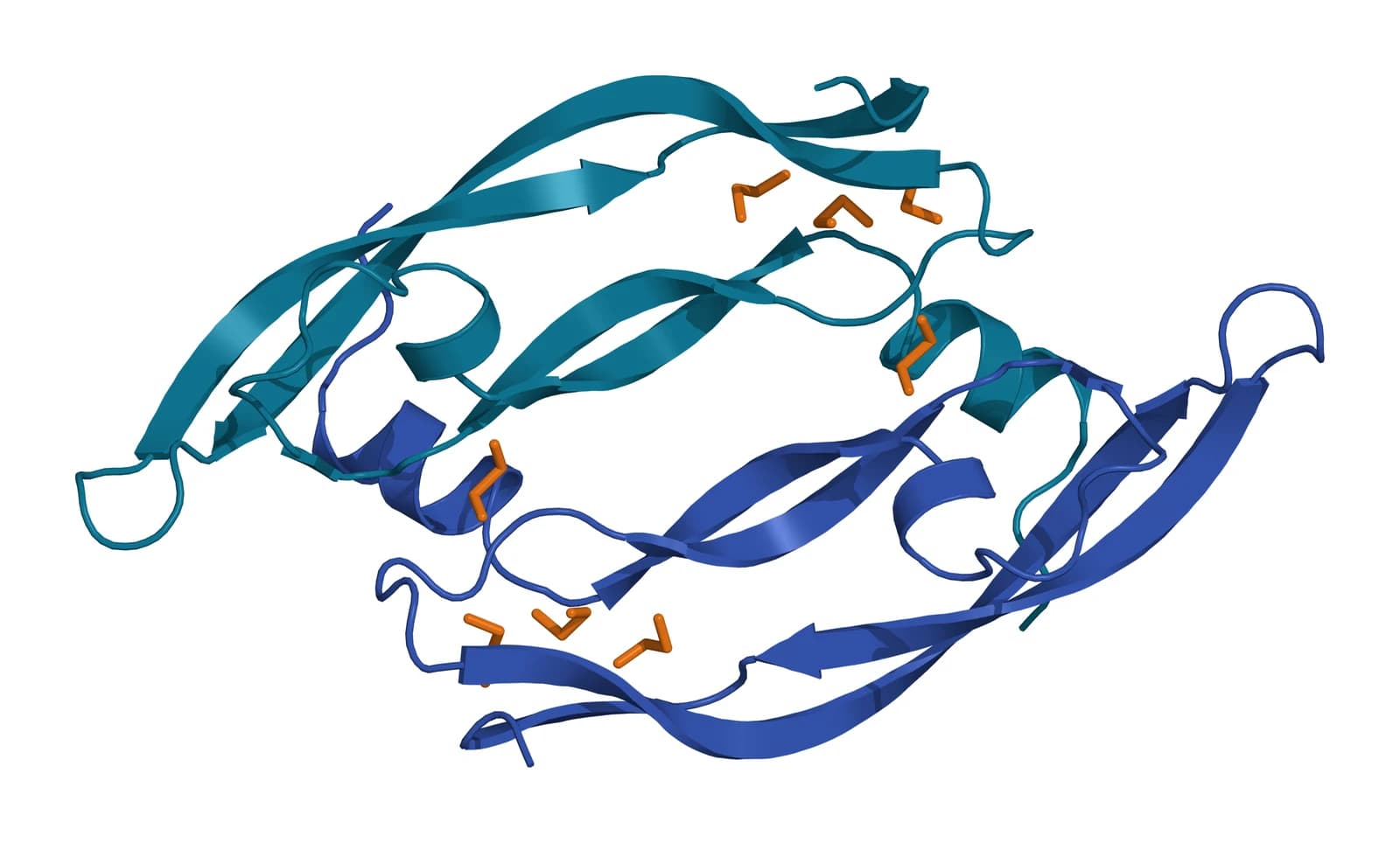
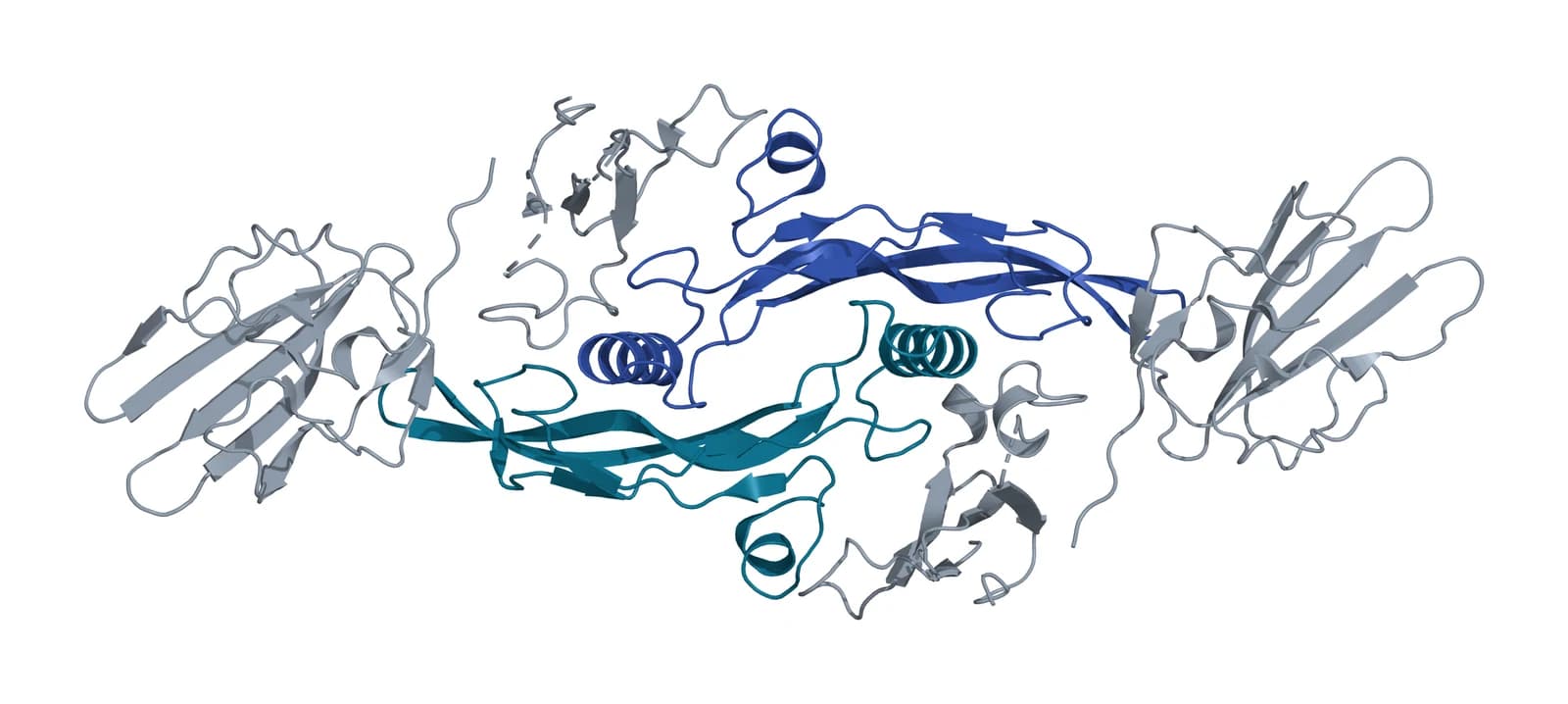
Fig. 2 | VEGF-A receptor-binding domain homodimer and TGF-beta1 bound to its type II and type I receptors.
MSC conditioned medium is produced through a defined culture workflow
Conditioned medium production starts with expanded cells, continues with a conditioning period in defined medium, and ends with harvest and clarification of the supernatant (Fig. 3).[17] Witwer and colleagues list the diversity of MSC preparations and of vesicle production and separation methods among the main sources of variation between studies.[3]
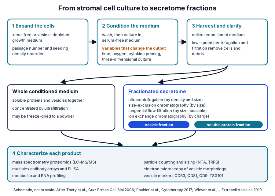
Expansion media and serum-free conditioning
Serum and platelet lysate supplements carry vesicles of their own. Pachler and colleagues depleted vesicles from pooled human platelet lysate medium by ultracentrifugation, and bone marrow MSCs grown in it retained their surface markers, morphology, viability and osteogenic and adipogenic differentiation in vitro.[18]
Vesicles from cells in that depleted medium showed a more clearly defined pattern of vesicle-associated proteins. Reducing the platelet lysate from 10 to 2 percent, or removing it, strongly altered MSC characteristics and the RNA content of released vesicles.[18]
For the conditioning step itself, Sze and colleagues used a chemically defined serum-free medium.[9] Mitchell and colleagues incubated adipose-derived MSCs in a saline solution to obtain a secretome free of exogenous molecules, and Kehl and colleagues collected secretomes under xeno-free conditions.[6][19]
Harvest, clarification and concentration
Culture supernatants contain several types of lipid membranes, and Théry and colleagues describe sequential purification steps and purity checks for isolating exosomes from them.[17] Clarified medium can then be concentrated and kept whole, or fractionated into vesicle and soluble components.[6][20]
Bari and colleagues purified secretome from culture supernatants by ultrafiltration in a validated good manufacturing practice cell factory, added a cryoprotectant and freeze-dried it. The powder contained a non-aggregated vesicle population by nanoparticle tracking analysis, and infrared spectra showed protein and lipid together.[20]
Passage number
Serra and colleagues collected conditioned media from adipose-derived cells at passages 3, 6, 9 and 12. Proteomic analysis found that secretome composition did not vary significantly across that range.[21]
Culture conditions and source tissue reshape secretome composition
Oxygen tension, inflammatory signals, culture format and source tissue each change what MSCs secrete. Paquet and colleagues compared conditioned media from cells at 21 percent, 5 percent and near-anoxic 0.1 percent oxygen.[22]
Near anoxia, the cells increased secretion of VEGF-A, VEGF-C, IL-8, RANTES and MCP-1 and decreased several inflammatory and immunomodulatory mediators. Media from those cells differed in chemotactic and angiogenic activity in the authors’ assays.[22]
Inflammatory priming affects soluble and vesicle fractions differently
Gorgun and colleagues stimulated MSCs with hypoxia and pro-inflammatory cytokines together and tested each secretome fraction separately. Inflammatory priming inhibited the pro-angiogenic capacity of the soluble fraction at every oxygen level, while the vesicle fraction kept its activity.[23]
Primed cells released proteins involved in interaction with innate immune cells and in tissue remodeling. The microRNA cargo of the vesicle fractions was less sensitive to the external stimuli than the soluble proteins.[23]
Three-dimensional culture raises vesicle yield
Haraszti and colleagues found that umbilical cord-derived MSCs gave the highest exosome yield of the sources they tested. Microcarrier-based three-dimensional culture produced 20-fold more exosomes than two-dimensional culture, and tangential flow filtration added a further 7-fold gain over ultracentrifugation.[24]
Source tissue shapes the secreted proteome
Comparative proteomic studies report distinct secretome profiles for each tissue source, with shared functional themes. The table summarizes three such comparisons by liquid chromatography and mass spectrometry.[10][19][25]
| Study | MSC sources compared | Reported finding |
|---|---|---|
| Kehl et al. 2019 | Adipose tissue, bone marrow, Wharton’s jelly | Wharton’s jelly secretomes showed the most complete network of angiogenesis-related proteins, followed by bone marrow |
| Shin et al. 2021 | Adipose tissue, bone marrow, placenta, Wharton’s jelly | Each source had a distinct profile; placental and Wharton’s jelly secretomes were the most diverse |
| Pires et al. 2016 | Bone marrow, adipose tissue, umbilical cord perivascular cells | Sources differed in neurotrophic and axon guidance proteins and shared neuroregulatory molecules |
Shin and colleagues found that, despite compositional differences, secretomes from all four sources shared functional annotations such as cell migration.[10] Pires and colleagues likewise reported that all three cell populations secreted neuroregulatory molecules.[25]
Purified EV fractions separate vesicles from the soluble secretome
Whole conditioned medium holds soluble proteins and vesicles together, and separation methods divide it into a vesicle fraction and a soluble fraction. Mitchell and colleagues reported that bioactive factors were distributed across both, and that each fraction affected different aspects of the processes they studied.[6]
Four separation principles
- Differential ultracentrifugation pellets vesicles by size and density after low-speed steps remove cells and debris.[17]
- Size-exclusion chromatography elutes vesicles ahead of smaller proteins and lipoproteins. Böing and colleagues recovered particles larger than 70 nm in early fractions with less than 5 percent of HDL and less than 1 percent of protein.[26]
- Tangential flow filtration retains vesicles while medium passes across a membrane, which scales to large culture volumes.[24]
- Ion exchange chromatography captures negatively charged vesicles on an anion exchange resin and elutes them with salt.[27]
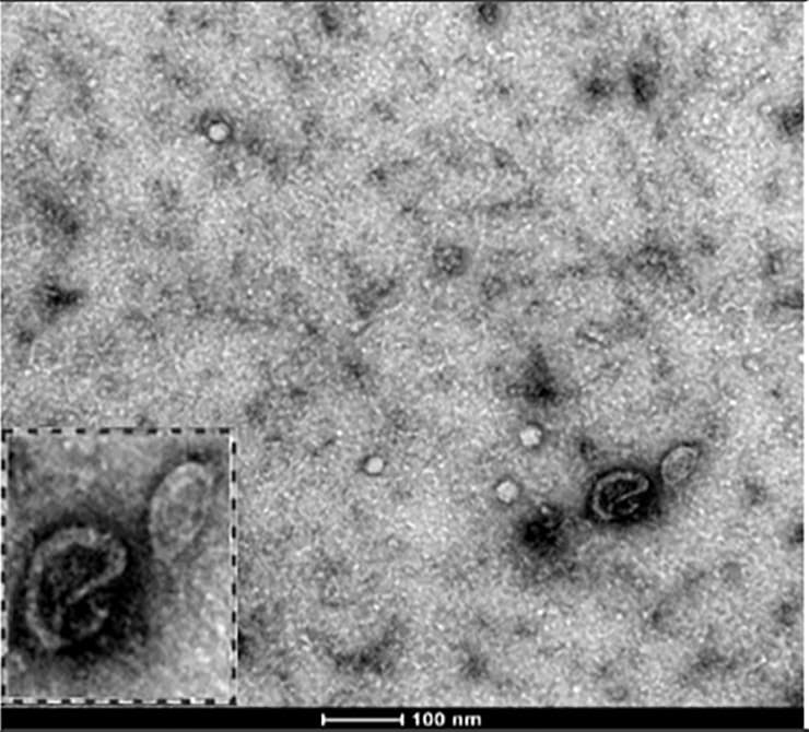
Malvicini and colleagues applied anion exchange chromatography to MSC conditioned media and recovered most vesicles in a single elution fraction. The vesicles measured under 150 nm on average and carried CD9, CD63, CD81 and TSG101 (Fig. 4).[27]
MISEV2023 terminology describes what a fraction contains
The International Society for Extracellular Vesicles updated its minimal information guidance as MISEV2023, covering production, separation and characterization of vesicles from cell culture, body fluids and tissues. The document addresses nomenclature and the separation of vesicles from non-vesicular extracellular particles.[28]
Applied to the secretome, this vocabulary names conditioned medium as the starting material and the separated product as an extracellular vesicle preparation with a stated separation method. The MISEV2023 compliance and exosome references on this site explain the terms in detail.[3][28]
Proteomics, immunoassays and particle counting characterize the secretome
Each fraction of the MSC secretome is measured with methods suited to its contents. Proteomics catalogs the proteins, immunoassays quantify named factors, and particle analysis counts and sizes the vesicles.[3]
Mass spectrometry proteomics
Kehl and colleagues analyzed secretomes by liquid chromatography tandem mass spectrometry and found biological processes related to angiogenesis enriched in the proteomic profiles.[19] Bari and colleagues used proteomic characterization to identify proteins involved in immune response, stress response, cytoskeleton and metabolism in their freeze-dried secretome.[20]
Multiplex immunoassays and ELISA
Antibody arrays measure many named factors in parallel, as in the IL-6, IL-8, MCP-1 and VEGF profile of Park and colleagues.[15] Targeted immunoassays quantify a single analyte, and Malvicini and colleagues used a CD63 ELISA to follow vesicles through their chromatography fractions.[27]
Particle counting and sizing
Nanoparticle tracking analysis follows the Brownian motion of individual particles in a light-scattering video and calculates their size and concentration. Dragovic and colleagues measured vesicles as small as about 50 nm, below the roughly 300 nm limit of conventional flow cytometry, and labeled them with antibody-conjugated quantum dots to determine phenotype.[29]
A characterization panel for a conditioned medium or vesicle preparation typically reports:
- Protein identity and relative abundance, from mass spectrometry proteomics.[9][19]
- Concentrations of named growth factors and cytokines, from antibody arrays or ELISA.[11][15]
- Particle concentration and size distribution, from nanoparticle tracking or resistive pulse sensing.[27][29]
- Vesicle morphology by electron microscopy and vesicle markers such as CD63, CD81, CD9 and TSG101.[27][28]
- Small RNA content by sequencing, where vesicle cargo is studied.[16]
Shared reporting standards make MSC-EV data comparable
Witwer and colleagues, on behalf of SOCRATES, ISEV, ISCT and ISBT, proposed harmonization criteria for small vesicles from MSCs. Preparations should be defined by quantifiable metrics for the cellular origin of the vesicles, the presence of lipid membrane vesicles and their physical and biochemical integrity.[3]
Gimona and colleagues describe MSC small vesicles as 50 to 200 nm in diameter and note that the cells producing them respond to changes in their microenvironment. They argue that identity and potency metrics must be specified during development, with potency assays built around the vesicle attribute most relevant to the intended biological activity.[30]
Together with MISEV2023, these papers give secretome studies a common vocabulary for source cells, culture conditions, separation methods and measurements.[3][28][30]
Where to go from here
The ISCT minimal criteria reference explains how the cells behind a secretome are defined, and the source tissues reference compares bone marrow, adipose and perinatal MSCs. The immunobiology reference follows HLA, IDO, PGE2 and TSG-6 into immune cell signaling, and the stem versus stromal cell reference explains the terminology.
The perinatal tissue donor eligibility reference covers how placental and umbilical cord tissue is screened under federal rules. The exosome and MISEV2023 compliance references describe vesicle biology and reporting, and the surface receptor chemistry reference covers the tetraspanins used as vesicle markers.
Verified practices can request lot documentation, and our team is glad to discuss the methods described on this page.
Referencias comentadas
Numeradas en el orden en que se citan en el texto. Cada fuente lleva una nota sobre lo que respalda en esta página; los artículos revisados por pares se listan con revista, volumen, número y páginas, y las fuentes primarias son regulaciones, documentos de agencias y bases de datos de referencia.
- 1.Peer-reviewedKonala VB, Mamidi MK, Bhonde R, et al. The current landscape of the mesenchymal stromal cell secretome: A new paradigm for cell-free regeneration. Cytotherapy. 2016;18(1):13-24. https://pubmed.ncbi.nlm.nih.gov/26631828/Annotation: Review of MSC secretome research. Cited for the definition of the secretome as soluble factors plus factors released in extracellular vesicles such as exosomes and microvesicles.
- 2.Peer-reviewedChen TS, Lai RC, Lee MM, et al. Mesenchymal stem cell secretes microparticles enriched in pre-microRNAs. Nucleic Acids Research. 2010;38(1):215-224. https://pubmed.ncbi.nlm.nih.gov/19850715/Annotation: Biochemical study of RNA in MSC conditioned medium. Cited for small RNAs enclosed in cholesterol-rich phospholipid vesicles and a high precursor to mature microRNA ratio.
- 3.Peer-reviewedWitwer KW, Van Balkom BWM, Bruno S, et al. Defining mesenchymal stromal cell (MSC)-derived small extracellular vesicles for therapeutic applications. Journal of Extracellular Vesicles. 2019;8(1):1609206. https://pubmed.ncbi.nlm.nih.gov/31069028/Annotation: Position paper from SOCRATES, ISEV, ISCT and ISBT members. Cited for harmonization criteria, quantifiable metrics of cellular origin, membrane vesicle presence and integrity, sources of variation between studies, and a biological reference preparation.
- 4.Peer-reviewedCaplan AI, Dennis JE. Mesenchymal stem cells as trophic mediators. Journal of Cellular Biochemistry. 2006;98(5):1076-1084. https://pubmed.ncbi.nlm.nih.gov/16619257/Annotation: Review that framed MSCs as sources of secreted cytokines and growth factors. Cited for paracrine and autocrine activity and for trophic effects as distinct from differentiation.
- 5.Peer-reviewedDi Nicola M, Carlo-Stella C, Magni M, et al. Human bone marrow stromal cells suppress T-lymphocyte proliferation induced by cellular or nonspecific mitogenic stimuli. Blood. 2002;99(10):3838-3843. https://pubmed.ncbi.nlm.nih.gov/11986244/Annotation: Co-culture study of bone marrow stromal cells and T cells. Cited for suppression across a Transwell membrane and for TGF-beta1 and HGF identified as mediators by neutralizing antibodies.
- 6.Peer-reviewedMitchell R, Mellows B, Sheard J, et al. Secretome of adipose-derived mesenchymal stem cells promotes skeletal muscle regeneration through synergistic action of extracellular vesicle cargo and soluble proteins. Stem Cell Research & Therapy. 2019;10(1):116. https://pubmed.ncbi.nlm.nih.gov/30953537/Annotation: Cell and mouse study comparing whole secretome with its vesicle fraction. Cited for a secretome collected in saline free of exogenous molecules and for bioactive factors distributed across both vesicle and soluble fractions.
- 7.Peer-reviewedShowalter MR, Wancewicz B, Fiehn O, et al. Primed mesenchymal stem cells package exosomes with metabolites associated with immunomodulation. Biochemical and Biophysical Research Communications. 2019;512(4):729-735. https://pubmed.ncbi.nlm.nih.gov/30926165/Annotation: Metabolomic profiling of primed MSCs and their exosomes. Cited for glycolytic reprogramming and exosomal metabolites associated with macrophage polarization and regulatory T cell induction.
- 8.Peer-reviewedLai RC, Tan SS, Yeo RW, et al. MSC secretes at least 3 EV types each with a unique permutation of membrane lipid, protein and RNA. Journal of Extracellular Vesicles. 2016;5:29828. https://pubmed.ncbi.nlm.nih.gov/26928672/Annotation: Affinity separation of MSC vesicle types with lipid-binding ligands. Cited for at least three vesicle types with distinct lipid, protein and RNA content and for EDA-containing fibronectin in one type.
- 9.Peer-reviewedSze SK, de Kleijn DP, Lai RC, et al. Elucidating the secretion proteome of human embryonic stem cell-derived mesenchymal stem cells. Molecular & Cellular Proteomics. 2007;6(10):1680-1689. https://pubmed.ncbi.nlm.nih.gov/17565974/Annotation: Proteomic and antibody array analysis of serum-free MSC conditioned medium. Cited for 201 unique gene products, 86 to 88 percent with detectable transcripts, and predicted signaling pathways.
- 10.Peer-reviewedShin S, Lee J, Kwon Y, et al. Comparative Proteomic Analysis of the Mesenchymal Stem Cells Secretome from Adipose, Bone Marrow, Placenta and Wharton’s Jelly. International Journal of Molecular Sciences. 2021;22(2):845. https://pubmed.ncbi.nlm.nih.gov/33467726/Annotation: Mass spectrometry of conditioned media from four MSC sources. Cited for distinct source-specific profiles, shared functions such as cell migration, and the more diverse composition of placental and Wharton’s jelly secretomes.
- 11.Peer-reviewedHsiao ST, Asgari A, Lokmic Z, et al. Comparative analysis of paracrine factor expression in human adult mesenchymal stem cells derived from bone marrow, adipose, and dermal tissue. Stem Cells and Development. 2012;21(12):2189-2203. https://pubmed.ncbi.nlm.nih.gov/22188562/Annotation: Expression and conditioned media protein comparison across MSC sources. Cited for VEGF-A, angiogenin, bFGF and NGF at comparable levels, higher IGF-1, VEGF-D and IL-8 in adipose-derived cells, and VEGF-A and VEGF-D in endothelial tube formation.
- 12.DatabaseNCBI Gene 7422, VEGFA vascular endothelial growth factor A (Homo sapiens). Accessed 20 October 2026. https://www.ncbi.nlm.nih.gov/gene/7422
- 13.DatabaseNCBI Gene 3082, HGF hepatocyte growth factor (Homo sapiens). Accessed 20 October 2026. https://www.ncbi.nlm.nih.gov/gene/3082
- 14.DatabaseNCBI Gene 7040, TGFB1 transforming growth factor beta 1 (Homo sapiens). Accessed 20 October 2026. https://www.ncbi.nlm.nih.gov/gene/7040
- 15.Peer-reviewedPark CW, Kim KS, Bae S, et al. Cytokine secretion profiling of human mesenchymal stem cells by antibody array. International Journal of Stem Cells. 2009;2(1):59-68. https://pubmed.ncbi.nlm.nih.gov/24855521/Annotation: Antibody array profiling of bone marrow MSC media. Cited for predominant IL-6, elevated IL-8, TIMP-2, MCP-1, VEGF and osteoprotegerin, and a profile shared across three donors.
- 16.Peer-reviewedBaglio SR, Rooijers K, Koppers-Lalic D, et al. Human bone marrow- and adipose-mesenchymal stem cells secrete exosomes enriched in distinctive miRNA and tRNA species. Stem Cell Research & Therapy. 2015;6(1):127. https://pubmed.ncbi.nlm.nih.gov/26129847/Annotation: Small RNA sequencing of MSCs and their exosomes. Cited for microRNAs at 2 to 5 percent of exosomal small RNA, selective microRNA enrichment and preferential release of tRNA species and tRNA halves.
- 17.Peer-reviewedThéry C, Amigorena S, Raposo G, et al. Isolation and characterization of exosomes from cell culture supernatants and biological fluids. Current Protocols in Cell Biology. 2006;Chapter 3:Unit 3.22. https://pubmed.ncbi.nlm.nih.gov/18228490/Annotation: Methods protocol for exosome purification. Cited for the several membrane types in culture supernatants and for sequential purification and purity checks, including differential ultracentrifugation.
- 18.Peer-reviewedPachler K, Lener T, Streif D, et al. A Good Manufacturing Practice-grade standard protocol for exclusively human mesenchymal stromal cell-derived extracellular vesicles. Cytotherapy. 2017;19(4):458-472. https://pubmed.ncbi.nlm.nih.gov/28188071/Annotation: Protocol study of vesicle-depleted human platelet lysate medium. Cited for retained MSC markers, morphology, viability and differentiation, a clearer vesicle protein pattern, and altered cells and vesicle RNA at reduced lysate.
- 19.Peer-reviewedKehl D, Generali M, Mallone A, et al. Proteomic analysis of human mesenchymal stromal cell secretomes: a systematic comparison of the angiogenic potential. NPJ Regenerative Medicine. 2019;4:8. https://pubmed.ncbi.nlm.nih.gov/31016031/Annotation: LC-MS/MS proteomics of xeno-free secretomes from adipose, bone marrow and Wharton’s jelly MSCs with cell and mouse assays. Cited for xeno-free collection, enrichment of angiogenesis-related processes and the most complete angiogenic network in Wharton’s jelly secretomes.
- 20.Peer-reviewedBari E, Perteghella S, Di Silvestre D, et al. Pilot Production of Mesenchymal Stem/Stromal Freeze-Dried Secretome for Cell-Free Regenerative Nanomedicine: A Validated GMP-Compliant Process. Cells. 2018;7(11):190. https://pubmed.ncbi.nlm.nih.gov/30380806/Annotation: Pilot manufacturing and characterization of freeze-dried secretome. Cited for ultrafiltration, lyophilization, non-aggregated vesicles by nanoparticle tracking, protein and lipid by infrared spectra, and proteomic characterization.
- 21.Peer-reviewedSerra SC, Costa JC, Assunção-Silva RC, et al. Influence of passage number on the impact of the secretome of adipose tissue stem cells on neural survival, neurodifferentiation and axonal growth. Biochimie. 2018;155:119-128. https://pubmed.ncbi.nlm.nih.gov/30342112/Annotation: Proteomic and functional comparison of conditioned media from passages 3 to 12. Cited for secretome composition that did not vary significantly with passage number.
- 22.Peer-reviewedPaquet J, Deschepper M, Moya A, et al. Oxygen Tension Regulates Human Mesenchymal Stem Cell Paracrine Functions. Stem Cells Translational Medicine. 2015;4(7):809-821. https://pubmed.ncbi.nlm.nih.gov/25979862/Annotation: Cell and animal study of conditioned media at 21, 5 and 0.1 percent oxygen. Cited for increased VEGF-A, VEGF-C, IL-8, RANTES and MCP-1 near anoxia, reduced inflammatory mediators and enhanced chemotactic and proangiogenic activity.
- 23.Peer-reviewedGorgun C, Ceresa D, Lesage R, et al. Dissecting the effects of preconditioning with inflammatory cytokines and hypoxia on the angiogenic potential of mesenchymal stromal cell (MSC)-derived soluble proteins and extracellular vesicles (EVs). Biomaterials. 2021;269:120633. https://pubmed.ncbi.nlm.nih.gov/33453634/Annotation: Fraction-by-fraction study of MSC priming with hypoxia and cytokines. Cited for inhibited angiogenic capacity of the soluble fraction, a stable vesicle fraction, shifted protein secretion and less sensitive vesicle microRNA cargo.
- 24.Peer-reviewedHaraszti RA, Miller R, Stoppato M, et al. Exosomes Produced from 3D Cultures of MSCs by Tangential Flow Filtration Show Higher Yield and Improved Activity. Molecular Therapy. 2018;26(12):2838-2847. https://pubmed.ncbi.nlm.nih.gov/30341012/Annotation: Comparison of MSC sources, culture formats and isolation methods. Cited for the highest yield from umbilical cord MSCs, 20-fold more exosomes from microcarrier 3D culture and a further 7-fold gain with tangential flow filtration.
- 25.Peer-reviewedPires AO, Mendes-Pinheiro B, Teixeira FG, et al. Unveiling the Differences of Secretome of Human Bone Marrow Mesenchymal Stem Cells, Adipose Tissue-Derived Stem Cells, and Human Umbilical Cord Perivascular Cells: A Proteomic Analysis. Stem Cells and Development. 2016;25(14):1073-1083. https://pubmed.ncbi.nlm.nih.gov/27226274/Annotation: Mass spectrometry comparison of three MSC secretomes. Cited for source differences in neurotrophic and axon guidance proteins and for neuroregulatory molecules shared by all three.
- 26.Peer-reviewedBöing AN, van der Pol E, Grootemaat AE, et al. Single-step isolation of extracellular vesicles by size-exclusion chromatography. Journal of Extracellular Vesicles. 2014;3:23430. https://pubmed.ncbi.nlm.nih.gov/25279113/Annotation: Size-exclusion chromatography of platelet concentrate supernatant on Sepharose CL-2B. Cited for early fractions holding particles above 70 nm with under 5 percent of HDL and under 1 percent of protein.
- 27.Peer-reviewedMalvicini R, Santa-Cruz D, Tolomeo AM, et al. Ion exchange chromatography as a simple and scalable method to isolate biologically active small extracellular vesicles from conditioned media. PLoS One. 2023;18(9):e0291589. https://pubmed.ncbi.nlm.nih.gov/37713424/Annotation: Anion exchange isolation of MSC small vesicles from conditioned media. Cited for single-fraction elution, mean size under 150 nm, CD9, CD63, CD81 and TSG101, CD63 ELISA, resistive pulse sensing and electron microscopy.
- 28.Peer-reviewedWelsh JA, Goberdhan DCI, O’Driscoll L, et al. Minimal information for studies of extracellular vesicles (MISEV2023): From basic to advanced approaches. Journal of Extracellular Vesicles. 2024;13(2):e12404. https://pubmed.ncbi.nlm.nih.gov/38326288/Annotation: ISEV minimal information guidance. Cited for approaches to vesicle production, separation and characterization from cell culture, nomenclature, and separation from non-vesicular extracellular particles.
- 29.Peer-reviewedDragovic RA, Gardiner C, Brooks AS, et al. Sizing and phenotyping of cellular vesicles using Nanoparticle Tracking Analysis. Nanomedicine: Nanotechnology, Biology, and Medicine. 2011;7(6):780-788. https://pubmed.ncbi.nlm.nih.gov/21601655/Annotation: Methods study of fluorescence nanoparticle tracking analysis. Cited for size and concentration from Brownian motion, detection down to about 50 nm against about 300 nm for flow cytometry, and phenotyping with quantum dot labels.
- 30.Peer-reviewedGimona M, Brizzi MF, Choo ABH, et al. Critical considerations for the development of potency tests for therapeutic applications of mesenchymal stromal cell-derived small extracellular vesicles. Cytotherapy. 2021;23(5):373-380. https://pubmed.ncbi.nlm.nih.gov/33934807/Annotation: Expert review of quality metrics for MSC small vesicles. Cited for the 50 to 200 nm size range, the sensitivity of producing cells to their microenvironment, and potency assays built on a quantified vesicle attribute.
Cite this page
ExaVeyra Sciences Editorial Team. The MSC Secretome: Conditioned Medium, Growth Factors and EVs. ExaVeyra Sciences; 20 October 2026. Available from: https://exaveyra.com/science/stem-cells/msc-secretome-conditioned-medium. Accessed [date accessed].
Fuentes de las figuras
- 31.PDB 2VPF, VEGF-A receptor-binding domain homodimer. ExaVeyra Sciences, rendered in PyMOL from PDB 2VPF. Structure data: RCSB Protein Data Bank (rcsb.org), CC0. Publicación de la estructura: Muller YA, Christinger HW, Keyt BA, de Vos AM. The crystal structure of vascular endothelial growth factor (VEGF) refined to 1.93 A resolution: multiple copy flexibility and receptor binding. Structure. 1997;5(10):1325-1338.
- 32.PDB 3KFD, TGF-beta1 ternary complex with TGFBR2 and TGFBR1. ExaVeyra Sciences, rendered in PyMOL from PDB 3KFD. Structure data: RCSB Protein Data Bank (rcsb.org), CC0. Publicación de la estructura: Radaev S, Zou Z, Huang T, Lafer EM, Hinck AP, Sun PD. Ternary complex of transforming growth factor-beta1 reveals isoform-specific ligand recognition and receptor recruitment in the superfamily. J Biol Chem. 2010;285(19):14806-14814.