Esta referencia está disponible actualmente en inglés. La versión en español está en preparación.
MSC Licensing and Preconditioning: IFN-γ, TNF-α and Hypoxia
Por Benn Bluestein-Veyra, M.Sc. en Química Orgánica · Actualizado el 20 de octubre de 2026
ExaVeyra Sciences es un proveedor B2B con sede en Miami que suministra exosomas para uso estético tópico a clínicas con licencia, y remite a los prescriptores con licencia a una farmacia de preparados compuestos 503A que prepara formulaciones según prescripciones individuales.
| Pregunta | Respuesta |
|---|---|
| Qué cubre esta página | How inflammatory cytokines, low oxygen and three-dimensional culture reprogram stromal cell gene expression and the cargo of their extracellular vesicles. |
| Quién puede hacer pedidos | Clínicas y profesionales con licencia profesional activa y NPI verificados por ExaVeyra. Los pedidos de exosomas requieren además un reconocimiento firmado de uso profesional. |
| Qué se envía y cómo | El material de exosomas para uso estético tópico, caracterizado por lote, se envía en cadena de frío por exprés nocturno. La entrega puede tardar 1 a 3 días hábiles. Se emite un certificado de análisis para cada lote y se entrega a solicitud. |
MSC licensing is the exposure of mesenchymal stromal cells to a defined signal that switches on an inducible gene program before the cells are assayed. The best-studied signals are IFN-gamma and TNF-alpha, low oxygen culture and aggregation into three-dimensional spheroids.[1][2][3]
This reference covers each priming condition, the genes and secreted factors it induces, and the assays that measure the response. The molecular actions of IDO1, PGE2 and TSG-6 belong to the MSC immunobiology reference, and this page centers on the culture conditions that set them in motion.
Defining MSC licensing as a response to the culture environment
Krampera and colleagues showed in 2006 that bone marrow MSCs suppressed the proliferation of CD4+ and CD8+ T cells and NK cells, and that this suppression required IFN-gamma produced by the activated lymphocytes. IFN-gamma acted by inducing IDO activity in the stromal cells.[1]
Ren and colleagues reached a parallel result in mouse MSCs. IFN-gamma together with TNF-alpha, IL-1alpha or IL-1beta induced chemokines and inducible nitric oxide synthase, and the suppressive effect required the IFN-gamma receptor IFNGR1.[4]
In the Krampera study, suppression worked without cell contact, and added IFN-gamma made even activated B cells susceptible to it.[1] In the Ren study, the induced chemokines drew T cells close to the stromal cells, where nitric oxide held their responsiveness down.[4]
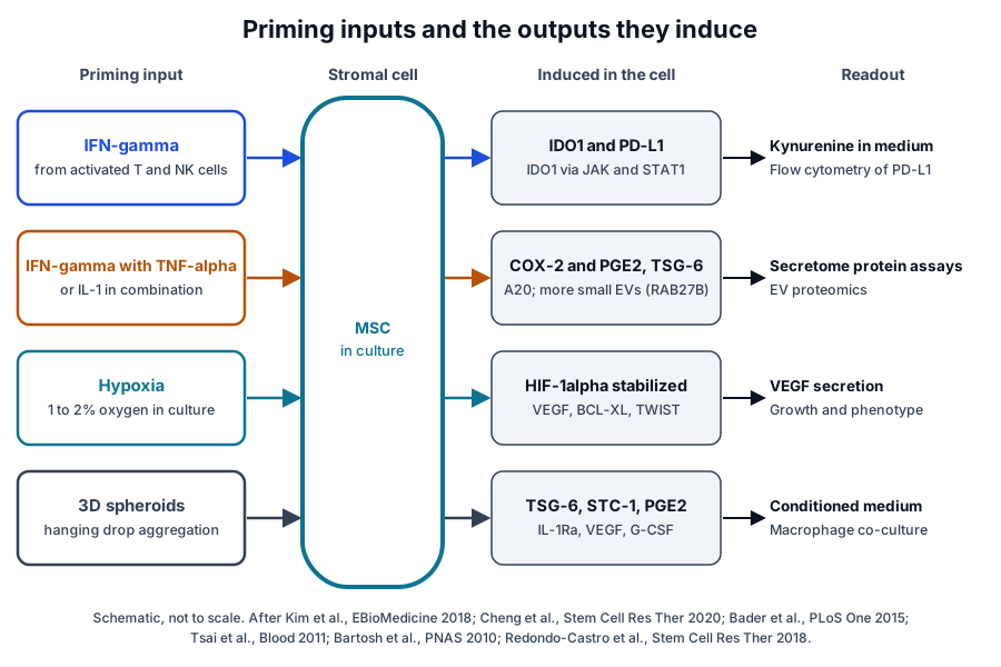
These studies framed MSC immune activity as an inducible state set by the culture environment (Fig. 1). An ISCT working proposal led by Krampera then called for standardized immune assays that yield shared, reproducible and consistent data on that state.[5]
Licensing, priming and preconditioning describe one practice
Three overlapping terms describe this practice. Licensing and priming usually refer to cytokine exposure, and preconditioning often refers to a period of hypoxia before a test; Cheng and colleagues group cytokines, hypoxia and chemicals together as priming approaches.[2][6][7]
IFN-gamma and TNF-alpha priming induces a defined set of genes
IFN-gamma signals as a homodimer that engages two chains of its high-affinity receptor. Walter and colleagues solved this complex in 1995 and found one IFN-gamma homodimer bound to two receptor molecules held 27 Å apart.[8] The human genes IFNG and IFNGR1 lie on chromosomes 12q15 and 6q23.3.[9][10]
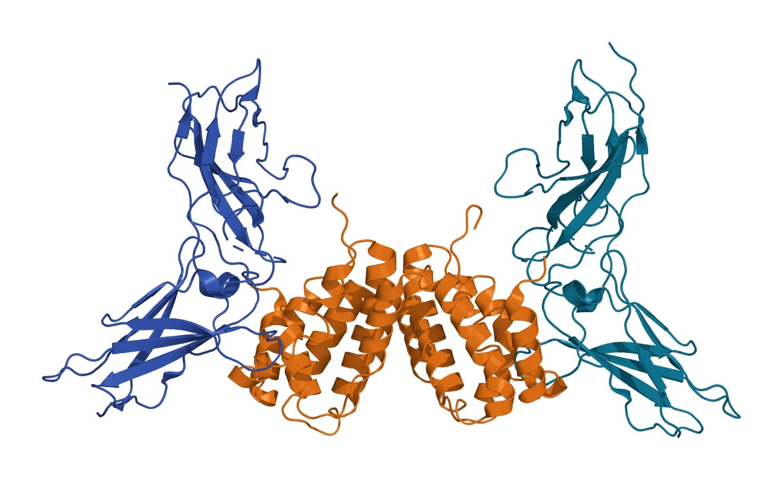
Thiel and colleagues solved the human complex with glycosylated receptor at 2.9 Å resolution (Fig. 2). Beside the expected 2:1 complex, the crystal held a third receptor molecule packed against the edge strands of the receptor C-terminal domains.[11]
Kim and colleagues traced IDO expression in IFN-gamma-primed human MSCs to the JAK and STAT1 pathway. In their account, IFN-gamma from activated T cells induces IDO in the stromal cells, which in turn suppresses T cell proliferation.[12]
Crop and colleagues profiled the full genome of adipose MSCs exposed to IFN-gamma, TNF-alpha and IL-6 or to a mixed lymphocyte reaction. The cytokines mainly raised IDO, the mixed lymphocyte reaction raised COX-2, and both conditions enhanced suppression with differentiation capacity preserved.[13]
Roemeling-van Rhijn and colleagues found that IFN-gamma with TNF-alpha strongly raised IDO and PD-L1 in adipose MSCs. L-kynurenine accumulated in the medium as a direct measure of IDO activity, and the same induction appeared at 1% and 20% oxygen.[14]
Hypoxic preconditioning stabilizes HIF-1alpha
Wang, Semenza and colleagues identified HIF-1 in 1995 as a heterodimer of two basic helix-loop-helix PAS proteins, HIF-1alpha and HIF-1beta, the latter encoded by ARNT. Both subunits rose in cells held at 1% oxygen and decayed rapidly on return to 20%.[15]
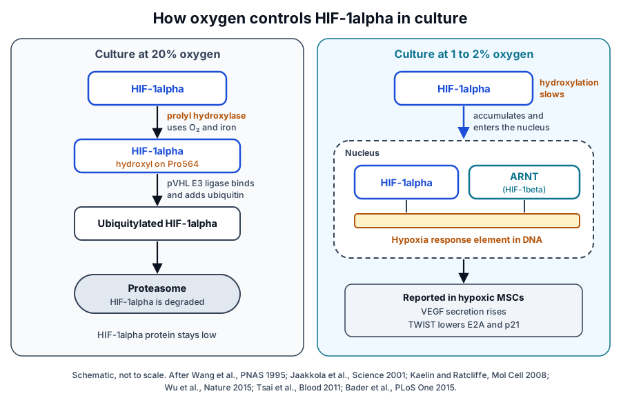
Jaakkola and colleagues showed that oxygen controls HIF-1alpha through hydroxylation of proline 564, which lets the pVHL E3 ligase complex bind and ubiquitylate it (Fig. 3). The hydroxylase needs dioxygen and iron, so it acts as a direct cellular oxygen sensor.[16]
Kaelin and Ratcliffe describe the PHD family of prolyl hydroxylases as the oxygen sensors of this pathway, with asparagine hydroxylation by FIH1 tuning HIF transcriptional activity further.[17] The human HIF1A gene lies on chromosome 14q23.2.[18]
Crystal structures show ARNT wrapped around HIF-1alpha
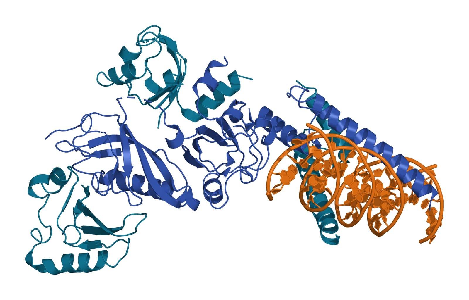
Wu and colleagues solved mouse HIF-1alpha and HIF-2alpha in complex with ARNT, including HIF-1alpha and ARNT on hypoxia response element DNA (Fig. 4). ARNT spirals around the outside of each HIF-alpha subunit, and the DNA-reading head cooperates with a distal PAS domain to bind the element.[19]
HIF-1alpha and HIF-2alpha share this highly integrated quaternary architecture with ARNT. The structures also revealed five pockets able to bind small molecules, including sites inside the PAS domains and a cavity formed at the heterodimer interface.[19]
Low oxygen culture changes growth, survival signaling and immune function
Roemeling-van Rhijn and colleagues note that oxygen levels within tissues are typically much lower than those of standard culture.[14] Hypoxic preconditioning sets the culture oxygen closer to that tissue range before the cells are tested.[2]
Grayson and colleagues kept human MSCs at 2% oxygen for up to seven passages and measured about 30-fold higher expansion over 6 weeks, with multilineage differentiation retained. Hypoxic cells also expressed more Oct-4, HIF-2alpha and connexin-43 and built more extracellular matrix.[20]
Tsai and colleagues found that low-density hypoxic culture preserved MSC properties through a HIF-1alpha and TWIST axis that lowered E2A and p21. Hypoxic cells reached up to 100 population doublings with telomerase activity, stable telomere length and a normal karyotype.[21]
Bader and colleagues preconditioned umbilical cord blood MSCs in hypoxia for 24 h with immunophenotype and karyotype unchanged. Preconditioned cells showed more AKT and BAD phosphorylation, higher BCL-XL, BAG1 and VEGF expression and more VEGF secretion, and they raised endothelial cell proliferation and migration.[2]
Hypoxia preserves the cytokine licensing response
At 1% oxygen, adipose MSCs kept their proliferation and colony-forming efficiency, with CD105 the one marker that fell. IDO and PD-L1 induction by IFN-gamma and TNF-alpha, kynurenine output and suppression of CD4+ and CD8+ T cell proliferation all held steady.[14]
Three-dimensional spheroid culture self-activates stromal cells
Bartosh and colleagues aggregated human MSCs into spheroids by hanging drop culture and found high expression of TSG-6 and stanniocalcin-1, with properties that depended critically on the culture conditions. Spheroid cells were about one-fourth the volume of cells from adherent monolayers.[22]
In co-culture with LPS-activated macrophages, spheroid MSCs suppressed inflammatory responses more effectively than monolayer MSCs.[22] Ylöstalo and colleagues traced the activity of spheroid conditioned medium to PGE2, which required COX-2 and acted on macrophages through the EP4 receptor.[23]
Spheroid medium lowered TNF-alpha, IL-6 and IL-12p40 secretion by stimulated macrophages and raised IL-10, IL-1Ra and CD206. PGE2 production depended on caspase activity and NF-kappaB activation, and fibroblast spheroids produced little PGE2.[23]
Spheroid format and cytokine priming interact
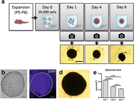
Redondo-Castro and colleagues formed spheroids from 20,000 human MSCs and followed their compaction over 6 d (Fig. 5). Spheroid culture raised IL-1Ra, VEGF and G-CSF secretion compared with monolayers, and IL-1 priming acted more strongly on monolayers than on spheroids.[24]
In the same study, spheroid conditioned medium lowered TNF-alpha secretion by LPS-stimulated BV2 microglial cells, and whole spheroids in co-culture raised IL-6. Protein arrays showed a stronger immune profile after priming, which the spheroid format partly offset through higher IL-6.[24]
Cesarz and Tamama review the wider changes in spheroid MSCs: cytoskeletal reorganization, higher expression of pluripotency marker genes, delayed replicative senescence and broad shifts in gene expression measured by microarray. The authors identify the signaling pathways that trigger and sustain these changes as an open question for the field.[3]
Priming reshapes the secretome and extracellular vesicle cargo
Harting and colleagues compared extracellular vesicles from naive and inflammation-stimulated MSCs, isolated with a tangential flow filtration system compliant with current Good Manufacturing Practice. The two preparations matched in size and common surface markers and differed in protein composition, cytokine profile and RNA content.[25]
Vesicles from stimulated cells lowered pro-inflammatory cytokine release in vitro further than vesicles from naive cells. Activated leukocyte subsets took them up in a distinct pattern, and part of their activity traced to the COX-2 and PGE2 pathway.[25]
Cheng and colleagues primed adipose MSCs with IFN-gamma and TNF-alpha and found higher RAB27B expression and more small vesicles under 200 nm. These vesicles carried A20 and TSG-6 and inhibited T cell proliferation in a dose-dependent manner.[7]
In the same study, MSCs from pediatric donors expressed more RAB27B and secreted more small vesicles than adult MSCs.[7] Peltzer and colleagues primed bone marrow MSCs from five donors with IFN-gamma or hypoxia and found vesicle amount and size unchanged.[26]
Of 81 miRNAs detected in those vesicles, 41 appeared in every sample. Hypoxia lowered miR-34a-3p and IFN-gamma raised five miRNAs, including miR-126-3p and miR-451a, and the authors described the overall change as limited, with high variability between donors.[26]
Gene panels and secretome assays measure the licensing response
Galipeau and colleagues, reporting an ISCT workshop, proposed that MSC potency be measured by licensing the cells in vitro and reading selected markers with a matrix of assays. The preferred methods were quantitative RNA analysis, flow cytometry of functional surface markers and protein assays of the secretome.[6]
Chinnadurai and colleagues tested two such systems on MSCs from human donors: secretome analysis and a quantitative RNA array of immunomodulatory and homing genes. A cytokine signature in the secretome correlated with T cell suppression, and IFN-gamma served as a surrogate for responder blood mononuclear cells.[27]
Single studies often read one licensed culture several ways. Roemeling-van Rhijn and colleagues measured IDO and PD-L1 expression, L-kynurenine in the medium and suppression of T cells stimulated with anti-CD3 and anti-CD28 antibodies.[14]
Crop and colleagues paired whole-genome expression with functional assays of morphology, proliferation, differentiation and immune suppression in the same adipose MSCs.[13] Kim and colleagues confirmed IDO’s role with knockdown experiments.[12]
| Priming condition | Typical readouts | Example studies |
|---|---|---|
| IFN-gamma | IDO transcript, kynurenine in medium, PD-L1 by flow cytometry | Kim 2018; Roemeling-van Rhijn 2013 |
| IFN-gamma with TNF-alpha or IL-6 | Genome-wide expression, secretome cytokines, vesicle proteome | Crop 2010; Cheng 2020 |
| Hypoxia | Expansion, HIF pathway transcripts, VEGF secretion | Grayson 2007; Bader 2015 |
| Three-dimensional spheroids | TSG-6, stanniocalcin-1 and PGE2; macrophage co-culture | Bartosh 2010; Ylöstalo 2012 |
| Vesicles from primed cells | Vesicle count and size, protein and miRNA cargo | Harting 2018; Peltzer 2020 |
Where to go from here
The MSC immunobiology reference follows IDO1, PGE2 and TSG-6 from licensing to their molecular actions, and the secretome reference covers the soluble factors and vesicles that MSCs release into conditioned medium. The single-cell heterogeneity reference explains why donors and subpopulations answer priming differently.
The source tissues and ISCT minimal criteria references describe the cells these protocols start from, and the exosome biochemistry pages cover vesicle composition in depth. Verified practices can request lot documentation, and our team is glad to discuss the methods described on this page.
Referencias comentadas
Numeradas en el orden en que se citan en el texto. Cada fuente lleva una nota sobre lo que respalda en esta página; los artículos revisados por pares se listan con revista, volumen, número y páginas, y las fuentes primarias son regulaciones, documentos de agencias y bases de datos de referencia.
- 1.Peer-reviewedKrampera M, Cosmi L, Angeli R, et al. Role for interferon-gamma in the immunomodulatory activity of human bone marrow mesenchymal stem cells. Stem Cells. 2006;24(2):386-398. https://pubmed.ncbi.nlm.nih.gov/16123384/Annotation: Co-culture study of lymphocyte subsets. Cited for suppression of CD4+, CD8+ and NK cell proliferation that required IFN-gamma from activated cells and acted through IDO.
- 2.Peer-reviewedBader AM, Klose K, Bieback K, et al. Hypoxic Preconditioning Increases Survival and Pro-Angiogenic Capacity of Human Cord Blood Mesenchymal Stromal Cells In Vitro. PLoS One. 2015;10(9):e0138477. https://pubmed.ncbi.nlm.nih.gov/26380983/Annotation: Hypoxic preconditioning of cord blood MSCs for 24 h. Cited for unchanged phenotype and karyotype, AKT and BAD phosphorylation, BCL-XL, BAG1 and VEGF, and endothelial proliferation and migration.
- 3.Peer-reviewedCesarz Z, Tamama K. Spheroid Culture of Mesenchymal Stem Cells. Stem Cells International. 2016;2016:9176357. https://pubmed.ncbi.nlm.nih.gov/26649054/Annotation: Review of MSC spheroid culture. Cited for cytoskeletal reorganization, pluripotency marker genes, delayed replicative senescence and microarray-level gene expression changes.
- 4.Peer-reviewedRen G, Zhang L, Zhao X, et al. Mesenchymal stem cell-mediated immunosuppression occurs via concerted action of chemokines and nitric oxide. Cell Stem Cell. 2008;2(2):141-150. https://pubmed.ncbi.nlm.nih.gov/18371435/Annotation: Mouse MSC study with knockout cells. Cited for IFN-gamma plus TNF-alpha, IL-1alpha or IL-1beta inducing chemokines and iNOS, and the requirement for IFNGR1.
- 5.Peer-reviewedKrampera M, Galipeau J, Shi Y, et al. Immunological characterization of multipotent mesenchymal stromal cells--The International Society for Cellular Therapy (ISCT) working proposal. Cytotherapy. 2013;15(9):1054-1061. https://pubmed.ncbi.nlm.nih.gov/23602578/Annotation: ISCT MSC Committee working proposal. Cited for the call for standardized immune assays yielding shared, reproducible and consistent data.
- 6.Peer-reviewedGalipeau J, Krampera M, Barrett J, et al. International Society for Cellular Therapy perspective on immune functional assays for mesenchymal stromal cells as potency release criterion for advanced phase clinical trials. Cytotherapy. 2016;18(2):151-159. https://pubmed.ncbi.nlm.nih.gov/26724220/Annotation: ISCT workshop perspective on MSC potency assays. Cited for in vitro licensing read with a matrix of RNA, flow cytometry and secretome protein assays.
- 7.Peer-reviewedCheng A, Choi D, Lora M, et al. Human multipotent mesenchymal stromal cells cytokine priming promotes RAB27B-regulated secretion of small extracellular vesicles with immunomodulatory cargo. Stem Cell Research & Therapy. 2020;11(1):539. https://pubmed.ncbi.nlm.nih.gov/33317598/Annotation: IFN-gamma and TNF-alpha priming of adipose MSCs. Cited for RAB27B induction, more small vesicles carrying A20 and TSG-6, dose-dependent T cell inhibition and donor age effects.
- 8.Peer-reviewedWalter MR, Windsor WT, Nagabhushan TL, et al. Crystal structure of a complex between interferon-gamma and its soluble high-affinity receptor. Nature. 1995;376(6537):230-235. https://pubmed.ncbi.nlm.nih.gov/7617032/Annotation: First crystal structure of a class-2 cytokine receptor complex. Cited for one IFN-gamma homodimer binding two receptor molecules separated by 27 Å.
- 9.DatabaseNCBI Gene 3458, IFNG interferon gamma (Homo sapiens). Accessed 20 October 2026. https://www.ncbi.nlm.nih.gov/gene/3458
- 10.DatabaseNCBI Gene 3459, IFNGR1 interferon gamma receptor 1 (Homo sapiens). Accessed 20 October 2026. https://www.ncbi.nlm.nih.gov/gene/3459
- 11.Peer-reviewedThiel DJ, le Du MH, Walter RL, et al. Observation of an unexpected third receptor molecule in the crystal structure of human interferon-gamma receptor complex. Structure. 2000;8(9):927-936. https://pubmed.ncbi.nlm.nih.gov/10986460/Annotation: Crystal structure of human IFN-gamma with glycosylated receptor alpha chains (PDB 1FG9). Cited for the 2.9 Å 2:1 complex and the third receptor molecule.
- 12.Peer-reviewedKim DS, Jang IK, Lee MW, et al. Enhanced Immunosuppressive Properties of Human Mesenchymal Stem Cells Primed by Interferon-γ. EBioMedicine. 2018;28:261-273. https://pubmed.ncbi.nlm.nih.gov/29366627/Annotation: IFN-gamma priming study of human MSCs with IDO knockdown. Cited for IDO induction through JAK and STAT1 and enhanced suppression of T cell proliferation.
- 13.Peer-reviewedCrop MJ, Baan CC, Korevaar SS, et al. Inflammatory conditions affect gene expression and function of human adipose tissue-derived mesenchymal stem cells. Clinical and Experimental Immunology. 2010;162(3):474-486. https://pubmed.ncbi.nlm.nih.gov/20846162/Annotation: Full genome expression study of adipose MSCs. Cited for IDO raised by IFN-gamma, TNF-alpha and IL-6, COX-2 raised by a mixed lymphocyte reaction, enhanced suppression and preserved differentiation.
- 14.Peer-reviewedRoemeling-van Rhijn M, Mensah FK, Korevaar SS, et al. Effects of Hypoxia on the Immunomodulatory Properties of Adipose Tissue-Derived Mesenchymal Stem cells. Frontiers in Immunology. 2013;4:203. https://pubmed.ncbi.nlm.nih.gov/23882269/Annotation: Culture of adipose MSCs at 1% and 20% oxygen. Cited for IDO and PD-L1 induction by IFN-gamma and TNF-alpha, kynurenine output, CD105 reduction and T cell suppression maintained under hypoxia.
- 15.Peer-reviewedWang GL, Jiang BH, Rue EA, et al. Hypoxia-inducible factor 1 is a basic-helix-loop-helix-PAS heterodimer regulated by cellular O2 tension. Proceedings of the National Academy of Sciences of the United States of America. 1995;92(12):5510-5514. https://pubmed.ncbi.nlm.nih.gov/7539918/Annotation: Cloning and characterization of HIF-1. Cited for the bHLH-PAS heterodimer of HIF-1alpha and ARNT and subunit levels that rose at 1% oxygen and decayed at 20%.
- 16.Peer-reviewedJaakkola P, Mole DR, Tian YM, et al. Targeting of HIF-alpha to the von Hippel-Lindau ubiquitylation complex by O2-regulated prolyl hydroxylation. Science. 2001;292(5516):468-472. https://pubmed.ncbi.nlm.nih.gov/11292861/Annotation: Biochemical study of HIF-1alpha recognition by pVHL. Cited for hydroxylation of proline 564 and the dioxygen and iron requirement of the hydroxylase.
- 17.Peer-reviewedKaelin WG Jr, Ratcliffe PJ. Oxygen sensing by metazoans: the central role of the HIF hydroxylase pathway. Molecular Cell. 2008;30(4):393-402. https://pubmed.ncbi.nlm.nih.gov/18498744/Annotation: Review of the HIF hydroxylase pathway. Cited for PHD prolyl hydroxylases as oxygen sensors and FIH1 asparagine hydroxylation tuning HIF activity.
- 18.DatabaseNCBI Gene 3091, HIF1A hypoxia inducible factor 1 subunit alpha (Homo sapiens). Accessed 20 October 2026. https://www.ncbi.nlm.nih.gov/gene/3091
- 19.Peer-reviewedWu D, Potluri N, Lu J, et al. Structural integration in hypoxia-inducible factors. Nature. 2015;524(7565):303-308. https://pubmed.ncbi.nlm.nih.gov/26245371/Annotation: Crystal structures of mouse HIF-1alpha and HIF-2alpha with ARNT (PDB 4ZPR). Cited for ARNT spiraling around HIF-alpha, DNA binding by the reading head and PAS domain, and five ligand pockets.
- 20.Peer-reviewedGrayson WL, Zhao F, Bunnell B, et al. Hypoxia enhances proliferation and tissue formation of human mesenchymal stem cells. Biochemical and Biophysical Research Communications. 2007;358(3):948-953. https://pubmed.ncbi.nlm.nih.gov/17521616/Annotation: Long-term culture of human MSCs at 2% oxygen. Cited for about 30-fold higher expansion over 6 weeks, retained multilineage differentiation, and higher Oct-4, HIF-2alpha and connexin-43.
- 21.Peer-reviewedTsai CC, Chen YJ, Yew TL, et al. Hypoxia inhibits senescence and maintains mesenchymal stem cell properties through down-regulation of E2A-p21 by HIF-TWIST. Blood. 2011;117(2):459-469. https://pubmed.ncbi.nlm.nih.gov/20952688/Annotation: Mechanistic study of low-density hypoxic MSC culture. Cited for the HIF-1alpha and TWIST axis lowering E2A and p21, up to 100 population doublings, telomerase activity and normal karyotype.
- 22.Peer-reviewedBartosh TJ, Ylöstalo JH, Mohammadipoor A, et al. Aggregation of human mesenchymal stromal cells (MSCs) into 3D spheroids enhances their antiinflammatory properties. Proceedings of the National Academy of Sciences of the United States of America. 2010;107(31):13724-13729. https://pubmed.ncbi.nlm.nih.gov/20643923/Annotation: Hanging drop spheroid study of human MSCs. Cited for TSG-6 and stanniocalcin-1 expression, dependence on culture conditions, smaller cell volume and effects in macrophage co-culture.
- 23.Peer-reviewedYlöstalo JH, Bartosh TJ, Coble K, et al. Human mesenchymal stem/stromal cells cultured as spheroids are self-activated to produce prostaglandin E2 that directs stimulated macrophages into an anti-inflammatory phenotype. Stem Cells. 2012;30(10):2283-2296. https://pubmed.ncbi.nlm.nih.gov/22865689/Annotation: Spheroid conditioned medium and macrophage study. Cited for PGE2 as the active factor, COX-2 and EP4 dependence, the macrophage cytokine shifts, and caspase and NF-kappaB dependence.
- 24.Peer-reviewedRedondo-Castro E, Cunningham CJ, Miller J, et al. Changes in the secretome of tri-dimensional spheroid-cultured human mesenchymal stem cells in vitro by interleukin-1 priming. Stem Cell Research & Therapy. 2018;9(1):11. https://pubmed.ncbi.nlm.nih.gov/29343288/Annotation: Comparison of monolayer and spheroid MSC secretomes with IL-1 priming. Cited for spheroid formation and compaction, higher IL-1Ra, VEGF and G-CSF, and stronger IL-1 priming in monolayers.
- 25.Peer-reviewedHarting MT, Srivastava AK, Zhaorigetu S, et al. Inflammation-Stimulated Mesenchymal Stromal Cell-Derived Extracellular Vesicles Attenuate Inflammation. Stem Cells. 2018;36(1):79-90. https://pubmed.ncbi.nlm.nih.gov/29076623/Annotation: Characterization of vesicles from naive and inflammation-stimulated MSCs. Cited for matched size and markers, differing protein, cytokine and RNA content, leukocyte uptake and the COX-2 and PGE2 contribution.
- 26.Peer-reviewedPeltzer J, Lund K, Goriot ME, et al. Interferon-γ and Hypoxia Priming Have Limited Effect on the miRNA Landscape of Human Mesenchymal Stromal Cells-Derived Extracellular Vesicles. Frontiers in Cell and Developmental Biology. 2020;8:581436. https://pubmed.ncbi.nlm.nih.gov/33384991/Annotation: Five-donor miRNA profiling of vesicles from primed bone marrow MSCs. Cited for unchanged vesicle amount and size, 81 detected miRNAs, the specific changes and high donor variability.
- 27.Peer-reviewedChinnadurai R, Rajan D, Qayed M, et al. Potency Analysis of Mesenchymal Stromal Cells Using a Combinatorial Assay Matrix Approach. Cell Reports. 2018;22(9):2504-2517. https://pubmed.ncbi.nlm.nih.gov/29490284/Annotation: Assay matrix study of MSC potency. Cited for secretome analysis and a quantitative RNA array, a cytokine signature correlated with T cell suppression, and IFN-gamma as a surrogate for responder cells.
Cite this page
ExaVeyra Sciences Editorial Team. MSC Licensing and Preconditioning: IFN-γ, TNF-α and Hypoxia. ExaVeyra Sciences; 20 October 2026. Available from: https://exaveyra.com/science/stem-cells/msc-licensing-preconditioning. Accessed [date accessed].
Fuentes de las figuras
- 28.PDB 1FG9, Human IFN-gamma bound to its receptor alpha chains. ExaVeyra Sciences, rendered in PyMOL from PDB 1FG9. Structure data: RCSB Protein Data Bank (rcsb.org), CC0. Publicación de la estructura: Thiel DJ, le Du MH, Walter RL, et al. Observation of an unexpected third receptor molecule in the crystal structure of human interferon-gamma receptor complex. Structure. 2000;8(9):927-936.
- 29.PDB 4ZPR, HIF-1alpha and ARNT on hypoxia response element DNA. ExaVeyra Sciences, rendered in PyMOL from PDB 4ZPR. Structure data: RCSB Protein Data Bank (rcsb.org), CC0. Publicación de la estructura: Wu D, Potluri N, Lu J, Kim Y, Rastinejad F. Structural integration in hypoxia-inducible factors. Nature. 2015;524(7565):303-308.