Esta referencia está disponible actualmente en inglés. La versión en español está en preparación.
Platelet-Rich Fibrin and Fibrin Matrix Biology
Por Benn Bluestein-Veyra, M.Sc. en Química Orgánica · Actualizado el 21 de octubre de 2026
ExaVeyra Sciences es un proveedor B2B con sede en Miami que suministra exosomas para uso estético tópico a clínicas con licencia, y remite a los prescriptores con licencia a una farmacia de preparados compuestos 503A que prepara formulaciones según prescripciones individuales.
| Pregunta | Respuesta |
|---|---|
| Qué cubre esta página | How fibrinogen polymerizes into a fibrin network, how platelet-rich fibrin preparations form, and how the fibrin matrix holds and releases platelet proteins. |
| Quién puede hacer pedidos | Clínicas y profesionales con licencia profesional activa y NPI verificados por ExaVeyra. Los pedidos de exosomas requieren además un reconocimiento firmado de uso profesional. |
| Qué se envía y cómo | El material de exosomas para uso estético tópico, caracterizado por lote, se envía en cadena de frío por exprés nocturno. La entrega puede tardar 1 a 3 días hábiles. Se emite un certificado de análisis para cada lote y se entrega a solicitud. |
Platelet-rich fibrin (PRF) is a fibrin clot that forms when blood collected without anticoagulant is centrifuged, gathering platelets and leukocytes into the fibrin matrix.[1][2] Its biology is the biology of fibrin itself: thrombin converts fibrinogen into monomers that polymerize into a branched, cross-linked network.[3]
This reference follows that sequence from the fibrinogen molecule to the finished network, then describes how preparation variables shape a platelet-rich fibrin clot and how the matrix holds and releases platelet proteins in vitro. Dohan Ehrenfest and colleagues proposed classifying platelet concentrates by their leukocyte and fibrin content, and that framework anchors the terms used here.[4]
Understanding the molecular architecture of fibrinogen
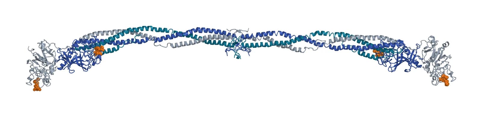
Fibrinogen is a large, 340 kDa hexameric homodimer that circulates in plasma and is secreted by the liver.[5] Each half carries one A-alpha, one B-beta and one gamma chain, and disulfide bridges hold the two halves together.[6]
The three chains are encoded by neighboring genes: FGA and FGB at 4q31.3 and FGG at 4q32.1.[7][8][9] Mosesson describes the folded molecule as two outer D domains joined to a central E domain by coiled-coil segments.[6]
Kollman and colleagues solved the crystal structure of human fibrinogen purified from plasma, resolving the twisted coiled-coil regions and the carbohydrate groups on the beta chains, with all 11 sugar residues positioned (Fig. 1).[10] As in earlier structures, the flexible alphaC domains gave no resolvable electron density.[10]
Fibrinogen also binds partners that matter for a platelet-rich matrix. Mosesson lists binding to the platelet integrin alphaIIb-beta3 and to fibronectin, and notes that bound growth factors such as FGF-2 and VEGF enhance the activities of the fibrin beta15-42 sequence.[6]
Thrombin converts fibrinogen into fibrin through ordered knob-hole binding
Thrombin cleaves fibrinopeptide A from the A-alpha chains, which starts polymerization.[6] Weisel and Litvinov describe an ordered sequence of fibrinopeptide release, A first and B later, that drives the knob-hole interactions behind oligomer formation (Fig. 2).[11]
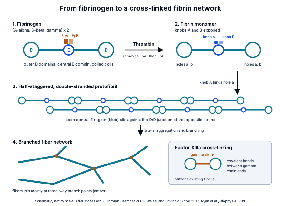
Removing the fibrinopeptides exposes knobs A and B in the central region, and these bind holes a and b that are always exposed at the ends of other molecules.[3] Kononova and colleagues modeled the A:a and B:b bonds with the knob mimetic peptides GPRP and GHRP and found the two complexes of roughly equal strength, with dissociation kinetics sensitive to pH and temperature.[12]
The bound monomers form half-staggered, double-stranded oligomers that lengthen into protofibrils. Protofibrils then aggregate laterally into fibers that branch into a three-dimensional network.[3]
Lateral aggregation is the least understood step. The alphaC region and B:b knob-hole binding appear to enhance it, and calcium ions bound to fibrin(ogen) at strong and weak sites have been located.[11]
Factor XIIIa cross-links the assembled network
Factor XIII is a protransglutaminase activated in the last stage of coagulation, and it works on an insoluble substrate.[13] Its A subunit is encoded by F13A1 at 6p25.1, and the same subunit occurs in the cytoplasm of platelets and monocytes.[13][14]
Fibrin assembly aligns the C-terminal ends of gamma chains from neighboring molecules in antiparallel pairs, and factor XIIIa joins them covalently into gamma dimers.[6] Ryan and colleagues found that cross-linking changed network morphology very little and raised clot rigidity, most likely by stiffening existing fibers.[15]
Thrombin concentration shapes fiber thickness and network density
Wolberg reviews in vitro work showing that the thrombin concentration present at gelation strongly influences fibrin clot structure. Clots formed at low thrombin consist of thick fibers, and clots formed at high thrombin consist of thin fibers (Fig. 3).[16]
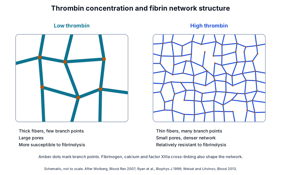
Ryan and colleagues varied fibrinogen, thrombin and calcium and measured fiber length, diameter and branching from stereo scanning electron micrographs. Large fiber diameters and lengths appeared only when branching was minimal, and three-way junctions were the dominant branch point.[15]
In the same study, maximal rigidity came from a balance between large fibers and extensive branching, and higher fibrinogen raised rigidity by increasing fiber and branch point density.[15] Pieters and Wolberg note that genetic, biochemical and environmental factors all regulate fibrin formation, structure and stability.[5]
Fibrin combines high elasticity with very low protein content
Litvinov and Weisel describe fibrin as both elastic and viscous, with remarkable elasticity and stability for a gel of very low protein content. Like many filamentous protein polymers, it stiffens under shear, tension or compression.[17]
The mechanics of the whole gel follow from its single fibers, through changes in their orientation, stretching, bending and buckling. Fiber properties in turn depend on how many protofibrils each fiber holds and how they pack.[17]
Platelets and fibrin together contract the clot
Platelets bind fibrinogen through alphaIIb-beta3, which incorporates them into a developing clot.[6] Cines and colleagues found that contracted blood clots carry a meshwork of fibrin and platelet aggregates on the outside and a close-packed array of compressed, polyhedral erythrocytes inside.[18]
Contraction required platelets, with their cytoskeletal motility proteins, and fibrin(ogen) as the substrate bridging them. The same structure formed with several clotting activators and in reconstituted human blood.[18] The authors also produced the polyhedral erythrocyte shape with the forces of centrifugation alone, in unclotted blood.[18]
Platelet-rich fibrin forms from blood collected without anticoagulant
Dohan and colleagues described PRF in 2006 as a second-generation platelet concentrate prepared without biochemical blood handling (Fig. 4). They traced it from fibrin glues and concentrated platelet-rich plasma, and proposed that slow polymerization during PRF preparation gives a fibrin network close to the natural one.[1]
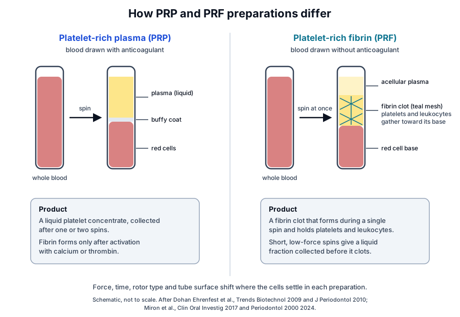
The two preparation routes differ in the anticoagulant and in when fibrin forms. Platelet-rich plasma is collected as a liquid from anticoagulated blood, and PRF clots during the spin itself.[1][19]
The Dohan Ehrenfest classification sorts platelet concentrates into four families by leukocyte and fibrin content.[4] The sibling reference on study reporting compares this framework with later reporting systems.
- Pure platelet-rich plasma (P-PRP): a liquid platelet concentrate without leukocytes.[4]
- Leukocyte- and platelet-rich plasma (L-PRP): a liquid platelet concentrate that keeps leukocytes.[4]
- Pure platelet-rich fibrin (P-PRF): a fibrin matrix without leukocytes.[4]
- Leukocyte- and platelet-rich fibrin (L-PRF): a fibrin matrix that holds platelets and leukocytes, the family that includes the original PRF protocol.[4]
Dohan Ehrenfest and colleagues analyzed the residual plasma left after collecting PRF clots and estimated that about 97% of platelets and more than 50% of leukocytes were concentrated in the clot. Platelets and fibrin formed large clusters in the first millimeters of the clot above the red cell base, within a mature, dense fibrin network.[2]
In that study, glass and glass-coated plastic tubes, and forcible or soft compression, gave clots of similar architecture.[2]
Relative centrifugal force and time set where cells settle in the clot
Miron and colleagues distinguish revolutions per minute from relative centrifugal force, the g-force a sample experiences, as the starting point for describing a protocol.[20] Their overview also covers fixed-angle and horizontal rotors, tube surface chemistry and temperature control of clotting.[20]
Ghanaati and colleagues compared a standard protocol with a lower-speed, longer-time protocol in clots from four donors. Platelets appeared throughout both clots, more of them in the distal part with the lower-speed protocol, and neutrophils shifted from the red cell interface toward the distal clot.[19]
Miron and colleagues introduced a method that pipettes sequential 1-milliliter layers from the top of a centrifuged tube and counts each with a hematology analyzer. Standard L-PRF settings concentrated platelets and leukocytes within the buffy coat, and slower settings distributed platelets more evenly through the upper layers.[21]
In the same comparison, horizontal centrifugation yielded higher numbers and concentrations of platelets and leukocytes than fixed-angle rotors.[21] The group then compared 24 horizontal-rotor protocols across six forces, 100 to 1,200 g, and four times, 3 to 12 min, using 960 complete blood counts.[22]
Spin time appeared to affect layer separation more than force, and protocols at or below 200 g accumulated few platelets and leukocytes in the upper layers. Baseline cell counts and hematocrit of each donor sample also influenced separation, most clearly at low forces.[22]
Liquid platelet-rich fibrin is collected before the fibrinogen clots
Very short, low-force spins yield a liquid upper fraction that can be drawn off before it clots. Miron and colleagues prepared such a liquid PRF at about 60 g for 3 min, with no anticoagulant.[23]
Layer counts showed that liquid protocols reached the highest concentrations of platelets and leukocytes in the collected fraction, with lower total cell numbers because the collected volume was smaller.[21]
The fibrin matrix binds growth factors and releases them gradually in vitro
Fibrin binds growth factors directly. Sahni and colleagues measured specific, saturable binding of basic fibroblast growth factor to fibrinogen and to fibrin, with high-affinity dissociation constants between 0.13 and 1.3 nM and two to four FGF molecules per fibrinogen at saturation.[24]
Dohan Ehrenfest and colleagues measured release from PRF membranes in culture over seven days. The membranes released TGF-beta1, PDGF-AB, VEGF and thrombospondin-1 throughout that time, and the authors proposed that trapped leukocytes went on producing TGF-beta1 and VEGF.[25]
Kobayashi and colleagues compared PRP, PRF and advanced PRF (A-PRF) from six donors by ELISA at 15 min, 60 min, 8 h and days 1, 3 and 10.[26] PDGF-AA was the most abundant factor released, followed by PDGF-BB, TGF-beta1, VEGF and PDGF-AB.[26]
In that study PRP released more protein in the first hour, and the PRF preparations released growth factors steadily through day 10, with A-PRF releasing the largest totals.[26] Fujioka-Kobayashi and colleagues reported that lowering centrifugal force raised the release of PDGF, TGF-beta1, EGF and IGF from PRF clots in culture.[27]
The receptors that read these factors, including the PDGF, EGF, VEGF and TGF-beta receptors, are described in the sibling growth factor receptors reference.
Plasmin turns fibrin over through regulated fibrinolysis
Fibrin is both the main product of coagulation and the substrate of fibrinolysis.[28] The zymogen plasminogen, encoded by PLG at 6q26, binds fibrin together with tissue-type plasminogen activator, and the activated enzyme plasmin cleaves fibrin at specific lysine residues.[3][29]
Mosesson describes this as a ternary complex of tPA, plasminogen and fibrin that stimulates plasminogen activation, with alpha2-antiplasmin and plasminogen activator inhibitor-2 among the binding inhibitors.[6] Longstaff and Kolev review how fibrin structure, clot composition, cells and flow localize and regulate these reactions.[30]
Network structure sets the pace. Wolberg notes that thick-fiber clots formed at low thrombin are highly susceptible to fibrinolysis, and thin-fiber clots formed at high thrombin are relatively resistant.[16]
Risman and colleagues added protofibril packing as a further variable. Tissue factor clots with dense networks and loosely packed fibers lysed faster at first than thrombin clots with looser networks and densely packed fibers.[31] Cines and colleagues proposed that clot contraction itself slows fibrinolysis.[18]
Scanning electron microscopy, turbidity and rheology measure fibrin structure
Each method reads a different scale of the network. Scanning electron microscopy images fibers and cells directly, and Dohan Ehrenfest and colleagues used it with light microscopy to map the architecture of PRF clots.[2][15]
Turbidity reports average fiber size in a hydrated gel. Carr and Hermans showed that the wavelength dependence of turbidity gives the mass-length ratio of fibrin fibers and, for thicker fibers, their radius.[32]
Rheometers measure stiffness and viscous behavior, which Ryan and colleagues correlated with fiber dimensions and branching.[15] Risman and colleagues combined turbidimetry with confocal and scanning electron microscopy to follow clot formation, protofibril packing and lysis together.[31]
| Method | What it measures | Typical use |
|---|---|---|
| Scanning electron microscopy | Fiber diameter, branching and cell positions | Architecture of fixed clots and PRF membranes |
| Confocal microscopy | Network structure in hydrated samples | Pore size and fiber density |
| Turbidity | Optical density across wavelengths | Mass-length ratio and fiber radius |
| Rheometry | Elastic and viscous moduli | Clot stiffness and deformation |
| Layer-by-layer cell counts | Platelets, leukocytes and red cells per layer | Cell distribution after centrifugation |
| ELISA of conditioned medium | Growth factor concentration over time | Release kinetics from a matrix |
For preparations, cell counts and release assays complete the picture. The sequential layer method counts cells in each fraction of a centrifuged tube, and ELISA follows growth factors released into the medium.[21][26]
Where to go from here
The platelet granule biology reference covers the alpha granule proteins that a fibrin matrix holds, and the PRP preparation variables reference follows anticoagulant, force and time through liquid concentrates. The growth factor receptors reference explains how PDGF, TGF-beta, VEGF and EGF signal once released.
The study reporting reference shows how classification systems describe a preparation, the research methods library explains study designs, and the BMAC and SVF reference turns to marrow and adipose preparations. The MSC secretome reference, the HCT/P regulatory reference and the quality references round out the library, and our team is glad to discuss the methods on this page.
Referencias comentadas
Numeradas en el orden en que se citan en el texto. Cada fuente lleva una nota sobre lo que respalda en esta página; los artículos revisados por pares se listan con revista, volumen, número y páginas, y las fuentes primarias son regulaciones, documentos de agencias y bases de datos de referencia.
- 1.Peer-reviewedDohan DM, Choukroun J, Diss A, et al. Platelet-rich fibrin (PRF): a second-generation platelet concentrate. Part I: technological concepts and evolution. Oral Surgery, Oral Medicine, Oral Pathology, Oral Radiology, and Endodontics. 2006. https://pubmed.ncbi.nlm.nih.gov/16504849/Annotation: Concept paper on PRF. Cited for its preparation without biochemical blood handling, its lineage from fibrin glues and PRP, and slow polymerization during preparation.
- 2.Peer-reviewedDohan Ehrenfest DM, Del Corso M, Diss A, et al. Three-dimensional architecture and cell composition of a Choukroun's platelet-rich fibrin clot and membrane. Journal of Periodontology. 2010. https://pubmed.ncbi.nlm.nih.gov/20373539/Annotation: Blood counts, light and scanning electron microscopy of PRF clots. Cited for about 97% of platelets and over 50% of leukocytes in the clot, a dense fibrin network and the effect of tubes and compression.
- 3.Peer-reviewedWeisel JW, Litvinov RI. Fibrin Formation, Structure and Properties. Sub-Cellular Biochemistry. 2017. https://pubmed.ncbi.nlm.nih.gov/28101869/Annotation: Review of fibrin assembly. Cited for knob-hole binding, half-staggered protofibrils, lateral aggregation into a branched network and plasmin cleavage at lysine residues.
- 4.Peer-reviewedDohan Ehrenfest DM, Rasmusson L, Albrektsson T. Classification of platelet concentrates: from pure platelet-rich plasma (P-PRP) to leucocyte- and platelet-rich fibrin (L-PRF). Trends in Biotechnology. 2009. https://pubmed.ncbi.nlm.nih.gov/19187989/Annotation: Classification of platelet concentrates. Cited for the four families defined by leukocyte and fibrin content, used here as a reporting framework.
- 5.Peer-reviewedPieters M, Wolberg AS. Fibrinogen and fibrin: An illustrated review. Research and Practice in Thrombosis and Haemostasis. 2019. https://pubmed.ncbi.nlm.nih.gov/31011700/Annotation: Illustrated review of fibrinogen. Cited for its 340 kDa hexameric homodimer structure, secretion from the liver and the factors that regulate fibrin formation and structure.
- 6.Peer-reviewedMosesson MW. Fibrinogen and fibrin structure and functions. Journal of Thrombosis and Haemostasis. 2005. https://pubmed.ncbi.nlm.nih.gov/16102057/Annotation: Review of fibrinogen chains, D and E domains, fibrinopeptide A cleavage, gamma-dimer cross-linking by factor XIIIa, and binding to alphaIIb-beta3, fibronectin, growth factors and the tPA-plasminogen complex.
- 7.DatabaseNCBI Gene 2243, FGA fibrinogen alpha chain (Homo sapiens). Accessed 21 October 2026. https://www.ncbi.nlm.nih.gov/gene/2243
- 8.DatabaseNCBI Gene 2244, FGB fibrinogen beta chain (Homo sapiens). Accessed 21 October 2026. https://www.ncbi.nlm.nih.gov/gene/2244
- 9.DatabaseNCBI Gene 2266, FGG fibrinogen gamma chain (Homo sapiens). Accessed 21 October 2026. https://www.ncbi.nlm.nih.gov/gene/2266
- 10.Peer-reviewedKollman JM, Pandi L, Sawaya MR, et al. Crystal structure of human fibrinogen. Biochemistry. 2009. https://pubmed.ncbi.nlm.nih.gov/19296670/Annotation: X-ray crystal structure of plasma-purified human fibrinogen (PDB 3GHG). Cited for the coiled-coil regions, the positioned beta-chain carbohydrate and the unresolved alphaC domains.
- 11.Peer-reviewedWeisel JW, Litvinov RI. Mechanisms of fibrin polymerization and clinical implications. Blood. 2013. https://pubmed.ncbi.nlm.nih.gov/23305734/Annotation: Review of fibrin polymerization. Cited for the ordered release of fibrinopeptides, calcium binding sites and the roles of the alphaC region and B:b binding in lateral aggregation.
- 12.Peer-reviewedKononova O, Litvinov RI, Zhmurov A, et al. Molecular mechanisms, thermodynamics, and dissociation kinetics of knob-hole interactions in fibrin. The Journal of Biological Chemistry. 2013. https://pubmed.ncbi.nlm.nih.gov/23720752/Annotation: Molecular dynamics study of fragment D with the knob mimetics GPRP and GHRP. Cited for the comparable strength of A:a and B:b bonds and their sensitivity to pH and temperature.
- 13.Peer-reviewedMuszbek L, Bereczky Z, Bagoly Z, et al. Factor XIII: a coagulation factor with multiple plasmatic and cellular functions. Physiological Reviews. 2011. https://pubmed.ncbi.nlm.nih.gov/21742792/Annotation: Review of factor XIII. Cited for its activation as a protransglutaminase late in coagulation, its insoluble substrate and the A subunit in the cytoplasm of platelets and monocytes.
- 14.DatabaseNCBI Gene 2162, F13A1 coagulation factor XIII A chain (Homo sapiens). Accessed 21 October 2026. https://www.ncbi.nlm.nih.gov/gene/2162
- 15.Peer-reviewedRyan EA, Mockros LF, Weisel JW, et al. Structural origins of fibrin clot rheology. Biophysical Journal. 1999. https://pubmed.ncbi.nlm.nih.gov/10545379/Annotation: Scanning electron microscopy and rheometry of fibrin clots. Cited for fiber size and branching, three-way junctions, rigidity from fiber and branch density, and stiffening of fibers by factor XIIIa.
- 16.Peer-reviewedWolberg AS. Thrombin generation and fibrin clot structure. Blood Reviews. 2007. https://pubmed.ncbi.nlm.nih.gov/17208341/Annotation: Review of thrombin concentration and clot structure. Cited for thick, lysis-susceptible fibers at low thrombin and thin, lysis-resistant fibers at high thrombin.
- 17.Peer-reviewedLitvinov RI, Weisel JW. Fibrin mechanical properties and their structural origins. Matrix Biology. 2017. https://pubmed.ncbi.nlm.nih.gov/27553509/Annotation: Review of fibrin mechanics. Cited for high elasticity at low protein content, strain stiffening, and gel mechanics that follow from single fibers and protofibril packing.
- 18.Peer-reviewedCines DB, Lebedeva T, Nagaswami C, et al. Clot contraction: compression of erythrocytes into tightly packed polyhedra and redistribution of platelets and fibrin. Blood. 2014. https://pubmed.ncbi.nlm.nih.gov/24335500/Annotation: Microscopy study of contracted blood clots. Cited for the outer fibrin and platelet meshwork, inner polyhedral erythrocytes, the need for platelets and fibrin(ogen), and polyhedra formed by centrifugation alone.
- 19.Peer-reviewedGhanaati S, Booms P, Orlowska A, et al. Advanced platelet-rich fibrin: a new concept for cell-based tissue engineering by means of inflammatory cells. The Journal of Oral Implantology. 2014. https://pubmed.ncbi.nlm.nih.gov/24945603/Annotation: Histology of PRF clots from four donors under two spin protocols. Cited for PRF prepared without anticoagulant and the shift of platelets and neutrophils with a lower-speed, longer spin.
- 20.Peer-reviewedMiron RJ, Fujioka-Kobayashi M, Sculean A, et al. Optimization of platelet-rich fibrin. Periodontology 2000. 2024. https://pubmed.ncbi.nlm.nih.gov/37681522/Annotation: Overview of PRF preparation variables. Cited for the distinction between rpm and relative centrifugal force, rotor geometry, tube chemistry and temperature control of clotting.
- 21.Peer-reviewedMiron RJ, Chai J, Zheng S, et al. A novel method for evaluating and quantifying cell types in platelet rich fibrin and an introduction to horizontal centrifugation. Journal of Biomedical Materials Research. Part A. 2019. https://pubmed.ncbi.nlm.nih.gov/31148358/Annotation: Layer-by-layer cell count method for centrifuged blood. Cited for buffy coat concentration under standard settings, even platelet spread under slower settings, horizontal rotor yields and liquid fraction counts.
- 22.Peer-reviewedMiron RJ, Chai J, Fujioka-Kobayashi M, et al. Evaluation of 24 protocols for the production of platelet-rich fibrin. BMC Oral Health. 2020. https://pubmed.ncbi.nlm.nih.gov/33160335/Annotation: Layer counts across 24 horizontal-rotor protocols. Cited for the force and time ranges tested, the larger effect of time, poor accumulation at or below 200 g and the influence of baseline counts.
- 23.Peer-reviewedMiron RJ, Fujioka-Kobayashi M, Hernandez M, et al. Injectable platelet rich fibrin (i-PRF): opportunities in regenerative dentistry? Clinical Oral Investigations. 2017. https://pubmed.ncbi.nlm.nih.gov/28154995/Annotation: In vitro study of a liquid PRF. Cited for its preparation without anticoagulant by a short, low-force spin of about 60 g for 3 min.
- 24.Peer-reviewedSahni A, Odrljin T, Francis CW. Binding of basic fibroblast growth factor to fibrinogen and fibrin. The Journal of Biological Chemistry. 1998. https://pubmed.ncbi.nlm.nih.gov/9516457/Annotation: Binding study of bFGF with fibrinogen and fibrin. Cited for specific, saturable, high-affinity binding and a molar binding ratio of two to four.
- 25.Peer-reviewedDohan Ehrenfest DM, de Peppo GM, Doglioli P, et al. Slow release of growth factors and thrombospondin-1 in Choukroun's platelet-rich fibrin (PRF): a gold standard to achieve for all surgical platelet concentrates technologies. Growth Factors. 2009. https://pubmed.ncbi.nlm.nih.gov/19089687/Annotation: Release study of PRF membranes in culture. Cited for TGF-beta1, PDGF-AB, VEGF and thrombospondin-1 released over seven days and continued production by trapped leukocytes.
- 26.Peer-reviewedKobayashi E, Flückiger L, Fujioka-Kobayashi M, et al. Comparative release of growth factors from PRP, PRF, and advanced-PRF. Clinical Oral Investigations. 2016. https://pubmed.ncbi.nlm.nih.gov/26809431/Annotation: ELISA release study of three preparations from six donors. Cited for the sampling times, the rank order of released factors, early release from PRP and steady release from PRF through day 10.
- 27.Peer-reviewedFujioka-Kobayashi M, Miron RJ, Hernandez M, et al. Optimized Platelet-Rich Fibrin With the Low-Speed Concept: Growth Factor Release, Biocompatibility, and Cellular Response. Journal of Periodontology. 2017. https://pubmed.ncbi.nlm.nih.gov/27587367/Annotation: In vitro comparison of PRF spin settings. Cited for higher release of PDGF, TGF-beta1, EGF and IGF from clots prepared at lower centrifugal force.
- 28.Peer-reviewedChapin JC, Hajjar KA. Fibrinolysis and the control of blood coagulation. Blood Reviews. 2015. https://pubmed.ncbi.nlm.nih.gov/25294122/Annotation: Review of the fibrinolytic system. Cited for fibrin as both the primary product of coagulation and the substrate of fibrinolysis.
- 29.DatabaseNCBI Gene 5340, PLG plasminogen (Homo sapiens). Accessed 21 October 2026. https://www.ncbi.nlm.nih.gov/gene/5340
- 30.Peer-reviewedLongstaff C, Kolev K. Basic mechanisms and regulation of fibrinolysis. Journal of Thrombosis and Haemostasis. 2015. https://pubmed.ncbi.nlm.nih.gov/26149056/Annotation: Review of fibrinolysis. Cited for the influence of fibrin structure, clot composition, cells and flow on the localization and regulation of plasmin generation.
- 31.Peer-reviewedRisman RA, Percoco V, Paynter B, et al. Protofibril packing density of individual fibers alters fibrinolysis. Research and Practice in Thrombosis and Haemostasis. 2025. https://pubmed.ncbi.nlm.nih.gov/40212773/Annotation: Turbidimetry, confocal and scanning electron microscopy and modeling of plasma clots. Cited for faster initial lysis of loosely packed fibers in dense tissue factor networks.
- 32.Peer-reviewedCarr ME Jr, Hermans J. Size and density of fibrin fibers from turbidity. Macromolecules. 1978. https://pubmed.ncbi.nlm.nih.gov/621951/Annotation: Light-scattering study of fibrin gels. Cited for deriving fiber mass-length ratio and radius from the wavelength dependence of turbidity.
Cite this page
ExaVeyra Sciences Editorial Team. Platelet-Rich Fibrin and Fibrin Matrix Biology. ExaVeyra Sciences; 21 October 2026. Available from: https://exaveyra.com/science/platelet-biology/platelet-rich-fibrin-biology. Accessed [date accessed].
Fuentes de las figuras
- 33.PDB 3GHG, Crystal structure of human fibrinogen. ExaVeyra Sciences, rendered in PyMOL from PDB 3GHG. Structure data: RCSB Protein Data Bank (rcsb.org), CC0. Publicación de la estructura: Kollman JM, Pandi L, Sawaya MR, Riley M, Doolittle RF. Crystal structure of human fibrinogen. Biochemistry. 2009;48(18):3877-3886.