Esta referencia está disponible actualmente en inglés. La versión en español está en preparación.
PRP Preparation Variables: Spin, Platelet Dose and Leukocytes
Por Benn Bluestein-Veyra, M.Sc. en Química Orgánica · Actualizado el 21 de octubre de 2026
ExaVeyra Sciences es un proveedor B2B con sede en Miami que suministra exosomas para uso estético tópico a clínicas con licencia, y remite a los prescriptores con licencia a una farmacia de preparados compuestos 503A que prepara formulaciones según prescripciones individuales.
| Pregunta | Respuesta |
|---|---|
| Qué cubre esta página | How platelet-rich plasma preparations differ: spin protocols, platelet concentration, leukocyte and red cell content, activation, and classification systems. |
| Quién puede hacer pedidos | Clínicas y profesionales con licencia profesional activa y NPI verificados por ExaVeyra. Los pedidos de exosomas requieren además un reconocimiento firmado de uso profesional. |
| Qué se envía y cómo | El material de exosomas para uso estético tópico, caracterizado por lote, se envía en cadena de frío por exprés nocturno. La entrega puede tardar 1 a 3 días hábiles. Se emite un certificado de análisis para cada lote y se entrega a solicitud. |
PRP preparation concentrates platelets in a small volume of plasma above their whole blood baseline. Each protocol yields its own mix of platelets, leukocytes, red cells and growth factors, so preparations made by different methods differ in measurable ways.[1][2] Dohan Ehrenfest and colleagues noted that each method leads to a different product with different biology.[3]
The baseline itself varies. In records from 40,987 people in Italian population studies, Biino and colleagues found that women carried more platelets than men after age 14 and that counts fell in later life.[4] This reference follows each variable from the blood draw to the final count, and closes with the systems used to report them.
Centrifugation separates blood components by size and density
Centrifugation sorts the cells of whole blood by size and density. After a first spin, red cells pack at the bottom, a thin buffy coat of leukocytes and platelets lies above them, and plasma fills the upper tube with platelets concentrated just above the buffy coat (Fig. 1).[5][6]
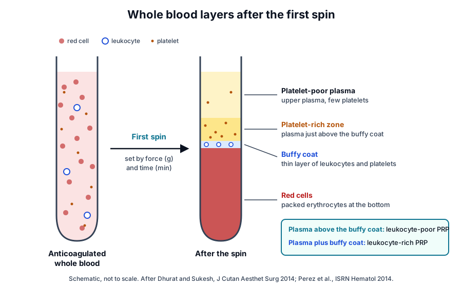
Perez and colleagues describe the driving force as the difference in size and density between the particles, opposed by a frictional force that grows with particle velocity and fluid viscosity in the Stokes regime. Force and time together drive the packing of red cells and the recovery of platelets in the upper layer.[6]
The same group modeled discontinuous centrifugation from the balance of forces on a particle, adding a factor for red cell packing. Their model predicted platelet and plasma recovery from blood volume, hematocrit and supernatant volume as functions of centrifugal acceleration and time, and it matched data from 10 donors.[7]
Relative centrifugal force makes protocols reproducible
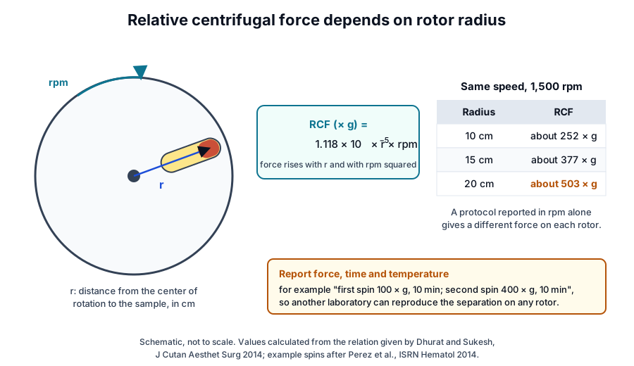
Relative centrifugal force (RCF), written in multiples of g, depends on the rotor radius and on the square of the speed in revolutions per minute. Dhurat and Sukesh give the relation RCF = 1.118 × 10⁻⁵ × r × rpm², with r in centimeters, and note that many studies report rpm alone, which complicates comparison (Fig. 2).[5]
Radius also changes within one tube. Perez and colleagues found that a larger blood volume in a single tube reduced platelet recovery and red cell packing, because the blood surface sat closer to the rotor axis and the mean force on the cells fell.[6] Kahn and colleagues showed in 1976 that several combinations of speed and time reached the same optimum platelet yield from whole blood units.[8]
Single-spin and double-spin protocols yield different platelet and leukocyte content
A single-spin protocol collects plasma after the first spin. A double-spin protocol spins that plasma again at higher force to pellet the platelets, removes the upper platelet-poor plasma and resuspends the pellet in the rest (Fig. 3).[5][6]
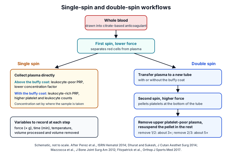
Perez and colleagues processed 3.5 milliliters of blood at 100 × g for 10 min, then at 400 × g for 10 min. Removing half the plasma gave about three times the baseline platelet concentration, removing two thirds gave about five times, and platelet recovery reached 70% to 80%.[6]
Amable and colleagues varied force, temperature and time and chose a two-step protocol that recovered 46.9% to 69.5% of platelets at 5.4 to 7.3 times baseline. Less than 0.3% of the starting red cells and leukocytes remained in the final preparation.[9]
The sampled layer sets the leukocyte content
Collecting the upper layer with the superficial buffy coat gives pure, leukocyte-poor PRP, and collecting the whole buffy coat with a few red cells gives leukocyte-rich PRP.[5] Fitzpatrick and colleagues found that three systems sampling the buffy coat layer reached 3 to 6 times baseline platelets with 3 to 6 times baseline leukocytes, and a plasma-sampling system reached 1.5 times.[10]
Mazzocca and colleagues compared two single-spin protocols and one double-spin protocol in eight donors. Platelet, leukocyte and growth factor concentrations differed between methods, and every method raised platelets above native blood.[1]
Spin force affects platelet integrity
Perez and colleagues measured soluble P-selectin as a marker of activation during processing. It rose when the second spin reached 800 × g and 1,200 × g, which the authors read as a loss of platelet integrity, and lower centrifugal accelerations favored platelet separation.[6] P-selectin is stored in platelet alpha-granules and moves to the plasma membrane on activation.[11]
Anticoagulant and temperature keep platelets at rest during processing
Blood for PRP is drawn into an anticoagulant, commonly citrate-based, so platelets stay unactivated until use.[5] Lei and colleagues compared heparin, citrate, acid citrate dextrose (ACD) and citrate-theophylline-adenosine-dipyridamole (CTAD) and found that ACD and CTAD preserved platelet structure and limited spontaneous activation more than heparin or citrate, measured as soluble P-selectin.[12]
In the same study, ACD and CTAD preparations released more TGF-beta1.[12] Dhurat and Sukesh note that ACD-A has a pH of 4.9 and 3.8% sodium citrate a pH of 7.8, with citrate ion at 15.6 and 24.4 milligrams per milliliter respectively.[5]
Temperature is a reported variable as well. Amable and colleagues varied it along with force and time, and Dhurat and Sukesh note that many authors centrifuge at 12 to 16 °C.[5][9] Fitzpatrick and colleagues measured a small drop in pH that they related to citrate, and glucose at 3 to 6 times baseline.[10]
Leukocyte and red cell content shape the molecular profile of PRP
Sundman and colleagues compared a leukocyte-poor preparation (1.99 times baseline platelets, 0.13 times leukocytes) with a leukocyte-rich one (4.69 and 4.26 times). TGF-beta1 and PDGF-AB tracked platelet counts, while MMP-9 tracked neutrophils and IL-1beta tracked neutrophils and monocytes.[13]
Castillo and colleagues found similar mean platelet, red cell, active TGF-beta1 and fibrinogen concentrations across three systems. Leukocyte-rich preparations carried more leukocytes, PDGF-AB, PDGF-BB and VEGF, and platelet capture efficiency differed between systems.[14] TGF-beta1 exists in a latent complex with its latency-associated peptide or as the active mature dimer.[15]
Magalon and colleagues found that leukocyte-rich preparations also held more red cells and neutrophils. Platelet dose correlated with every growth factor measured, while leukocyte dose correlated only with VEGF and EGF.[2] PDGF-B chains form homodimers, or heterodimers with PDGF-A, that bind PDGF receptor tyrosine kinases.[16]
| Component | How it is measured | Reported association |
|---|---|---|
| Platelets | Hematology analyzer count | TGF-beta1, PDGF-AB and other growth factors |
| Neutrophils | Analyzer differential count | MMP-9 and IL-1beta |
| Monocytes | Analyzer differential count | IL-1beta |
| Total leukocytes | Analyzer count | VEGF and EGF |
| Red cells | Analyzer count | Purity of the preparation |
Activation method sets the timing of growth factor release
Amable and colleagues detected no secreted cytokines before activation. After calcium or calcium with thrombin, platelets released high concentrations of PDGF and TGF together with IL-4, IL-8, IL-13, IL-17, TNF-alpha and IFN-alpha, and the clots retained a high concentration of several growth factors.[9]
Cavallo and colleagues activated PRP with calcium chloride, autologous thrombin, both together or collagen type I. Calcium and thrombin formed clots by the 15 min evaluation and collagen formed none; collagen gave lower overall release, and calcium chloride alone gave progressive release up to 24 h.[17]
Harrison and colleagues found that thrombin released TGF-beta1 and PDGF-AB at once, while collagen-activated clots released similar amounts each day through day 5. Concentrating platelets to three times blood level tripled TGF-beta1 release over whole blood clots.[18]
Activation routes differ in their enzyme dependence. In twelve donors, Jayaram and colleagues found that two weeks of low-dose aspirin, which blocks COX-1, reduced growth factor release triggered by arachidonic acid, left thrombin-triggered VEGF release unchanged and partly reduced thrombin-triggered PDGF-AB release.[19]
Donor age and sex contribute to variation between preparations
Variation begins with the donor. Mazzocca and colleagues drew blood three times from each donor and found wide intra-individual variation in platelet and cell numbers and in growth factor levels, whatever the separation method.[1]
In a landmark study of 115 specimens, Weibrich and colleagues measured platelets at five times donor blood, PDGF-AB at 117 ng/mL and TGF-beta1 at 169 ng/mL. Growth factor content correlated poorly with platelet count.[20]
Xiong and colleagues prepared leukocyte-poor PRP from 39 donors with one protocol. Platelet counts were similar across groups, men had higher IL-1beta, TNF-alpha, IL-1 receptor antagonist and TGF-beta1, and IGF-1 differed with age.[21] In 102 adults, Evanson and colleagues reported higher EGF, HGF, IGF-1 and PDGF-BB in women and higher levels of several factors at age 25 or younger.[22]
Analyzer counts and immunoassays characterize the final preparation
A hematology analyzer gives platelet, leukocyte and red cell counts in whole blood and in the preparation. Perez and colleagues define platelet recovery efficiency as the platelets in the collected layer over those in whole blood, and the concentration factor as the ratio of their concentrations.[6]
Platelets settle into a concentration gradient after each spin, so the preparation is mixed before counting.[6] Dhurat and Sukesh describe resuspending the final concentrate before counting so that platelets are evenly distributed.[5]
ELISA and multiplex assays quantify growth factors and cytokines, and flow cytometry reads platelet activation status.[2][21] Integrin alpha IIb, the CD41 antigen encoded by ITGA2B, forms the alpha IIb beta 3 receptor that mediates platelet aggregation.[23]
Classification systems give PRP preparations a shared reporting language
Dohan Ehrenfest and colleagues proposed four families in 2009, by leukocyte and fibrin content: pure PRP, leukocyte- and platelet-rich plasma, pure platelet-rich fibrin and leukocyte- and platelet-rich fibrin.[3] Later systems added more variables (Fig. 4).
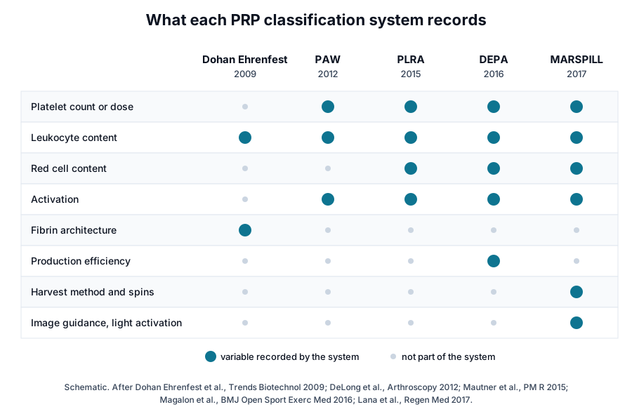
- PAW (DeLong and colleagues, 2012): the absolute number of Platelets, the manner of Activation and the presence of White cells.[25]
- PLRA (Mautner and colleagues, 2015): platelet concentration, leukocytes, red cells and activation, with minimal reporting standards.[26]
- DEPA (Magalon and colleagues, 2016): Dose of platelets, Efficiency of production, Purity and Activation, each graded A to D.[24]
- MARSPILL (Lana and colleagues, 2017): harvest method, activation, red cells, number of spins, platelet number, image guidance, leukocytes and light activation.[27]
DEPA grades platelet dose from A, above 5 billion, to D, below 1 billion, and production efficiency from A, above 90% recovery, to D, below 30%. Applied to 20 published preparations, platelet dose ranged from 0.21 to 5.43 billion and no method recovered more than 90% of blood platelets.[24]
The Platelet Physiology Subcommittee of the ISTH used the RAND consensus method on 45 statements and proposed its own classification.[28] Murray and colleagues built the MIBO checklist through Delphi rounds, reaching 23 reporting statements for PRP.[29]
Chahla and colleagues reviewed 105 orthopedic studies and found that 10% described a reproducible preparation protocol and 16% reported quantitative composition.[30] These frameworks give authors a common checklist for describing what they prepared.[26][29]
Where to go from here
The platelet granule reference describes the alpha-granule and dense-granule cargo that activation releases, and the growth factor receptor reference follows PDGF, TGF-beta and VEGF to their receptors. The platelet-rich fibrin reference covers anticoagulant-free preparations and fibrin architecture, and the BMAC and SVF reference compares marrow and adipose preparations.
The study reporting reference expands on PAW, PLRA, DEPA and MIBO, and the HCT/P reference explains the federal framework for cell and tissue preparations. Our team is glad to discuss the methods described on this page.
Referencias comentadas
Numeradas en el orden en que se citan en el texto. Cada fuente lleva una nota sobre lo que respalda en esta página; los artículos revisados por pares se listan con revista, volumen, número y páginas, y las fuentes primarias son regulaciones, documentos de agencias y bases de datos de referencia.
- 1.Peer-reviewedMazzocca AD, McCarthy MB, Chowaniec DM, et al. Platelet-rich plasma differs according to preparation method and human variability. The Journal of Bone and Joint Surgery. American Volume. 2012. https://pubmed.ncbi.nlm.nih.gov/22336969/Annotation: Laboratory comparison of two single-spin and one double-spin method in eight donors. Cited for method differences in platelets, leukocytes and growth factors and wide intra-individual variation across three draws.
- 2.Peer-reviewedMagalon J, Bausset O, Serratrice N, et al. Characterization and comparison of 5 platelet-rich plasma preparations in a single-donor model. Arthroscopy. 2014. https://pubmed.ncbi.nlm.nih.gov/24725317/Annotation: Single-donor comparison of five preparations with cell counts, flow cytometry and ELISA. Cited for red cell and neutrophil content of leukocyte-rich preparations and the correlations of platelet and leukocyte dose with growth factors.
- 3.Peer-reviewedDohan Ehrenfest DM, Rasmusson L, Albrektsson T. Classification of platelet concentrates: from pure platelet-rich plasma (P-PRP) to leucocyte- and platelet-rich fibrin (L-PRF). Trends in Biotechnology. 2009. https://pubmed.ncbi.nlm.nih.gov/19187989/Annotation: Classification of platelet concentrates by leukocyte and fibrin content. Cited for the four families P-PRP, L-PRP, P-PRF and L-PRF and for each method yielding a product with different biology.
- 4.Peer-reviewedBiino G, Santimone I, Minelli C, et al. Age- and sex-related variations in platelet count in Italy: a proposal of reference ranges based on 40987 subjects' data. PLoS One. 2013. https://pubmed.ncbi.nlm.nih.gov/23382888/Annotation: Population-based reference interval study. Cited for higher platelet counts in women after age 14 and lower counts in later life.
- 5.Peer-reviewedDhurat R, Sukesh M. Principles and Methods of Preparation of Platelet-Rich Plasma: A Review and Author's Perspective. Journal of Cutaneous and Aesthetic Surgery. 2014. https://pubmed.ncbi.nlm.nih.gov/25722595/Annotation: Review of PRP preparation principles. Cited for the layers after the first spin, P-PRP and L-PRP collection, the RCF relation, rpm reporting, citrate anticoagulants and their pH, centrifugation temperature and resuspension before counting.
- 6.Peer-reviewedPerez AG, Lana JF, Rodrigues AA, et al. Relevant aspects of centrifugation step in the preparation of platelet-rich plasma. ISRN Hematology. 2014. https://pubmed.ncbi.nlm.nih.gov/25006472/Annotation: Two-spin study of donor blood. Cited for the 100 and 400 × g protocol, three- and five-fold concentration by plasma removal, 70% to 80% recovery, the P-selectin rise at 800 and 1,200 × g, the processed-volume effect, the platelet gradient and the recovery and concentration-factor definitions.
- 7.Peer-reviewedPerez AG, Lichy R, Lana JF, et al. Prediction and modulation of platelet recovery by discontinuous centrifugation of whole blood for the preparation of pure platelet-rich plasma. BioResearch Open Access. 2013. https://pubmed.ncbi.nlm.nih.gov/23914337/Annotation: Analytical model of red cell packing validated with blood from 10 donors. Cited for predicting platelet and plasma recovery from volume, hematocrit, acceleration and time.
- 8.Peer-reviewedKahn RA, Cossette I, Friedman LI. Optimum centrifugation conditions for the preparation of platelet and plasma products. Transfusion. 1976. https://pubmed.ncbi.nlm.nih.gov/1258118/Annotation: Centrifugation study of whole blood units. Cited for several speed and time combinations reaching the same optimum platelet yield.
- 9.Peer-reviewedAmable PR, Carias RB, Teixeira MV, et al. Platelet-rich plasma preparation for regenerative medicine: optimization and quantification of cytokines and growth factors. Stem Cell Research & Therapy. 2013. https://pubmed.ncbi.nlm.nih.gov/23759113/Annotation: Optimization of a two-step protocol by force, temperature and time. Cited for platelet recovery and concentration, red cell and leukocyte depletion, no secretion before activation, the released cytokines and growth factors retained in clots.
- 10.Peer-reviewedFitzpatrick J, Bulsara MK, McCrory PR, et al. Analysis of Platelet-Rich Plasma Extraction: Variations in Platelet and Blood Components Between 4 Common Commercial Kits. Orthopaedic Journal of Sports Medicine. 2017. https://pubmed.ncbi.nlm.nih.gov/28210651/Annotation: Controlled laboratory study of four separation kits. Cited for buffy coat versus plasma sampling, platelet and leukocyte concentration factors, the pH drop related to citrate and raised glucose.
- 11.DatabaseNCBI Gene 6403, SELP selectin P (Homo sapiens). Accessed 21 October 2026. https://www.ncbi.nlm.nih.gov/gene/6403
- 12.Peer-reviewedLei H, Gui L, Xiao R. The effect of anticoagulants on the quality and biological efficacy of platelet-rich plasma. Clinical Biochemistry. 2009. https://pubmed.ncbi.nlm.nih.gov/19560449/Annotation: Comparison of heparin, citrate, ACD and CTAD. Cited for platelet structure, spontaneous activation by soluble P-selectin and TGF-beta1 release.
- 13.Peer-reviewedSundman EA, Cole BJ, Fortier LA. Growth factor and catabolic cytokine concentrations are influenced by the cellular composition of platelet-rich plasma. The American Journal of Sports Medicine. 2011. https://pubmed.ncbi.nlm.nih.gov/21846925/Annotation: Controlled laboratory study of leukocyte-poor and leukocyte-rich preparations. Cited for the concentration factors and the correlations of TGF-beta1 and PDGF-AB with platelets and of MMP-9 and IL-1beta with leukocytes.
- 14.Peer-reviewedCastillo TN, Pouliot MA, Kim HJ, et al. Comparison of growth factor and platelet concentration from commercial platelet-rich plasma separation systems. The American Journal of Sports Medicine. 2011. https://pubmed.ncbi.nlm.nih.gov/21051428/Annotation: Controlled laboratory study of three separation systems. Cited for similar platelet, red cell, active TGF-beta1 and fibrinogen levels and higher leukocytes, PDGF-AB, PDGF-BB and VEGF in leukocyte-rich preparations.
- 15.DatabaseNCBI Gene 7040, TGFB1 transforming growth factor beta 1 (Homo sapiens). Accessed 21 October 2026. https://www.ncbi.nlm.nih.gov/gene/7040
- 16.DatabaseNCBI Gene 5155, PDGFB platelet derived growth factor subunit B (Homo sapiens). Accessed 21 October 2026. https://www.ncbi.nlm.nih.gov/gene/5155
- 17.Peer-reviewedCavallo C, Roffi A, Grigolo B, et al. Platelet-Rich Plasma: The Choice of Activation Method Affects the Release of Bioactive Molecules. BioMed Research International. 2016. https://pubmed.ncbi.nlm.nih.gov/27672658/Annotation: Comparison of calcium chloride, thrombin, both and collagen type I activation. Cited for clot formation, lower release with collagen and progressive release with calcium chloride.
- 18.Peer-reviewedHarrison S, Vavken P, Kevy S, et al. Platelet activation by collagen provides sustained release of anabolic cytokines. The American Journal of Sports Medicine. 2011. https://pubmed.ncbi.nlm.nih.gov/21398575/Annotation: Release kinetics after thrombin or collagen activation. Cited for immediate release with thrombin, steady daily release with collagen and three-fold TGF-beta1 release at three-fold platelet concentration.
- 19.Peer-reviewedJayaram P, Yeh P, Patel SJ, et al. Effects of Aspirin on Growth Factor Release From Freshly Isolated Leukocyte-Rich Platelet-Rich Plasma in Healthy Men: A Prospective Fixed-Sequence Controlled Laboratory Study. The American Journal of Sports Medicine. 2019. https://pubmed.ncbi.nlm.nih.gov/30888847/Annotation: Controlled laboratory study of PRP from twelve donors before and after low-dose aspirin. Cited for reduced arachidonic acid-triggered release and unchanged thrombin-triggered VEGF and TGF-beta1 release.
- 20.Peer-reviewedWeibrich G, Kleis WK, Hafner G, et al. Growth factor levels in platelet-rich plasma and correlations with donor age, sex, and platelet count. Journal of Cranio-Maxillo-Facial Surgery. 2002. https://pubmed.ncbi.nlm.nih.gov/12069512/Annotation: Growth factor and platelet measurements in 115 PRP specimens. Cited for five-fold platelet concentration, PDGF-AB and TGF-beta1 levels and the weak correlation with platelet count.
- 21.Peer-reviewedXiong G, Lingampalli N, Koltsov JCB, et al. Men and Women Differ in the Biochemical Composition of Platelet-Rich Plasma. The American Journal of Sports Medicine. 2018. https://pubmed.ncbi.nlm.nih.gov/29211968/Annotation: Controlled laboratory study of PRP from 39 donors with multiplex assay and ELISA. Cited for sex differences in cytokines and growth factors, similar platelet counts and age differences in IGF-1.
- 22.Peer-reviewedEvanson JR, Guyton MK, Oliver DL, et al. Gender and age differences in growth factor concentrations from platelet-rich plasma in adults. Military Medicine. 2014. https://pubmed.ncbi.nlm.nih.gov/25003868/Annotation: Growth factor measurements in activated PRP from 102 adults. Cited for higher EGF, HGF, IGF-1 and PDGF-BB in women and higher levels at age 25 or younger.
- 23.DatabaseNCBI Gene 3674, ITGA2B integrin subunit alpha 2b (Homo sapiens). Accessed 21 October 2026. https://www.ncbi.nlm.nih.gov/gene/3674
- 24.Peer-reviewedMagalon J, Chateau AL, Bertrand B, et al. DEPA classification: a proposal for standardising PRP use and a retrospective application of available devices. BMJ Open Sport & Exercise Medicine. 2016. https://pubmed.ncbi.nlm.nih.gov/27900152/Annotation: Proposal of the DEPA classification applied to 20 published preparations. Cited for its four criteria, the A to D grade thresholds, the 0.21 to 5.43 billion platelet dose range and recovery below 90%.
- 25.Peer-reviewedDeLong JM, Russell RP, Mazzocca AD. Platelet-rich plasma: the PAW classification system. Arthroscopy. 2012. https://pubmed.ncbi.nlm.nih.gov/22738751/Annotation: Proposal of the PAW classification. Cited for its three components: absolute platelet number, manner of activation and presence of white cells.
- 26.Peer-reviewedMautner K, Malanga GA, Smith J, et al. A call for a standard classification system for future biologic research: the rationale for new PRP nomenclature. PM&R. 2015. https://pubmed.ncbi.nlm.nih.gov/25864661/Annotation: Proposal of a PRP classification and minimal reporting standards. Cited for platelet concentration, leukocytes, red cells and activation as reported characteristics.
- 27.Peer-reviewedLana JFSD, Purita J, Paulus C, et al. Contributions for classification of platelet rich plasma - proposal of a new classification: MARSPILL. Regenerative Medicine. 2017. https://pubmed.ncbi.nlm.nih.gov/28758836/Annotation: Proposal of the MARSPILL classification. Cited for its variables: harvest method, activation, red cells, number of spins, platelet number, image guidance, leukocytes and light activation.
- 28.Peer-reviewedHarrison P, Subcommittee on Platelet Physiology. The use of platelets in regenerative medicine and proposal for a new classification system: guidance from the SSC of the ISTH. Journal of Thrombosis and Haemostasis. 2018. https://pubmed.ncbi.nlm.nih.gov/30099839/Annotation: ISTH Scientific and Standardization Committee guidance. Cited for the RAND consensus on 45 statements and the proposed classification.
- 29.Peer-reviewedMurray IR, Geeslin AG, Goudie EB, et al. Minimum Information for Studies Evaluating Biologics in Orthopaedics (MIBO): Platelet-Rich Plasma and Mesenchymal Stem Cells. The Journal of Bone and Joint Surgery. American Volume. 2017. https://pubmed.ncbi.nlm.nih.gov/28509821/Annotation: Delphi consensus on minimum reporting requirements. Cited for the 23-statement PRP checklist.
- 30.Peer-reviewedChahla J, Cinque ME, Piuzzi NS, et al. A Call for Standardization in Platelet-Rich Plasma Preparation Protocols and Composition Reporting: A Systematic Review of the Clinical Orthopaedic Literature. The Journal of Bone and Joint Surgery. American Volume. 2017. https://pubmed.ncbi.nlm.nih.gov/29040132/Annotation: Systematic review of preparation reporting in 105 studies. Cited for the shares reporting a reproducible protocol (10%) and quantitative composition (16%).
Cite this page
ExaVeyra Sciences Editorial Team. PRP Preparation Variables: Spin, Platelet Dose and Leukocytes. ExaVeyra Sciences; 21 October 2026. Available from: https://exaveyra.com/science/platelet-biology/prp-preparation-variables. Accessed [date accessed].