EGFR: How Ligand Binding Opens the Receptor
By Benn Bluestein-Veyra, M.Sc. Organic Chemistry · Updated 9 November 2026
ExaVeyra Sciences is a Miami-based B2B supplier of exosomes for topical aesthetic use to licensed clinics, and it works with a 503A compounding pharmacy that prepares formulations for licensed prescribers against individual prescriptions.
| Question | Answer |
|---|---|
| What this page covers | How EGFR is built and activated: the tethered ectodomain, the domain II dimerization arm, the asymmetric kinase dimer, seven ligands and endocytic sorting. |
| Who can order | Licensed clinics and practitioners whose active professional license and NPI ExaVeyra has verified. Exosome orders also require a signed professional-use acknowledgment. |
| What ships, and how | Exosome material for topical aesthetic use, characterized by lot, ships overnight in a cold chain. Delivery can take 1 to 3 business days. A certificate of analysis is issued for every lot and provided on request. |
EGFR, the epidermal growth factor receptor, is a single-pass membrane protein of 1,210 amino acids whose extracellular region binds a growth factor and whose cytoplasmic region is a tyrosine kinase.[1] Ligand binding opens the receptor: it converts a tethered, closed extracellular region into an extended one that can pair with a second receptor, and that pairing brings two kinase domains together in an arrangement that switches one of them on.[2][3]
The mechanism is unusual among receptor tyrosine kinases, and Lemmon, Schlessinger and Ferguson have argued that EGFR is a poor prototype for the family.[4] This page follows the receptor from its four extracellular domains to the endosome, and ends with what the receptor does in developing skin.
Understanding the four extracellular domains of EGFR
The 621-residue extracellular region is built from four domains. Domains I and III are leucine-rich beta-helical solenoids that together form the ligand binding site, and domains II and IV are cysteine-rich rods made of small disulfide-bonded modules.[2][4][5] A single transmembrane helix follows, then a juxtamembrane segment, the kinase domain and a tail of about 230 residues that carries the autophosphorylation sites.[4]
Ferguson and colleagues solved the whole extracellular region in an unactivated state, crystallized at low pH with EGF bound weakly to domain I alone, and found it folded back on itself. A loop that protrudes from domain II, the dimerization arm seen in the liganded structures, was packed against domain IV, and domains I and III were held too far apart to bind one ligand between them.[2][6] Reaching the extended form requires domains I and II to rotate by about 130 degrees relative to domains III and IV.[6]
That tethered conformation is autoinhibited because the arm that two receptors use to pair is buried in an intramolecular contact. Kovacs and colleagues review the evidence that unliganded EGFR exists as both monomers and preformed dimers, so the tether restrains productive pairing without preventing all receptor association.[6][7]
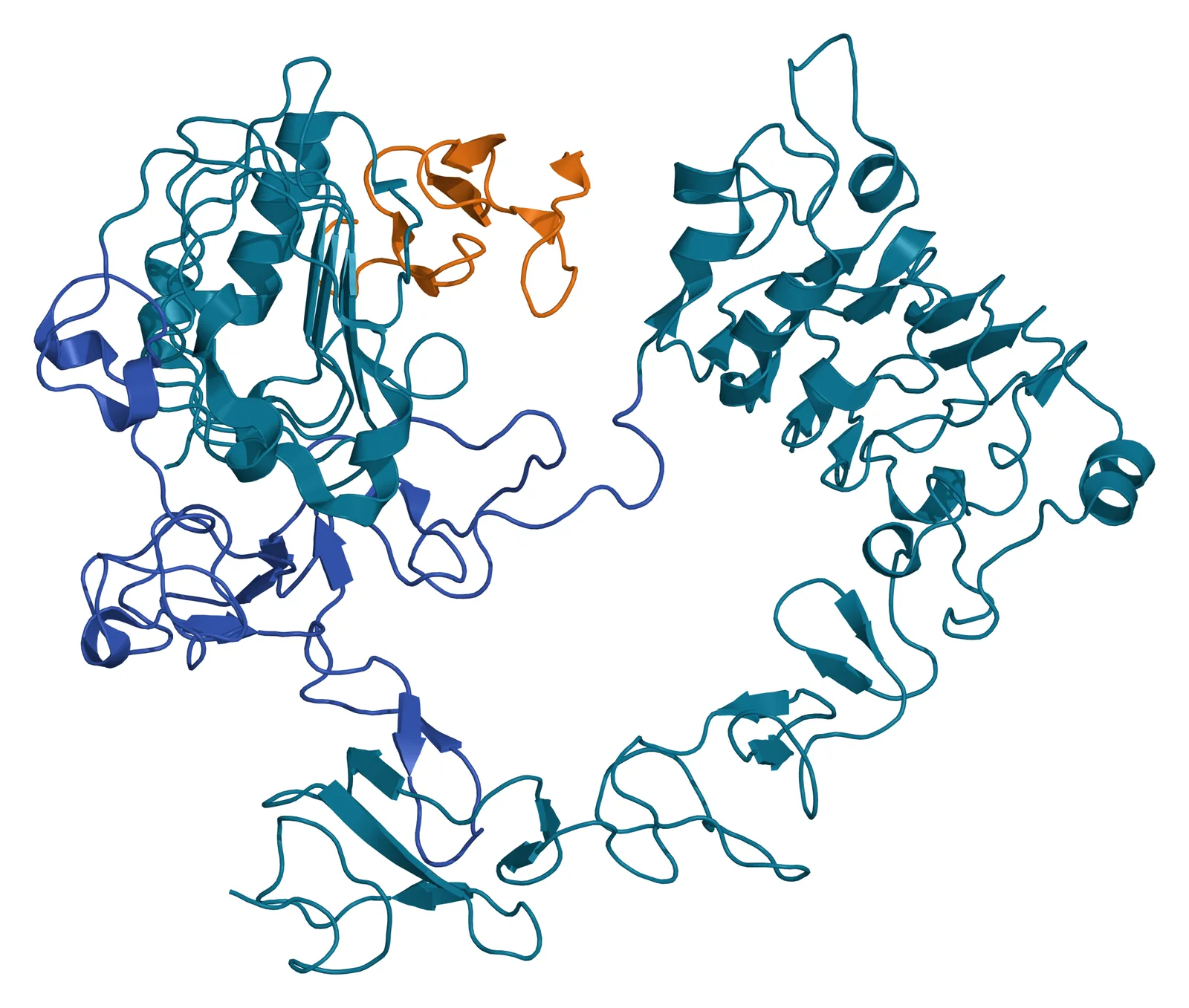
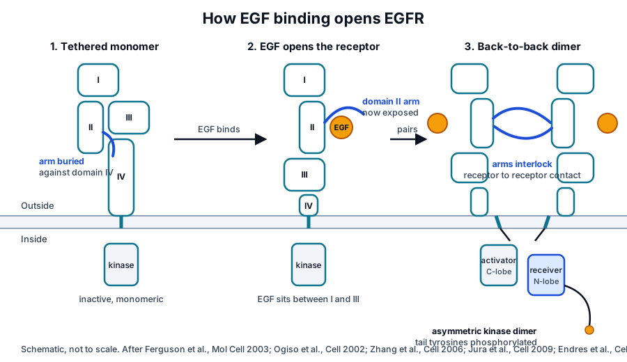
EGF binding opens the receptor and exposes the dimerization arm
Two crystal structures published together in 2002 showed what the ligand does. Ogiso and colleagues solved a 2:2 complex of EGF with the EGFR extracellular region and found domains I to III arranged in a C shape with EGF docked between domains I and III.[5] Garrett and colleagues solved a truncated receptor bound to transforming growth factor alpha and saw the same arrangement, with the ligand contacting both domains I and III.[8]
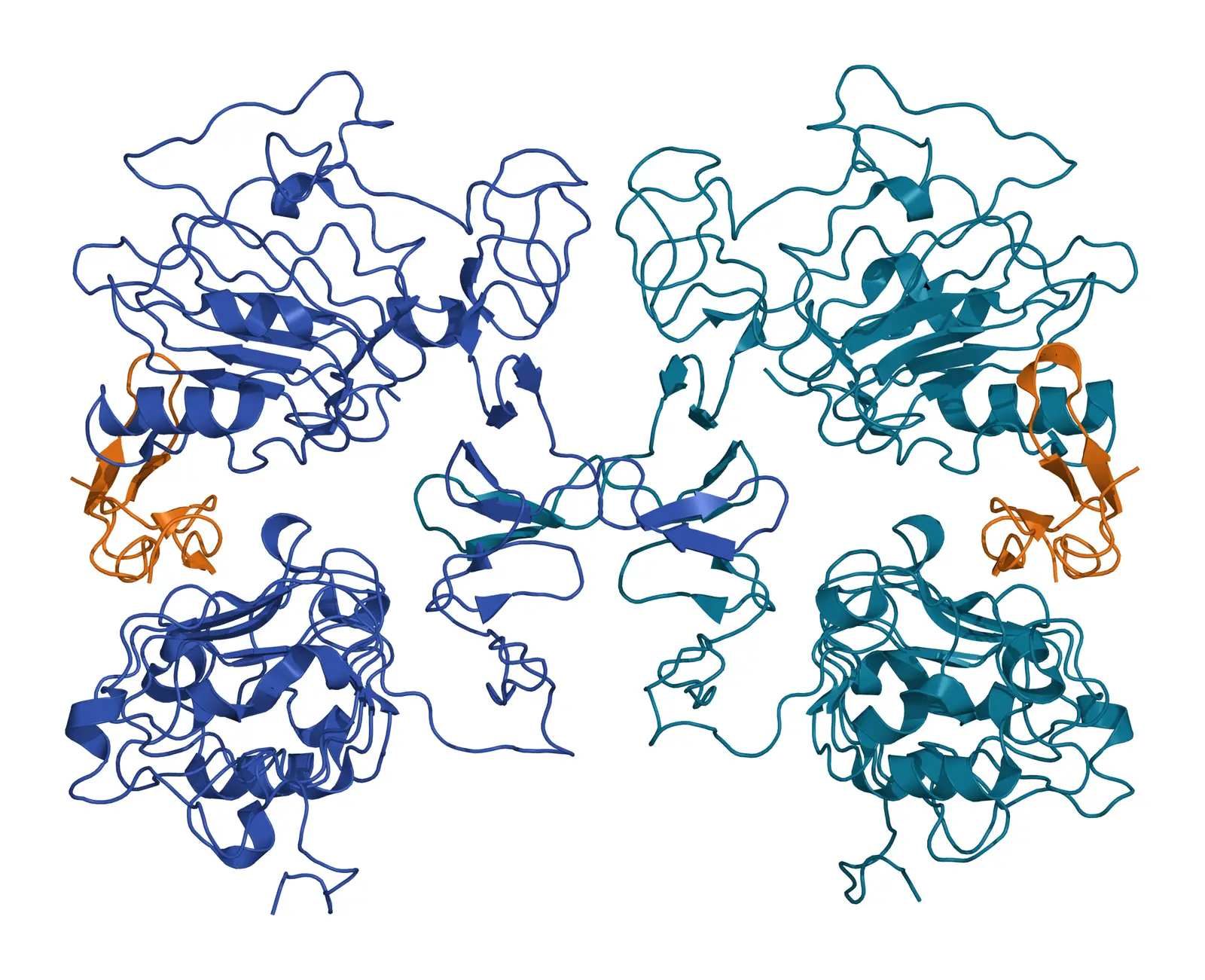
In both structures each ligand touches only one receptor. The two receptors pair through a direct receptor to receptor contact in which the beta-hairpin arm of each domain II reaches across and holds the body of the other, and mutagenesis confirmed that this contact mediates dimerization.[5][8] Ogiso called this receptor-mediated dimerization, and it is the opposite of the ligand-bridged dimers that KIT and the VEGF receptors form with their ligands.[9]
Comparing the liganded and tethered structures explains the activation step. Ligand binding to domains I and III can only happen when the region straightens, and straightening pulls the domain II arm away from domain IV and exposes it for pairing.[2][4] Bessman and colleagues later showed that ligand binding and dimerization compete with each other in the isolated extracellular region, and that forcing receptors into dimers without ligand gives ill-defined structures, so ligand binding is what organizes the dimer.[10]
Cryo-electron microscopy of the full-length receptor has added motion to this picture. Huang and colleagues found that EGF-bound EGFR samples an ensemble of dimeric conformations whose membrane-proximal tips are either together or apart, and that EGF and transforming growth factor alpha hold those two states to different degrees.[11] At physiological EGF concentrations the dimers go on to assemble into larger oligomers, which a structural model suggests are organized by the unoccupied ligand binding sites.[12]
Seven ligands tune how long the dimer lasts
Seven growth factors activate mammalian EGFR: EGF, transforming growth factor alpha, heparin-binding EGF-like growth factor, betacellulin, amphiregulin, epiregulin and epigen. The first four bind with high affinity and the last three with lower affinity.[13] Each is made as a membrane-anchored precursor that is released by a protease, so the supply of ligand is regulated at the cell surface.[13][14]
The ligands do more than open the receptor at different strengths. Freed and colleagues crystallized epiregulin and epigen bound to the extracellular region and found that they stabilize a different dimer geometry from EGF, producing less stable dimers.[15] Those weaker dimers signaled for longer, so the identity of the ligand is encoded in how long the dimer lasts and how the signal develops in time.[15]
The ligand also sets what happens to the receptor afterward. Roepstorff and colleagues compared six ligands and found that heparin-binding EGF-like growth factor and betacellulin send all receptors, and EGF most receptors, to the lysosome, while transforming growth factor alpha and epiregulin return them to the surface.[16] The endocytosis section below explains the sorting step that makes that difference.
An asymmetric kinase dimer switches the receptor on
Most receptor tyrosine kinases are activated when the two kinase domains in a dimer phosphorylate each other’s activation loops.[4] EGFR is activated differently. Zhang and colleagues found that the isolated kinase domain is intrinsically autoinhibited, in a conformation resembling inactive Src and cyclin-dependent kinases, and that it becomes active when its local concentration rises.[3]
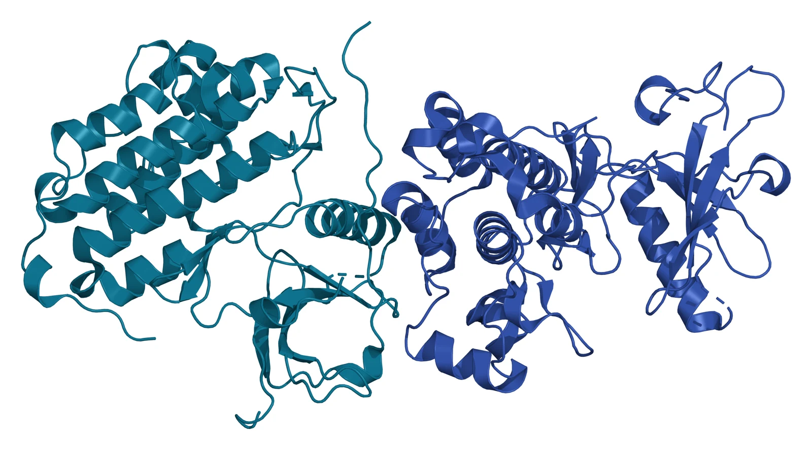
Their crystal structure showed why. Two kinase domains form an asymmetric dimer in which the C-terminal lobe of one, the activator, packs against the N-terminal lobe of the other, the receiver, in the same way cyclin packs against a cyclin-dependent kinase. The contact pushes the receiver into its active conformation, and phosphorylation of its activation loop is not required.[3]
The juxtamembrane segment between the membrane and the kinase holds this dimer together. Jura and colleagues showed that the C-terminal half of the segment latches the receiver kinase onto the activator, and that the N-terminal half most likely forms an antiparallel helical dimer that engages the transmembrane helices.[17] Red Brewer and colleagues reached the same conclusion by alanine scanning: mutations in the last 19 residues of the segment abolished activation, and their structure showed the segment of the receiver wrapped across the C-lobe of the activator.[18]
The extracellular and intracellular halves are coupled across the membrane. Endres and colleagues measured autophosphorylation as a function of receptor density and found that the intracellular module is inhibited by the membrane when tethered on its own, and that activation needs the transmembrane helices to pair near their N termini, which lets the juxtamembrane segments pair and releases that inhibition.[19] Molecular dynamics simulations by Arkhipov and colleagues gave the same answer: ligand-bound extracellular dimers favor the N-terminal helix crossing, and ligand-free dimers hold the helix N termini apart and favor a crossing that keeps the kinases inactive.[7]
Phosphotyrosines dock Grb2, Shc and PLC-gamma
The active receiver kinase phosphorylates tyrosines on the C-terminal tail of its partner, and each phosphotyrosine in its sequence context becomes a docking site for a protein with an SH2 or PTB domain.[9] Batzer and colleagues mapped the hierarchy for Grb2: it binds directly to phosphorylated tyrosine 1068 and to a lesser extent tyrosine 1086, and indirectly, through Shc, at tyrosine 1173.[20]
Grb2 brings the exchange factor SOS to the membrane, which activates Ras and the ERK cascade, and Shc recruits Grb2 to the same cascade from a second site.[9] Phospholipase C-gamma binds the receptor through its own SH2 domains and is phosphorylated by it, which turns on the production of inositol trisphosphate and diacylglycerol.[9]
Because the tail tyrosines are phosphorylated in trans within a dimer, the signal depends on the dimer’s lifetime, which helps explain why epiregulin and epigen produce different outputs from EGF.[15] Needham and colleagues argue that the higher-order oligomers formed at physiological ligand concentrations place neighboring dimers so that their tails are phosphorylated across dimers as well.[12]
Endocytosis sorts EGFR to recycling or degradation
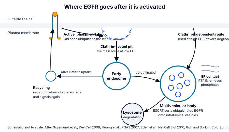
Activated EGFR leaves the surface within minutes.[21] Clathrin-mediated endocytosis is the main route at low ligand concentrations, and a slower clathrin-independent route contributes more as ligand concentration rises.[21][22] Goh and Sorkin review how activation, autophosphorylation, adaptor recruitment and ubiquitination are coupled to this uptake and to the sorting that follows.[21]
The route chosen decides the receptor’s fate. Sigismund and colleagues found that receptors taken in through clathrin are mostly recycled to the surface and keep signaling, while receptors taken in by the clathrin-independent route are committed to degradation.[22] The ubiquitin ligase Cbl attaches ubiquitin to lysines in the kinase domain, and Huang, Goh and Sorkin showed that a receptor with fifteen of those lysines mutated was internalized at the normal rate, so ubiquitination directs lysosomal sorting but is not needed for uptake itself.[23]
Receptors bound for degradation are sorted by the ESCRT machinery onto the intraluminal vesicles of multivesicular bodies, and Eden and colleagues found that the phosphatase PTP1B, which sits on the endoplasmic reticulum, reaches the receptor at direct membrane contacts between the multivesicular body and the endoplasmic reticulum.[24] The lysosome then degrades both receptor and ligand, which is how the cell ends a signal that recycling would otherwise sustain.[21]
ErbB heterodimers extend the family’s signaling
EGFR is the first of four ErbB receptors; the others are ErbB2, ErbB3 and ErbB4. Because dimerization is receptor-mediated and the ligand touches only its own receptor, a liganded EGFR can pair with any family member that can present a dimerization arm, and ErbB2, which binds no ligand, holds its arm permanently exposed.[4][6]
The asymmetric kinase dimer explains the same flexibility inside the cell. ErbB3 has a kinase domain with very little catalytic activity, but it can serve as the activator in an asymmetric dimer, and in ErbB2 and ErbB3 heterodimers ErbB2 takes the receiver position.[4][6] Heterodimers therefore combine the ligand preferences of one receptor with the docking sites of another, and the same structural rules that govern the EGFR homodimer govern them all.[9]
EGFR supports epidermal and hair follicle development
Mice carrying a null Egfr allele show how broadly the receptor acts in development. Threadgill and colleagues found that the outcome depended on genetic background: on a 129/Sv background the embryos did not survive mid-gestation because of placental defects, and on a CD-1 background they lived up to three weeks with abnormalities in skin, kidney, brain, liver and gut.[25]
The skin phenotype is the most instructive. Hansen and colleagues found disorganized hair follicles and a fuzzy coat in the null mice, and grafts of null skin showed wavy, flattened hair fibers and follicles that failed to progress through the hair cycle and differentiated prematurely.[26] Schneider and colleagues review the mouse models in which EGFR, its ligands and the proteases that release them have been altered, and the consistent theme is that the receptor keeps keratinocyte proliferation and differentiation in balance in the interfollicular epidermis and the follicle.[14]
That role continues into adult tissue maintenance. Nanba and colleagues showed that EGFR activation drives the motility of human keratinocyte stem cells by inducing TIMP-1 secretion, which protects the hemidesmosome protein collagen XVII from proteolysis, and their imaging and modeling linked this motility to self-renewal and epidermal regeneration.[27]
Advancing the research question
The growth factor receptors overview sets EGFR beside PDGFR-beta, VEGFR-2 and the TGF-beta receptors, and the sibling pages on PDGFR-beta and on VEGFR-2 and FGFR show the ligand-bridged dimers that EGFR avoids. The surface receptor chemistry page explains the SH2 and PTB docking that this page assumes.
Our team can explain how receptor structures such as these are read and what a given PDB entry does and does not show.
Annotated References
Numbered in the order they are cited in the text. Each source is annotated with what it supports on this page; peer-reviewed articles are listed with journal, volume, issue and pages, and primary sources are regulations, agency documents and reference databases.
- 1.DatabaseNCBI Gene 1956, EGFR epidermal growth factor receptor (Homo sapiens). Accessed 8 November 2026. https://www.ncbi.nlm.nih.gov/gene/1956
- 2.Peer-reviewedFerguson KM, Berger MB, Mendrola JM, et al. EGF activates its receptor by removing interactions that autoinhibit ectodomain dimerization. Molecular Cell. 2003;11(2):507-517. https://pubmed.ncbi.nlm.nih.gov/12620237/Annotation: Crystal structure of the unactivated EGFR extracellular region at 2.8 Å, crystallized at low pH with EGF bound weakly to domain I (PDB 1NQL). Cited for the tethered, autoinhibited conformation in which the domain II arm is buried against domain IV and domains I and III are held apart.
- 3.Peer-reviewedZhang X, Gureasko J, Shen K, et al. An allosteric mechanism for activation of the kinase domain of epidermal growth factor receptor. Cell. 2006;125(6):1137-1149. https://pubmed.ncbi.nlm.nih.gov/16777603/Annotation: Crystallography and mutagenesis of the EGFR kinase domain (PDB 2GS6). Cited for the intrinsically autoinhibited kinase, the asymmetric dimer in which the activator C-lobe switches on the receiver, and activation without activation loop phosphorylation.
- 4.Peer-reviewedLemmon MA, Schlessinger J, Ferguson KM. The EGFR family: not so prototypical receptor tyrosine kinases. Cold Spring Harbor Perspectives in Biology. 2014;6(4):a020768. https://pubmed.ncbi.nlm.nih.gov/24691965/Annotation: Review of EGFR family mechanisms. Cited for the domain layout, the exposed dimerization arm, allosteric kinase activation, the activating juxtamembrane segment, the receiver phosphorylating the activator tail and the impaired ErbB3 kinase.
- 5.Peer-reviewedOgiso H, Ishitani R, Nureki O, et al. Crystal structure of the complex of human epidermal growth factor and receptor extracellular domains. Cell. 2002;110(6):775-787. https://pubmed.ncbi.nlm.nih.gov/12297050/Annotation: Crystal structure of the 2:2 EGF and EGFR extracellular region complex at 3.3 Å (PDB 1IVO). Cited for EGF docked between domains I and III, the C-shaped arrangement and receptor-mediated dimerization through the domain II arm.
- 6.Peer-reviewedKovacs E, Zorn JA, Huang Y, et al. A structural perspective on the regulation of the epidermal growth factor receptor. Annual Review of Biochemistry. 2015;84:739-764. https://pubmed.ncbi.nlm.nih.gov/25621509/Annotation: Structural review of EGFR regulation. Cited for the mix of monomers and weak dimers in the unliganded receptor, the coupling of extracellular and kinase dimers through the transmembrane helices, heterodimerization without direct ligand involvement, the roughly 130 degree domain rotation on ligand binding and the activator and receiver roles of ErbB3 and ErbB2.
- 7.Peer-reviewedArkhipov A, Shan Y, Das R, et al. Architecture and membrane interactions of the EGF receptor. Cell. 2013;152(3):557-569. https://pubmed.ncbi.nlm.nih.gov/23374350/Annotation: Molecular dynamics simulations of membrane-embedded EGFR. Cited for inactive receptors existing as monomers and dimers, and for ligand-bound and ligand-free dimers favoring different crossings of the transmembrane helices.
- 8.Peer-reviewedGarrett TP, McKern NM, Lou M, et al. Crystal structure of a truncated epidermal growth factor receptor extracellular domain bound to transforming growth factor alpha. Cell. 2002;110(6):763-773. https://pubmed.ncbi.nlm.nih.gov/12297049/Annotation: Crystal structure of a truncated EGFR extracellular region with transforming growth factor alpha at 2.5 Å. Cited for the ligand contacting domains I and III, each ligand touching one receptor, and the back-to-back dimer through domain II.
- 9.Peer-reviewedLemmon MA, Schlessinger J. Cell signaling by receptor tyrosine kinases. Cell. 2010;141(7):1117-1134. https://pubmed.ncbi.nlm.nih.gov/20602996/Annotation: Review of receptor tyrosine kinase activation. Cited for the contrast between receptor-mediated and ligand-bridged dimerization, phosphotyrosine docking of SH2 and PTB proteins, Grb2 and SOS recruitment, PLC-gamma activation and the shared rules of ErbB heterodimers.
- 10.Peer-reviewedBessman NJ, Bagchi A, Ferguson KM, Lemmon MA. Complex relationship between ligand binding and dimerization in the epidermal growth factor receptor. Cell Reports. 2014;9(4):1306-1317. https://pubmed.ncbi.nlm.nih.gov/25453753/Annotation: Binding and dimerization studies of the isolated extracellular region. Cited for ligand binding and dimerization competing with each other and for forced ligand-free dimers being ill-defined.
- 11.Peer-reviewedHuang Y, Ognjenovic J, Karandur D, et al. A molecular mechanism for the generation of ligand-dependent differential outputs by the epidermal growth factor receptor. eLife. 2021;10:e73218. https://pubmed.ncbi.nlm.nih.gov/34846302/Annotation: Cryo-electron microscopy of full-length EGFR with EGF and transforming growth factor alpha. Cited for the ensemble of dimeric conformations with membrane-proximal tips together or apart and the ligands holding those states to different degrees.
- 12.Peer-reviewedNeedham SR, Roberts SK, Arkhipov A, et al. EGFR oligomerization organizes kinase-active dimers into competent signalling platforms. Nature Communications. 2016;7:13307. https://pubmed.ncbi.nlm.nih.gov/27796308/Annotation: Single-molecule imaging and modeling of EGFR oligomers. Cited for dimers assembling into oligomers at physiological EGF concentrations, organized by unoccupied ligand binding sites, with phosphorylation across neighboring dimers.
- 13.Peer-reviewedSingh B, Carpenter G, Coffey RJ. EGF receptor ligands: recent advances. F1000Research. 2016;5:2270. https://pubmed.ncbi.nlm.nih.gov/27635238/Annotation: Review of the seven EGFR ligands. Cited for the ligand list, the high-affinity and low-affinity groups and the membrane-anchored precursors released by proteolysis.
- 14.Peer-reviewedSchneider MR, Werner S, Paus R, Wolf E. Beyond wavy hairs: the epidermal growth factor receptor and its ligands in skin biology and pathology. American Journal of Pathology. 2008;173(1):14-24. https://pubmed.ncbi.nlm.nih.gov/18556782/Annotation: Review of the EGFR system in skin. Cited for the precursor processing of the ligands and for the mouse models showing that the receptor balances keratinocyte proliferation and differentiation in the epidermis and hair follicle.
- 15.Peer-reviewedFreed DM, Bessman NJ, Kiyatkin A, et al. EGFR Ligands Differentially Stabilize Receptor Dimers to Specify Signaling Kinetics. Cell. 2017;171(3):683-695.e18. https://pubmed.ncbi.nlm.nih.gov/28988771/Annotation: Crystal structures of epiregulin and epigen bound to the EGFR extracellular region. Cited for the different dimer geometry, the less stable dimers and the longer signaling they produce, and for dimer lifetime encoding ligand identity.
- 16.Peer-reviewedRoepstorff K, Grandal MV, Henriksen L, et al. Differential effects of EGFR ligands on endocytic sorting of the receptor. Traffic. 2009;10(8):1115-1127. https://pubmed.ncbi.nlm.nih.gov/19531065/Annotation: Comparison of six ligands on EGFR trafficking. Cited for EGF, heparin-binding EGF-like growth factor and betacellulin directing receptors to the lysosome while transforming growth factor alpha and epiregulin recycle them.
- 17.Peer-reviewedJura N, Endres NF, Engel K, et al. Mechanism for activation of the EGF receptor catalytic domain by the juxtamembrane segment. Cell. 2009;137(7):1293-1307. https://pubmed.ncbi.nlm.nih.gov/19563760/Annotation: Structural and biochemical study of the juxtamembrane segment. Cited for its C-terminal half latching the receiver kinase onto the activator and its N-terminal half forming an antiparallel helical dimer near the transmembrane helices.
- 18.Peer-reviewedRed Brewer M, Choi SH, Alvarado D, et al. The juxtamembrane region of the EGF receptor functions as an activation domain. Molecular Cell. 2009;34(6):641-651. https://pubmed.ncbi.nlm.nih.gov/19560417/Annotation: Alanine scanning and crystallography of the juxtamembrane region. Cited for mutations in its last 19 residues abolishing activation and for the receiver segment contacting the activator C-lobe in the asymmetric dimer.
- 19.Peer-reviewedEndres NF, Das R, Smith AW, et al. Conformational coupling across the plasma membrane in activation of the EGF receptor. Cell. 2013;152(3):543-556. https://pubmed.ncbi.nlm.nih.gov/23374349/Annotation: Cell-based, NMR and fluorescence studies of EGFR activation. Cited for membrane inhibition of the intracellular module, the N-terminal pairing of the transmembrane helices and the antiparallel juxtamembrane dimer that releases inhibition.
- 20.Peer-reviewedBatzer AG, Rotin D, Ureña JM, et al. Hierarchy of binding sites for Grb2 and Shc on the epidermal growth factor receptor. Molecular and Cellular Biology. 1994;14(8):5192-5201. https://pubmed.ncbi.nlm.nih.gov/7518560/Annotation: Mutagenesis, peptide competition and binding measurements of Grb2 docking. Cited for direct Grb2 binding at tyrosine 1068 and to a lesser extent tyrosine 1086, and indirect binding through Shc at tyrosine 1173.
- 21.Peer-reviewedGoh LK, Sorkin A. Endocytosis of receptor tyrosine kinases. Cold Spring Harbor Perspectives in Biology. 2013;5(5):a017459. https://pubmed.ncbi.nlm.nih.gov/23637288/Annotation: Review of receptor tyrosine kinase endocytosis. Cited for the coupling of autophosphorylation, adaptor recruitment and ubiquitination to uptake and sorting, and for lysosomal degradation of receptor and ligand ending the signal.
- 22.Peer-reviewedSigismund S, Argenzio E, Tosoni D, et al. Clathrin-mediated internalization is essential for sustained EGFR signaling but dispensable for degradation. Developmental Cell. 2008;15(2):209-219. https://pubmed.ncbi.nlm.nih.gov/18694561/Annotation: Endocytic route studies in cultured cells. Cited for clathrin-mediated uptake being the main route at low EGF and recycling the receptor, while clathrin-independent uptake commits it to degradation.
- 23.Peer-reviewedHuang F, Goh LK, Sorkin A. EGF receptor ubiquitination is not necessary for its internalization. Proceedings of the National Academy of Sciences of the United States of America. 2007;104(43):16904-16909. https://pubmed.ncbi.nlm.nih.gov/17940017/Annotation: Lysine mutagenesis of the EGFR kinase domain. Cited for a receptor lacking fifteen ubiquitination-site lysines being internalized at the normal rate, so ubiquitination directs sorting without being required for uptake.
- 24.Peer-reviewedEden ER, White IJ, Tsapara A, Futter CE. Membrane contacts between endosomes and ER provide sites for PTP1B-epidermal growth factor receptor interaction. Nature Cell Biology. 2010;12(3):267-272. https://pubmed.ncbi.nlm.nih.gov/20118922/Annotation: Electron microscopy and biochemistry of endosome and endoplasmic reticulum contacts. Cited for PTP1B reaching EGFR at direct membrane contacts with multivesicular bodies and for ESCRT sorting of the receptor onto intraluminal vesicles.
- 25.Peer-reviewedThreadgill DW, Dlugosz AA, Hansen LA, et al. Targeted disruption of mouse EGF receptor: effect of genetic background on mutant phenotype. Science. 1995;269(5221):230-234. https://pubmed.ncbi.nlm.nih.gov/7618084/Annotation: Egfr null mice on three genetic backgrounds. Cited for the placental defects on 129/Sv and for the skin, kidney, brain, liver and gut abnormalities in CD-1 mice that survived up to three weeks.
- 26.Peer-reviewedHansen LA, Alexander N, Hogan ME, et al. Genetically null mice reveal a central role for epidermal growth factor receptor in the differentiation of the hair follicle and normal hair development. American Journal of Pathology. 1997;150(6):1959-1975. https://pubmed.ncbi.nlm.nih.gov/9176390/Annotation: Skin and hair follicle analysis of Egfr null mice and skin grafts. Cited for disorganized follicles, wavy flattened hair fibers, follicles that failed to progress through the hair cycle and premature differentiation.
- 27.Peer-reviewedNanba D, Toki F, Asakawa K, et al. EGFR-mediated epidermal stem cell motility drives skin regeneration through COL17A1 proteolysis. Journal of Cell Biology. 2021;220(11):e202012073. https://pubmed.ncbi.nlm.nih.gov/34550317/Annotation: Live imaging and culture studies of human keratinocyte stem cells. Cited for EGFR activation driving stem cell motility coupled to self-renewal, through TIMP-1 secretion and stabilization of collagen XVII.
Cite this page
ExaVeyra Sciences Editorial Team. EGFR: Structure, Ligand Binding and Activation. ExaVeyra Sciences; 9 November 2026. Available from: https://exaveyra.com/science/surface-receptors/egfr. Accessed [date accessed].
Figure sources
- 28.PDB 1NQL, EGFR extracellular domain in an inactive (low pH) complex with EGF. ExaVeyra Sciences, rendered in PyMOL from PDB 1NQL. Structure data: RCSB Protein Data Bank (rcsb.org), CC0. Structure publication: Ferguson KM, Berger MB, Mendrola JM, Cho HS, Leahy DJ, Lemmon MA. EGF activates its receptor by removing interactions that autoinhibit ectodomain dimerization. Mol Cell. 2003;11(2):507-517.
- 29.PDB 1IVO, Human EGF in complex with the EGFR extracellular domains. ExaVeyra Sciences, rendered in PyMOL from PDB 1IVO. Structure data: RCSB Protein Data Bank (rcsb.org), CC0. Structure publication: Ogiso H, Ishitani R, Nureki O, et al. Crystal structure of the complex of human epidermal growth factor and receptor extracellular domains. Cell. 2002;110(6):775-787.
- 30.PDB 2GS6, active EGFR kinase domain in complex with an ATP analog-peptide conjugate. ExaVeyra Sciences, rendered in PyMOL from PDB 2GS6. Structure data: RCSB Protein Data Bank (rcsb.org), CC0. Structure publication: Zhang X, Gureasko J, Shen K, Cole PA, Kuriyan J. An allosteric mechanism for activation of the kinase domain of epidermal growth factor receptor. Cell. 2006;125(6):1137-1149.
Image credits
- Fig. 2ExaVeyra Sciences, original figure. After Ferguson et al., Mol Cell 2003; Ogiso et al., Cell 2002; Zhang et al., Cell 2006; Jura et al., Cell 2009; Endres et al., Cell 2013. ↩
- Fig. 5ExaVeyra Sciences, original figure. After Sigismund et al., Dev Cell 2008; Huang et al., Proc Natl Acad Sci U S A 2007; Eden et al., Nat Cell Biol 2010; Goh and Sorkin, Cold Spring Harb Perspect Biol 2013. ↩