The Tetraspanin Family: Structure and Membrane Organization
Reviewed by ExaVeyra Sciences editorial team · Updated 19 October 2026
ExaVeyra Sciences is a Miami-based B2B supplier of exosomes for topical aesthetic use to licensed clinics, and it refers licensed prescribers to a 503A compounding pharmacy that prepares formulations against individual prescriptions.
| Question | Answer |
|---|---|
| What this page covers | The four-pass tetraspanin fold, the large extracellular loop and its disulfides, the cholesterol pocket, palmitoylation, and tetraspanin-enriched microdomains. |
| Who can order | Licensed clinics and practitioners whose active professional license and NPI ExaVeyra has verified. Exosome orders also require a signed professional-use acknowledgment. |
| What ships, and how | Exosome material for topical aesthetic use, characterized by lot, ships overnight in a cold chain. Delivery can take 1 to 3 business days. A certificate of analysis is issued for every lot and provided on request. |
Tetraspanins are small membrane proteins that cross the lipid bilayer four times and spend most of their working lives organizing other proteins.1 Mammals carry 33 of them, and three, CD9, CD63 and CD81, are among the most widely studied proteins on cells and on extracellular vesicles.2,3,4,5
Their biology follows from their architecture. This page explains the fold the family shares, what the first full-length crystal structures revealed, and how tetraspanins gather partner receptors into nanoscale domains.
Understanding the tetraspanin fold
Every tetraspanin has four transmembrane helices, a short cytoplasmic amino terminus and carboxyl terminus, and two loops outside the cell. The small extracellular loop, called EC1, lies between the first two helices, and the large extracellular loop, EC2, lies between the third and fourth (Fig. 1).6
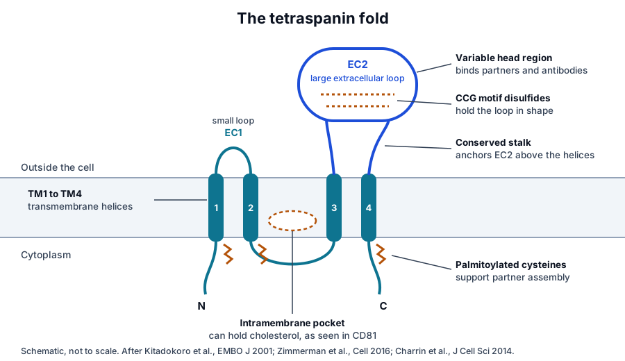
EC2 carries the family signature. A conserved cysteine, cysteine, glycine motif forms disulfide bonds that hold the loop in shape, and Kitadokoro and colleagues showed in 2001 that the large loop of CD81 folds into a mushroom-like structure of five helices with a stalk and a head.7
Sequence analysis of 160 tetraspanins in that study found the same structural features conserved across the family.7 The head region varies most between members, and it is where most partner proteins and antibodies bind.6
Full-length structures revealed a cone and a cholesterol pocket
The first crystal structure of a complete tetraspanin, human CD81, was reported by Zimmerman and colleagues in 2016. Its four helices pack as two separated pairs capped by EC2 at the outer leaflet, and they converge toward the inner leaflet to form a pocket that held a bound cholesterol molecule (Fig. 2a).8
Umeda and colleagues then solved CD9 (Fig. 2b) and found the same reversed cone shape. In crystalline lipid layers that shape generated membrane curvature, which explains why CD9 concentrates where the membrane is most strongly curved.9
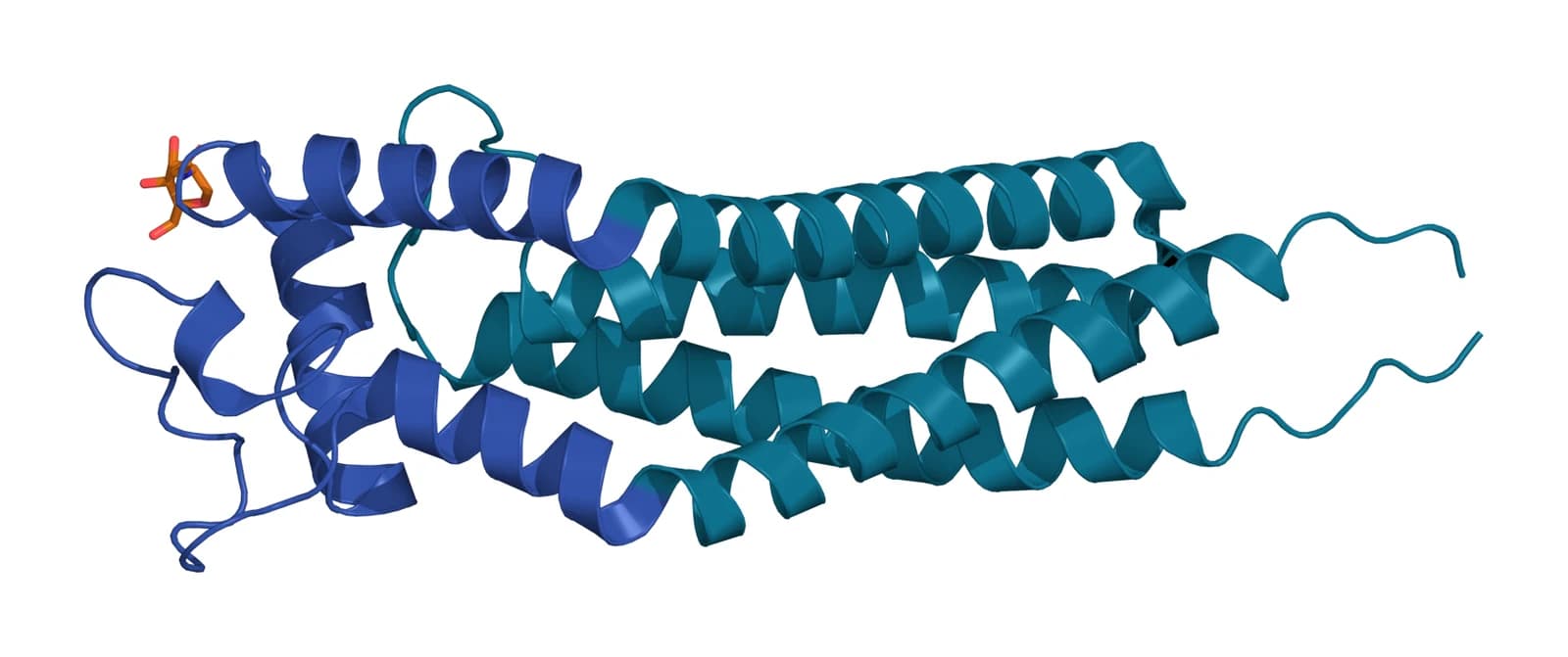
Later work showed the fold is dynamic. The CD53 structure reported by Yang and colleagues captured an open conformation in which EC2 stands away from the membrane (Fig. 3),10 and Susa and colleagues found that CD81 opens in the same way when it binds its partner CD19.11
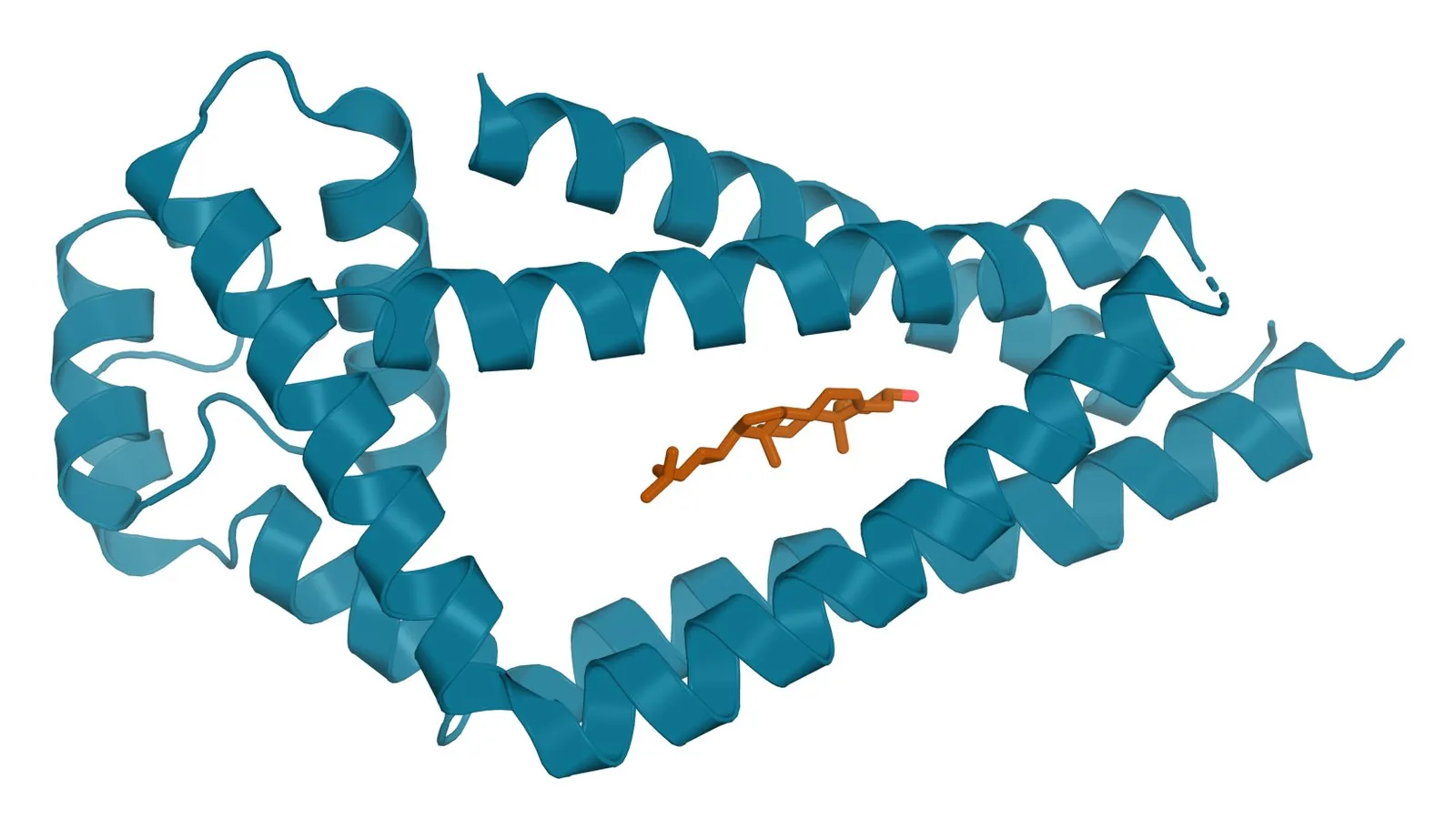
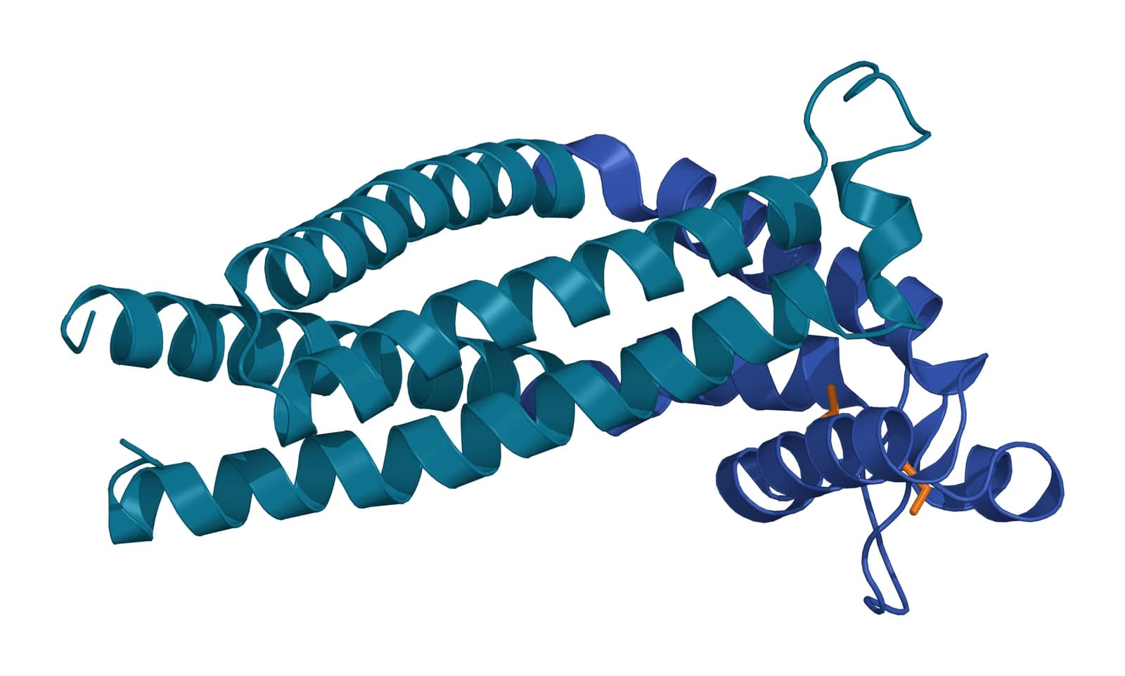
Fig. 2 | Tetraspanin CD81 with cholesterol and Tetraspanin CD9.
Tetraspanins sense and shape membrane curvature
The cone shape has a measurable consequence. Using membrane tubes pulled from cell-derived vesicles, Dharan and colleagues showed that CD9 and tetraspanin 4 prefer positively curved membranes, the geometry of microvilli and retraction fibers.12
Reviews by Dharan and Sorkin and by Rubinstein, Théry and Zimmermann connect this to the structures where tetraspanins collect. Microvilli, tunneling nanotubes, migrasomes and extracellular vesicles are all small, highly curved membranes.2,13
Palmitoylation and partner proteins build the tetraspanin web
Tetraspanins carry palmitate on cysteines next to their membrane helices. Yang and colleagues showed that palmitoylated integrins and the tetraspanins CD9, CD81 and CD63 coexist in overlapping complexes, and that palmitoylation supports these secondary associations.14
Each tetraspanin binds a small set of direct partners, and tetraspanins also bind one another.15,16 Charrin and colleagues described the result as a hierarchical network, the tetraspanin web, in which partner receptors are positioned next to the molecules they work with.17,18
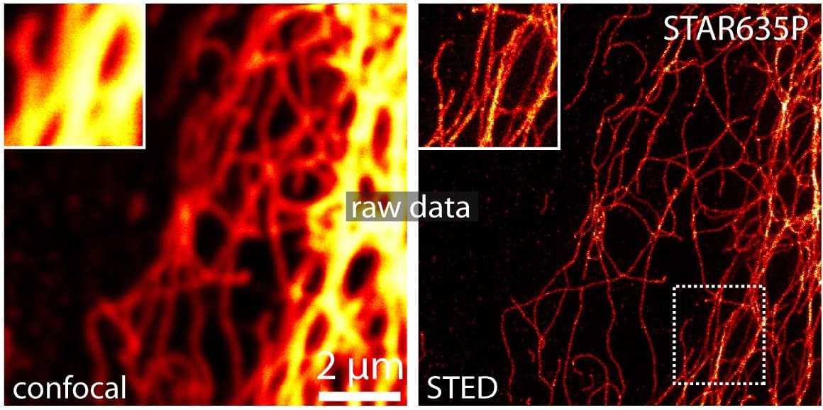
Super-resolution microscopy (Fig. 4) has since measured the units of that web. Zuidscherwoude and colleagues found that tetraspanins form individual nanoclusters smaller than 120 nm, each containing fewer than ten copies of CD53, sitting close to their partners MHC class II and CD19.19
Tetraspanins regulate signaling through the partners they hold
Because they gather adhesion molecules, receptors and signaling enzymes into the same domain, tetraspanins influence how those partners signal. Termini and Gillette reviewed this role across integrin, growth factor receptor and kinase pathways.20
The same reviews describe how palmitoylation, N-linked glycosylation and ubiquitination of the tetraspanins themselves tune these complexes.20 Susa, Kruse and Blacklow summarized the structural principles that let one fold recognize remarkably different partner families.6
An ancient family with vertebrate additions
Tetraspanins are found in plants, fungi and single-celled organisms as well as animals. Huang and colleagues proposed from their phylogeny that the family took part in the transition from single cells to multicellular life.21
Garcia-España and colleagues divided the superfamily into four families, the CD family, the CD63 family, the uroplakins and the RDS family.22 CD9 and CD81 sit together in one closely related cluster, which fits the overlapping functions the two proteins share.22
Why tetraspanins mark extracellular vesicles
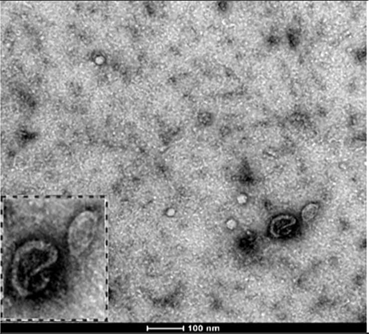
Small extracellular vesicles (Fig. 5) are highly curved membranes built from plasma membrane and endosomes, both compartments where tetraspanins are abundant. CD9, CD63 and CD81 are therefore among the most consistently detected proteins on them.2
Rubinstein, Théry and Zimmermann note that their enrichment on vesicles is well established, while their exact role in vesicle formation is still being worked out.2 The individual pages on CD9, CD63 and CD81 describe what each protein contributes.
Where to go from here
The CD9, CD63 and CD81 references cover each protein’s partners and biological roles. The quality reference on marker panels explains how the fraction of vesicles carrying each tetraspanin is measured and reported.
Verified practices can request lot-specific characterization data with the certificate of analysis, and our team can explain how to read the tetraspanin section of a panel.
References
Numbered in the order they are cited in the text. Each study carries its design and, where people were enrolled, how many, with a line saying what it supports on this page. Primary sources are regulations, agency documents and reference databases.
- 1.Hemler ME. Tetraspanin functions and associated microdomains. Nature Reviews Molecular Cell Biology. 2005. doi:10.1038/nrm1736. PMID: 16314869.Narrative reviewLandmarkReference 1 supports: Foundational review of tetraspanin microdomains. Cited for tetraspanins as small proteins present on nearly all cell types.
- 2.Rubinstein E, Théry C, Zimmermann P. Tetraspanins affect membrane structures and the trafficking of molecular partners: what impact on extracellular vesicles? Biochemical Society Transactions. 2025. doi:10.1042/BST20240523. PMID: 40135387.Narrative reviewReference 2 supports: Review of tetraspanins on curved structures and vesicles. Cited for the family size of 33 in mammals and for CD9, CD81 and CD63 as vesicle markers.
- 3.NCBI Gene 928, CD9 molecule (Homo sapiens). Accessed 19 October 2026.Primary source
- 4.NCBI Gene 967, CD63 molecule (Homo sapiens). Accessed 19 October 2026.Primary source
- 5.NCBI Gene 975, CD81 molecule (Homo sapiens). Accessed 19 October 2026.Primary source
- 6.Susa KJ, Kruse AC, Blacklow SC. Tetraspanins: structure, dynamics, and principles of partner-protein recognition. Trends in Cell Biology. 2024. doi:10.1016/j.tcb.2023.09.003. PMID: 37783654.Narrative reviewReference 6 supports: Current review of tetraspanin structures and complexes. Cited for the structural plasticity that lets one fold recognize very different partner families.
- 7.Kitadokoro K, Bordo D, Galli G, et al. CD81 extracellular domain 3D structure: insight into the tetraspanin superfamily structural motifs. EMBO Journal. 2001. doi:10.1093/emboj/20.1.12. PMID: 11226150.In vitroLandmarkReference 7 supports: The first structure of a tetraspanin large extracellular loop. Cited for the five-helix stalk and head fold conserved across 160 tetraspanin sequences.
- 8.Zimmerman B, Kelly B, McMillan BJ, et al. Crystal Structure of a Full-Length Human Tetraspanin Reveals a Cholesterol-Binding Pocket. Cell. 2016. doi:10.1016/j.cell.2016.09.056. PMID: 27881302.In vitroReference 8 supports: The first full-length tetraspanin structure, human CD81. Cited for the two helix pairs, the EC2 cap and the intramembrane cholesterol pocket.
- 9.Umeda R, Satouh Y, Takemoto M, et al. Structural insights into tetraspanin CD9 function. Nature Communications. 2020. doi:10.1038/s41467-020-15459-7. PMID: 32231207.In vitroReference 9 supports: Crystal structure of CD9 and cryo-EM of CD9 with EWI-2. Cited for the reversed cone shape that generates membrane curvature.
- 10.Yang Y, Liu XR, Greenberg ZJ, et al. Open conformation of tetraspanins shapes interaction partner networks on cell membranes. EMBO Journal. 2020. doi:10.15252/embj.2020105246. PMID: 32974937.In vitroReference 10 supports: Crystal structure of CD53 in an open conformation. Cited for EC2 standing away from the membrane to expose a partner-binding region.
- 11.Susa KJ, Rawson S, Kruse AC, et al. Cryo-EM structure of the B cell co-receptor CD19 bound to the tetraspanin CD81. Science. 2021. doi:10.1126/science.abd9836. PMID: 33446559.In vitroReference 11 supports: Cryo-EM structure of CD81 bound to CD19. Cited for the opening of EC2 when a tetraspanin binds its partner.
- 12.Dharan R, Goren S, Cheppali SK, et al. Transmembrane proteins tetraspanin 4 and CD9 sense membrane curvature. Proceedings of the National Academy of Sciences of the United States of America. 2022. doi:10.1073/pnas.2208993119. PMID: 36252000.In vitroReference 12 supports: Membrane tube experiments with CD9 and tetraspanin 4. Cited for the preference of both proteins for positively curved membrane.
- 13.Dharan R, Sorkin R. Tetraspanin proteins in membrane remodeling processes. Journal of Cell Science. 2024. doi:10.1242/jcs.261532. PMID: 39051897.Narrative reviewReference 13 supports: Review of tetraspanins in membrane remodeling. Cited for the link between curvature, tetraspanin distribution and membrane domain formation.
- 14.Yang X, Kovalenko OV, Tang W, et al. Palmitoylation supports assembly and function of integrin-tetraspanin complexes. Journal of Cell Biology. 2004. doi:10.1083/jcb.200404100. PMID: 15611341.In vitroLandmarkReference 14 supports: Biochemistry of palmitoylated integrin and tetraspanin complexes. Cited for palmitoylation supporting the secondary associations of CD9, CD81 and CD63.
- 15.van Deventer SJ, Dunlock VE, van Spriel AB. Molecular interactions shaping the tetraspanin web. Biochemical Society Transactions. 2017. doi:10.1042/BST20160284. PMID: 28620035.Narrative reviewReference 15 supports: Review classifying tetraspanin interactions by function. Cited for the intramolecular, tetraspanin and partner interactions that build the web.
- 16.Oosterheert W, Xenaki KT, Neviani V, et al. Implications for tetraspanin-enriched microdomain assembly based on structures of CD9 with EWI-F. Life Science Alliance. 2020. doi:10.26508/lsa.202000883. PMID: 32958604.In vitroReference 16 supports: Structures of CD9 with its partner EWI-F. Cited for the tetrameric complex that suggests how microdomains are concatenated.
- 17.Charrin S, le Naour F, Silvie O, et al. Lateral organization of membrane proteins: tetraspanins spin their web. Biochemical Journal. 2009. doi:10.1042/BJ20082422. PMID: 19426143.Narrative reviewLandmarkReference 17 supports: Review introducing the tetraspanin web model. Cited for tetraspanin function arising from association with one another and with partner proteins.
- 18.Charrin S, Jouannet S, Boucheix C, et al. Tetraspanins at a glance. Journal of Cell Science. 2014. doi:10.1242/jcs.154906. PMID: 25128561.Narrative reviewReference 18 supports: Overview of tetraspanin assemblies. Cited for the hierarchical network of direct partner and tetraspanin-tetraspanin interactions.
- 19.Zuidscherwoude M, Göttfert F, Dunlock VM, et al. The tetraspanin web revisited by super-resolution microscopy. Scientific Reports. 2015. doi:10.1038/srep12201. PMID: 26183063.In vitroReference 19 supports: STED microscopy of tetraspanins on B cells and dendritic cells. Cited for nanoclusters under 120 nm containing fewer than ten CD53 molecules.
- 20.Termini CM, Gillette JM. Tetraspanins Function as Regulators of Cellular Signaling. Frontiers in Cell and Developmental Biology. 2017. doi:10.3389/fcell.2017.00034. PMID: 28428953.Narrative reviewReference 20 supports: Review of tetraspanins as signaling scaffolds. Cited for their influence on integrin, receptor and kinase signaling and for the modifications that tune it.
- 21.Huang S, Yuan S, Dong M, et al. The phylogenetic analysis of tetraspanins projects the evolution of cell-cell interactions from unicellular to multicellular organisms. Genomics. 2005. doi:10.1016/j.ygeno.2005.08.004. PMID: 16242907.In vitroLandmarkReference 21 supports: Phylogeny spanning animals, plants, fungi and amoebae. Cited for the family’s distribution beyond animals and its proposed role in multicellularity.
- 22.Garcia-España A, Chung PJ, Sarkar IN, et al. Appearance of new tetraspanin genes during vertebrate evolution. Genomics. 2008. doi:10.1016/j.ygeno.2007.12.005. PMID: 18291621.In vitroReference 22 supports: Whole-genome phylogeny of the tetraspanins. Cited for the four families and the close CD9 and CD81 cluster.
Figure sources
- 23.PDB 5TCX, Tetraspanin CD81 with cholesterol. ExaVeyra Sciences, rendered in PyMOL from PDB 5TCX. Structure data: RCSB Protein Data Bank (rcsb.org), CC0. Structure publication: Zimmerman B, Kelly B, McMillan BJ, et al. Crystal structure of a full-length human tetraspanin reveals a cholesterol-binding pocket. Cell. 2016;167(4):1041-1051.e11.
- 24.PDB 6K4J, Tetraspanin CD9. ExaVeyra Sciences, rendered in PyMOL from PDB 6K4J. Structure data: RCSB Protein Data Bank (rcsb.org), CC0. Structure publication: Umeda R, Satouh Y, Takemoto M, et al. Structural insights into tetraspanin CD9 function. Nat Commun. 2020;11(1):1606.
- 25.PDB 6WVG, Tetraspanin CD53 in the open conformation. ExaVeyra Sciences, rendered in PyMOL from PDB 6WVG. Structure data: RCSB Protein Data Bank (rcsb.org), CC0. Structure publication: Yang Y, Liu XR, Greenberg ZJ, et al. Open conformation of tetraspanins shapes interaction partner networks on cell membranes. EMBO J. 2020;39(18):e105246.