How cell surface receptors are built and organized
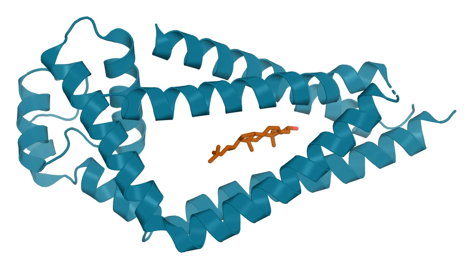
A cell reads its surroundings through the proteins on its surface. Their shape, their sugars and the lipids that hold them in the membrane (Fig. 1) decide what each one binds, and those same proteins are what an antibody panel detects when a cell or a vesicle is characterized.
These references explain that chemistry protein by protein, starting with the shared rules of membrane biochemistry and moving through the tetraspanins and the mesenchymal stromal cell marker panel. Every page cites at least fifteen peer-reviewed studies.
The references, in reading order
5 of 13 published. The remainder are in review and will appear as they are completed.
Receptor chemistry shared by every surface protein
Every receptor on these pages is assembled by the same cellular machinery. Its membrane topology, its sugars and its lipid anchors decide where it sits, what it binds and which partners it can reach.
Tetraspanins organize the membrane around them
Tetraspanins are four-pass membrane proteins that gather partner receptors into nanoscale domains. CD9, CD63 and CD81 are the three most studied members and the three most often measured on extracellular vesicles.
The Tetraspanin Family: Structure and Membrane Organization
The fold, the cone shape and the tetraspanin web.
CD9: Structure, Partners and Biological Functions
Structure, partners, gamete fusion and vesicles.
CD63 (LAMP-3): Endosomal Trafficking and Biological Roles
Lysosomal targeting, vesicle sorting and TIMP-1.
CD81: Tetraspanin Structure and the CD19 Co-Receptor
The cholesterol pocket, CD19 and the immune synapse.
The marker panel that defines a mesenchymal stromal cell
The International Society for Cell and Gene Therapy defines these cells partly by the receptors they carry and partly by the ones they lack. Each marker in the panel is a protein with its own chemistry and its own job.
The ISCT Marker Criteria for Mesenchymal Stromal Cells
The positive and negative panel, and why it exists.
In review
CD73: Ecto-5′-Nucleotidase and Adenosine Chemistry
AMP to adenosine, and the enzyme that does it.
In review
CD90 (Thy-1): A GPI-Anchored Adhesion Glycoprotein
The GPI anchor, integrins and adhesion.
In review
CD105 (Endoglin): A TGF-β and BMP Co-Receptor
BMP9 binding and TGF-β co-receptor chemistry.
In review
CD44: The Hyaluronan Receptor and Its Isoforms
Hyaluronan binding, isoforms and migration.
In review
CD29 (Integrin β1): Heterodimer Chemistry and Matrix Binding
Twelve partners, metal sites and adhesion.
In review
CD166 (ALCAM): Homophilic Adhesion in the Ig Superfamily
Self-binding and the CD6 partnership.
In review
The MSC Negative Marker Panel: CD34, CD45, CD14 and HLA-DR
What each exclusion demonstrates.
In review
Figure sources
- [1]PDB 5TCX, Tetraspanin CD81 with cholesterol. ExaVeyra Sciences, rendered in PyMOL from PDB 5TCX. Structure data: RCSB Protein Data Bank (rcsb.org), CC0. Structure publication: Zimmerman B, Kelly B, McMillan BJ, et al. Crystal structure of a full-length human tetraspanin reveals a cholesterol-binding pocket. Cell. 2016;167(4):1041-1051.e11.
These references describe protein biochemistry and normal cell biology for licensed practitioner education, and make no statement about the composition of any product. They are not medical advice or regulatory guidance. There are no FDA-approved exosome products. Biomolecular signaling vesicle products distributed by ExaVeyra Sciences are supplied for topical aesthetic use by licensed practitioners and for laboratory and IND-authorized clinical research, are not intended to diagnose, treat, cure, mitigate or prevent any disease, and are not tissue products as defined by FDA guidelines.