Cell Surface Receptor Chemistry: How Structure Sets Function
Reviewed by ExaVeyra Sciences editorial team · Updated 19 October 2026
ExaVeyra Sciences is a Miami-based B2B supplier of exosomes for topical aesthetic use to licensed clinics, and it refers licensed prescribers to a 503A compounding pharmacy that prepares formulations against individual prescriptions.
| Question | Answer |
|---|---|
| What this page covers | How surface receptors are built and anchored: membrane topology, glycosylation, GPI anchors, S-acylation and microdomains, and how the CD system names them. |
| Who can order | Licensed clinics and practitioners whose active professional license and NPI ExaVeyra has verified. Exosome orders also require a signed professional-use acknowledgment. |
| What ships, and how | Exosome material for topical aesthetic use, characterized by lot, ships overnight in a cold chain. Delivery can take 1 to 3 business days. A certificate of analysis is issued for every lot and provided on request. |
A cell surface receptor is a protein whose chemistry has been arranged to face two worlds at once. Part of it sits in the watery space outside the cell, part of it crosses the oily core of the membrane, and part of it reaches into the cytoplasm, and each part is built and decorated differently.
That arrangement decides what a receptor can bind, which partners it can reach, and what an antibody sees when a cell or a vesicle is characterized. This page sets out the shared chemistry once, so the pages on CD9, CD63, CD81 and the stromal cell marker panel can build on it.
Understanding the cell surface as a chemical interface
Roughly one quarter of human genes encode integral membrane proteins, and only part of that set reaches the plasma membrane.1,2 A machine learning survey of the human surfaceome by Bausch-Fluck and colleagues predicted 2,886 cell surface proteins, trained on proteins experimentally confirmed at the surface.3
The same group found that the surface repertoire is cell type specific and more dynamic than the rest of the proteome.3 Their Cell Surface Protein Atlas, built by capturing surface glycoproteins from 41 human cell types, concluded that cell types differ mainly in how much of each surface protein they carry.4
This is the practical reason surface chemistry matters. The proteins on the outside of a cell are the ones it uses to read its surroundings, and they are also the ones a laboratory can reach with an antibody without opening the cell.3
Membrane topology decides which part of a receptor faces outward
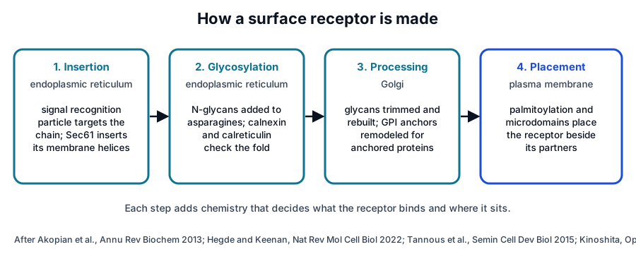
Most surface receptors are made on ribosomes bound to the endoplasmic reticulum.1 The signal recognition particle recognizes a hydrophobic signal sequence as it emerges from the ribosome and couples synthesis to delivery at the membrane, where the Sec61 translocon threads the chain across or into the bilayer (Fig. 1).5
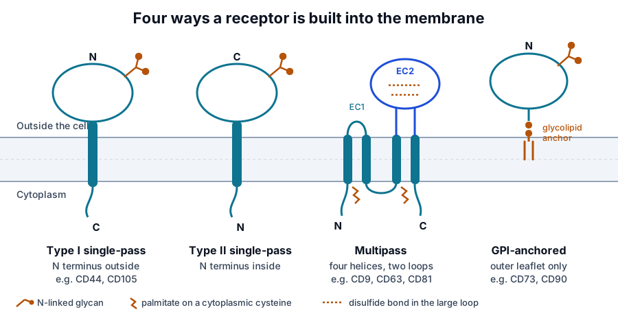
Each stretch of about twenty hydrophobic residues can become a membrane-spanning helix, and the order in which those stretches are inserted fixes the topology (Fig. 2). Flanking positive charges tend to stay on the cytoplasmic side, a pattern known as the positive-inside rule.6
- Type I single-pass proteins place the amino terminus outside the cell and cross the membrane once. CD44 and CD105 are built this way.6
- Type II single-pass proteins keep the amino terminus inside and display the carboxyl end outside.6
- Multipass proteins cross the membrane several times. The tetraspanins CD9, CD63 and CD81 cross it four times.7,8
- GPI-anchored proteins have no membrane-spanning segment and are held by a glycolipid. CD73 and CD90 are anchored this way.9,10
Not every protein uses the same insertion route. Guna and colleagues showed that the ER membrane protein complex acts as an insertase for tail-anchored proteins with moderately hydrophobic segments,11 one of several dedicated pathways reviewed by Hegde and Keenan.1
Glycosylation gives each receptor its outer chemistry
Glycosylation is the most abundant and diverse modification of proteins in eukaryotic cells.12 N-linked glycans are attached to asparagine within an asparagine, any residue, serine or threonine sequence as the protein enters the endoplasmic reticulum, and they are then trimmed and rebuilt as the protein moves through the Golgi.12
Those early glycans double as a folding checkpoint. Trimming of glucose residues engages the lectin chaperones calnexin and calreticulin, and only correctly folded proteins are released to continue toward the surface.13
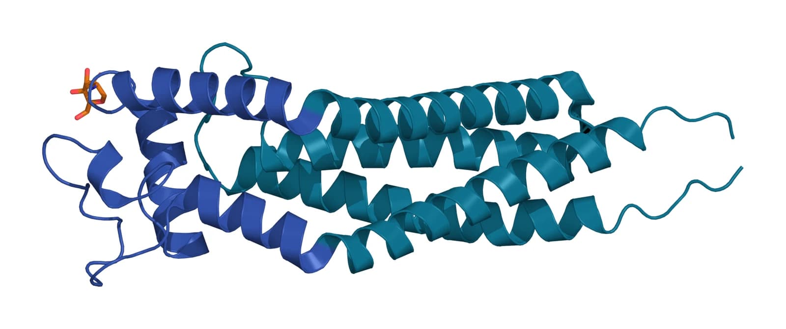
The finished sugars change what a receptor looks like to the outside world (Fig. 3). They add mass, shield parts of the protein surface, and can form or mask antibody epitopes, which is why heavily glycosylated proteins such as CD63 run far above their predicted size on a Western blot.12
Lipid anchors place receptors in particular membrane neighborhoods
At least 150 human proteins are attached to the membrane by a glycosylphosphatidylinositol anchor added to their carboxyl terminus.14 The anchor holds them in the outer leaflet of the bilayer only, and it associates them with ordered membrane domains often described as rafts.14
S-acylation works from the other side. It adds a long-chain fatty acid, usually palmitate, to cytoplasmic cysteines through a thioester bond, and it is the only fully reversible lipid modification of proteins.15 Tetraspanins are heavily palmitoylated, which helps them assemble with partner proteins.7
Rafts themselves are now understood as packed domains driven by the preferred association of cholesterol with saturated lipids.16 A receptor with the right anchor gathers into these domains with selected partners, which is one way the membrane concentrates signaling components.16
Receptors work in organized assemblies
Few surface proteins act as isolated units. Integrins pair into heterodimers and gather into adhesion complexes that sense the stiffness and composition of the surrounding matrix, transmitting signals in both directions across the membrane.17
Tetraspanins take organization further. They bind one another and partner proteins such as integrins and immunoglobulin superfamily members, assembling tetraspanin-enriched microdomains that position receptors next to the molecules they work with.7
The CD system gives each surface molecule a shared name
CD stands for cluster of differentiation. The Human Leukocyte Differentiation Antigens workshops grouped monoclonal antibodies that recognized the same molecule, and over ten workshops more than 400 molecules received formal CD designations.18
The nomenclature is approved by the International Union of Immunological Societies and sanctioned by the World Health Organization.18 A CD number therefore names both a protein and the antibodies that detect it, which is why the same numbers appear on research papers and characterization reports alike.
Surface chemistry is how cells and vesicles are identified
Mesenchymal stromal cells are a clear example. The 2006 International Society for Cellular Therapy criteria require expression of CD105, CD73 and CD90 (Fig. 4) and the absence of hematopoietic markers, in addition to plastic adherence and differentiation in culture.19
Proteomic surveys show how much more is present. Niehage and colleagues identified 888 proteins after biotin capture of the bone marrow stromal cell surface, including about 200 plasma membrane proteins and 41 CD markers,20 and Lv and colleagues reviewed how candidate markers vary with tissue source.21
Extracellular vesicles inherit membrane proteins from the cells that release them. The tetraspanins in particular are so consistently present that CD9, CD63 and CD81 have become the conventional vesicle markers,7 which is the subject of the next pages in this series.
Where to go from here
The tetraspanin family page explains the four-pass fold that CD9, CD63 and CD81 share, and each of those three proteins has its own reference. The quality references explain how a marker panel is measured and reported on a certificate of analysis.
Licensed practices that want characterization data for a specific lot can request the certificate of analysis once their account is verified. Our team can also walk through what each marker on a panel measures.
References
Numbered in the order they are cited in the text. Each study carries its design and, where people were enrolled, how many, with a line saying what it supports on this page. Primary sources are regulations, agency documents and reference databases.
- 1.Hegde RS, Keenan RJ. The mechanisms of integral membrane protein biogenesis. Nature Reviews Molecular Cell Biology. 2022. doi:10.1038/s41580-021-00413-2. PMID: 34556847.Narrative reviewReference 1 supports: The current account of how membrane proteins are targeted to and inserted into the endoplasmic reticulum. Cited for the share of genes encoding integral membrane proteins and for the multiple insertion pathways.
- 2.Uhlén M, Fagerberg L, Hallström BM, et al. Tissue-based map of the human proteome. Science. 2015. doi:10.1126/science.1260419. PMID: 25613900.In vitroReference 2 supports: The Human Protein Atlas tissue map, including the membrane proteome across 32 tissues. Cited as the reference resource for where surface proteins are expressed.
- 3.Bausch-Fluck D, Goldmann U, Müller S, et al. The in silico human surfaceome. Proceedings of the National Academy of Sciences of the United States of America. 2018. doi:10.1073/pnas.1808790115. PMID: 30373828.In vitroReference 3 supports: Machine learning prediction of the human surfaceome. Cited for the 2,886 predicted surface proteins and for surface repertoires that vary by cell type.
- 4.Bausch-Fluck D, Hofmann A, Bock T, et al. A mass spectrometric-derived cell surface protein atlas. PLoS One. 2015. doi:10.1371/journal.pone.0121314. PMID: 25894527.In vitroReference 4 supports: Cell surface capture across 41 human cell types. Cited for the finding that cell types differ mainly in the quantity of each surface protein rather than in which proteins are present.
- 5.Akopian D, Shen K, Zhang X, et al. Signal recognition particle: an essential protein-targeting machine. Annual Review of Biochemistry. 2013. doi:10.1146/annurev-biochem-072711-164732. PMID: 23414305.Narrative reviewReference 5 supports: Review of the signal recognition particle and its receptor. Cited for how synthesis of a nascent surface protein is coupled to its delivery at the membrane.
- 6.von Heijne G. Membrane-protein topology. Nature Reviews Molecular Cell Biology. 2006. doi:10.1038/nrm2063. PMID: 17139331.Narrative reviewLandmarkReference 6 supports: The classic account of how membrane protein topology is determined. Cited for hydrophobic membrane-spanning segments and the positive-inside rule.
- 7.Charrin S, Jouannet S, Boucheix C, et al. Tetraspanins at a glance. Journal of Cell Science. 2014. doi:10.1242/jcs.154906. PMID: 25128561.Narrative reviewReference 7 supports: Overview of tetraspanin structure and partners. Cited for tetraspanins as organizers that assemble partner receptors into microdomains.
- 8.NCBI Gene 928, CD9 molecule (Homo sapiens). Accessed 19 October 2026.Primary source
- 9.NCBI Gene 4907, NT5E (CD73), 5′-nucleotidase ecto (Homo sapiens). Accessed 19 October 2026.Primary source
- 10.NCBI Gene 7070, THY1 (CD90), Thy-1 cell surface antigen (Homo sapiens). Accessed 19 October 2026.Primary source
- 11.Guna A, Volkmar N, Christianson JC, et al. The ER membrane protein complex is a transmembrane domain insertase. Science. 2018. doi:10.1126/science.aao3099. PMID: 29242231.In vitroReference 11 supports: Reconstitution showing that the ER membrane protein complex inserts tail-anchored proteins. Cited as the example that membrane proteins use more than one insertion route.
- 12.Schjoldager KT, Narimatsu Y, Joshi HJ, et al. Global view of human protein glycosylation pathways and functions. Nature Reviews Molecular Cell Biology. 2020. doi:10.1038/s41580-020-00294-x. PMID: 33087899.Narrative reviewReference 12 supports: Map of the human glycosylation pathways. Cited for glycosylation as the most abundant and diverse protein modification in eukaryotic cells.
- 13.Tannous A, Pisoni GB, Hebert DN, et al. N-linked sugar-regulated protein folding and quality control in the ER. Seminars in Cell and Developmental Biology. 2015. doi:10.1016/j.semcdb.2014.12.001. PMID: 25534658.Narrative reviewReference 13 supports: Review of the calnexin and calreticulin folding cycle. Cited for the role of N-glycan trimming as a quality checkpoint before a glycoprotein leaves the endoplasmic reticulum.
- 14.Kinoshita T. Biosynthesis and biology of mammalian GPI-anchored proteins. Open Biology. 2020. doi:10.1098/rsob.190290. PMID: 32156170.Narrative reviewReference 14 supports: Review of GPI anchor biosynthesis. Cited for the figure of at least 150 human GPI-anchored proteins, their outer leaflet anchoring and their association with membrane rafts.
- 15.Chamberlain LH, Shipston MJ. The physiology of protein S-acylation. Physiological Reviews. 2015. doi:10.1152/physrev.00032.2014. PMID: 25834228.Narrative reviewReference 15 supports: Review of protein S-acylation. Cited for palmitate attachment to cysteines through a thioester bond and for S-acylation as the only fully reversible lipid modification of proteins.
- 16.Sezgin E, Levental I, Mayor S, et al. The mystery of membrane organization: composition, regulation and roles of lipid rafts. Nature Reviews Molecular Cell Biology. 2017. doi:10.1038/nrm.2017.16. PMID: 28356571.Narrative reviewReference 16 supports: Review of membrane raft biophysics. Cited for rafts as ordered domains driven by cholesterol and saturated lipids that selectively recruit proteins.
- 17.Kechagia JZ, Ivaska J, Roca-Cusachs P. Integrins as biomechanical sensors of the microenvironment. Nature Reviews Molecular Cell Biology. 2019. doi:10.1038/s41580-019-0134-2. PMID: 31182865.Narrative reviewReference 17 supports: Review of integrin mechanobiology. Cited for integrins as bidirectional signaling hubs that sense the rigidity and composition of the extracellular matrix.
- 18.Engel P, Boumsell L, Balderas R, et al. CD Nomenclature 2015: Human Leukocyte Differentiation Antigen Workshops as a Driving Force in Immunology. Journal of Immunology. 2015. doi:10.4049/jimmunol.1502033. PMID: 26546687.Consensus statementReference 18 supports: The workshop account of the CD system. Cited for the more than 400 designated molecules and for the nomenclature’s approval by the International Union of Immunological Societies and the World Health Organization.
- 19.Dominici M, Le Blanc K, Mueller I, et al. Minimal criteria for defining multipotent mesenchymal stromal cells. The International Society for Cellular Therapy position statement. Cytotherapy. 2006. doi:10.1080/14653240600855905. PMID: 16923606.Consensus statementLandmarkReference 19 supports: The ISCT minimal criteria. Cited for the positive markers CD105, CD73 and CD90 and the absence of hematopoietic markers.
- 20.Niehage C, Steenblock C, Pursche T, et al. The cell surface proteome of human mesenchymal stromal cells. PLoS One. 2011. doi:10.1371/journal.pone.0020399. PMID: 21637820.In vitroReference 20 supports: Surface biotinylation and mass spectrometry of bone marrow stromal cells. Cited for the 888 proteins identified, about 200 plasma membrane proteins and 41 CD markers.
- 21.Lv FJ, Tuan RS, Cheung KM, et al. Concise review: the surface markers and identity of human mesenchymal stem cells. Stem Cells. 2014. doi:10.1002/stem.1681. PMID: 24578244.Narrative reviewReference 21 supports: Review of candidate stromal cell surface markers across tissue sources. Cited for the variation in marker expression between sources.
Figure sources
- 22.PDB 6WVG, Tetraspanin CD53 in the open conformation. ExaVeyra Sciences, rendered in PyMOL from PDB 6WVG. Structure data: RCSB Protein Data Bank (rcsb.org), CC0. Structure publication: Yang Y, Liu XR, Greenberg ZJ, et al. Open conformation of tetraspanins shapes interaction partner networks on cell membranes. EMBO J. 2020;39(18):e105246.