Platelet Granules: Alpha Granules, Dense Granules and Release
By Benn Bluestein-Veyra, M.Sc. Organic Chemistry · Updated 21 October 2026
ExaVeyra Sciences is a Miami-based B2B supplier of exosomes for topical aesthetic use to licensed clinics, and it refers licensed prescribers to a 503A compounding pharmacy that prepares formulations against individual prescriptions.
| Question | Answer |
|---|---|
| What this page covers | How platelets store and release their cargo: alpha granule proteins, dense granule small molecules, lysosomes, the release machinery and what activation triggers. |
| Who can order | Licensed clinics and practitioners whose active professional license and NPI ExaVeyra has verified. Exosome orders also require a signed professional-use acknowledgment. |
| What ships, and how | Exosome material for topical aesthetic use, characterized by lot, ships overnight in a cold chain. Delivery can take 1 to 3 business days. A certificate of analysis is issued for every lot and provided on request. |
Platelet granules are the storage organelles that let a platelet release hundreds of proteins and small molecules within moments of activation. Each platelet carries three main types: alpha granules, dense granules and lysosomes.[1][2]
This reference describes what each granule type holds, how megakaryocytes build them, which receptors trigger their release and which SNARE proteins fuse them with the cell surface. It closes with the assays that measure release, from P-selectin flow cytometry to growth factor immunoassays.[3][4]
Megakaryocytes build platelet granules before platelets enter the blood
Platelets are anucleate, discoid cells roughly 2 to 3 micrometers across. They come from megakaryocytes, large nucleated cells of 30 to 100 micrometers that reside mainly in the bone marrow, and human adults carry nearly one trillion platelets in circulation.[5]
Machlus and Italiano describe megakaryocytes that become polyploid as they mature and accumulate large amounts of protein and membrane. They then extend long branching proplatelets into sinusoidal blood vessels, where fission releases platelets.[6]
Alpha granules draw their cargo from two routes in the megakaryocyte: proteins the cell synthesizes and sorts for regulated secretion, and proteins it takes up by endocytosis.[7] Both alpha and dense granules mature in multivesicular bodies, then travel through the proplatelet extensions and keep maturing there.[1]
The two granule types use distinct trafficking machinery. Sharda and Flaumenhaft note that VPS33B, VPS16B and NBEAL2 are required for alpha granule maturation, while dense granule formation depends on a different set of proteins.[1]
The finished platelet also carries an open canalicular system of surface membrane invaginations, a dense tubular system derived from endoplasmic reticulum, a spectrin-based membrane skeleton, an actin network and a peripheral band of microtubules.[5]
Understanding the three platelet granule types
Alpha granules are the most abundant, at 50 to 80 per platelet; they measure 200 to 500 nm and make up about 10% of platelet volume. Dense granules number 3 to 8 per platelet at about 150 nm, and each platelet holds about 1 to 3 lysosomes (Fig. 1).[1][7]
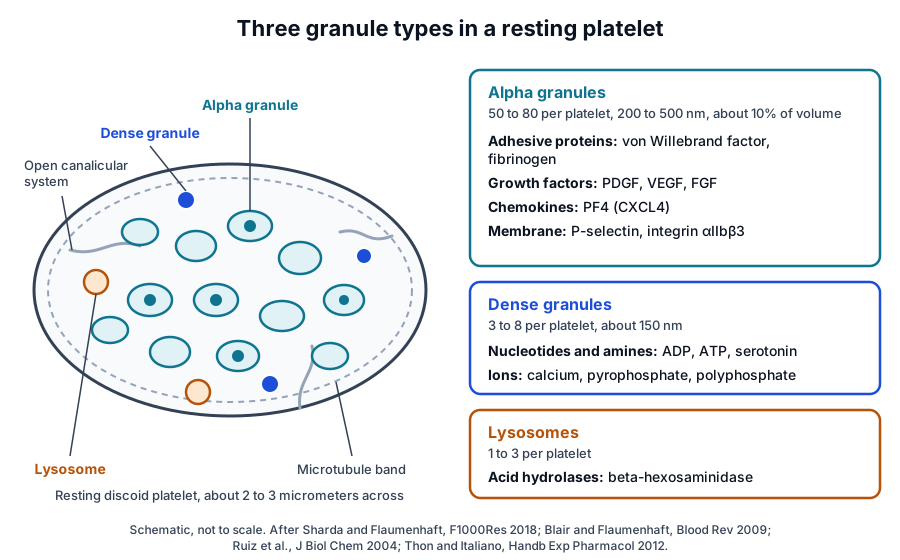
Pokrovskaya and colleagues reconstructed 30 resting human platelets from three donors by serial block face scanning electron microscopy. Alpha granule number rose linearly with platelet size, while dense granule and mitochondrial numbers showed little correlation with size.[8]
The same three-dimensional data showed limited spatial intermixing of the organelle classes. Almost 70% of alpha granules lay within 35 nm of one another, a distance associated in other cell systems with protein-mediated contact sites.[8]
| Granule type | Number and size | Representative contents | Common release readout |
|---|---|---|---|
| Alpha granule | 50 to 80 per platelet, 200 to 500 nm | von Willebrand factor, fibrinogen, PDGF, VEGF, PF4, P-selectin | Surface P-selectin by flow cytometry; PF4 or von Willebrand factor in the supernatant |
| Dense granule | 3 to 8 per platelet, about 150 nm | ADP, ATP, serotonin, calcium, pyrophosphate, polyphosphate | ADP and ATP luminescence; serotonin release; surface CD63 or LAMP2 |
| Lysosome | 1 to 3 per platelet | Acid hydrolases such as beta-hexosaminidase | Beta-hexosaminidase activity in the supernatant |
Alpha granules store adhesive proteins, growth factors and chemokines
Heijnen and van der Sluijs summarize alpha granule cargo as adhesive molecules such as von Willebrand factor and fibrinogen, growth factors, and inflammatory and angiogenic mediators.[2] Membrane proteins such as integrin αIIbβ3 and P-selectin sit in the granule membrane beside soluble cargo such as platelet factor 4.[1]
Proteomic surveys report more than 300 soluble alpha granule proteins, spanning hemostasis (von Willebrand factor, factor V), chemokine signaling (CXCL1, interleukin-8) and growth factors (VEGF, FGF).[1] Mice lacking serglycin, the main platelet glycosaminoglycan, fail to store basic proteins such as CXCL4, PDGF and NAP-2 in their alpha granules.[7]
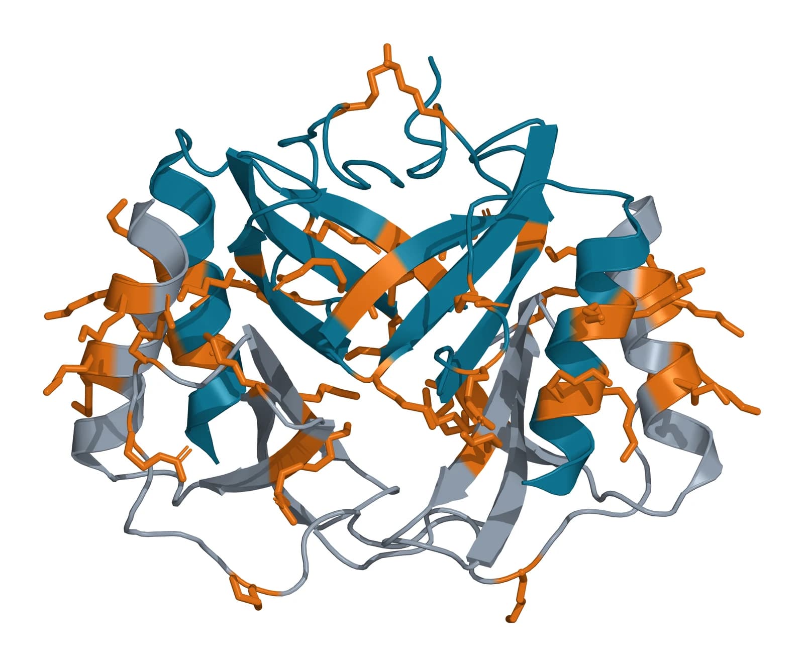
Platelet factor 4, also called CXCL4, is a chemokine encoded by the PF4 gene on chromosome 4q13.3.[9] Zhang and colleagues solved recombinant human PF4 at 2.4 Å as a tetramer ringed by lysine and arginine side chains that offer several heparin-binding sites (Fig. 2).[10]
Whether alpha granules form functional subpopulations is still debated
Italiano and colleagues reported that VEGF and endostatin, and basic FGF and thrombospondin-1, sit in separate alpha granules. In their experiments a PAR4 agonist released endostatin-containing granules and a PAR1 agonist released VEGF-containing granules.[11]
Kamykowski and colleagues compared 15 angiogenesis-related alpha granule proteins by quantitative immunofluorescence and found little functional coclustering, with a Gaussian pattern that suggests stochastic delivery.[12] Pokrovskaya and colleagues concluded that all alpha granules likely carry the same major protein set in varying amounts.[8]
Jonnalagadda and colleagues followed 28 alpha granule cargo molecules over time with micro-ELISA arrays. Thrombin gave the fastest and most extensive release, a PAR4 agonist slower release of fewer molecules, and cargo with opposing actions shared similar release profiles.[13]
Their kinetic analysis pointed to three classes of release events with different rates, and the authors described platelet secretion as a stochastic process shaped by factors such as cargo solubility, granule shape and fusion route.[13] Heijnen and van der Sluijs review the same question across granule subtypes and the open canalicular system.[2]
Dense granules and lysosomes complete the secretory repertoire
Dense granules belong to the lysosome-related organelles and hold bioactive amines such as serotonin, adenine nucleotides, pyrophosphate and polyphosphate, with high concentrations of cations, chiefly calcium. That cation load gives them their electron-dense look in whole mount electron microscopy.[1]
Ruiz and colleagues measured millimolar levels of short-chain polyphosphate in human platelets, about 70 to 75 phosphate residues long, concentrated in dense granules. Thrombin released it together with pyrophosphate and serotonin, and the authors likened dense granules to the acidocalcisomes of bacteria and unicellular eukaryotes.[14]
Morrissey and colleagues report that the medium-chain polyphosphate platelets secrete accelerates factor V activation, abrogates the anticoagulant function of tissue factor pathway inhibitor, enhances fibrin clot structure and speeds factor XI activation by thrombin.[15]
Secreted ADP acts back on platelets. Hollopeter and colleagues describe ADP released from activated platelets as positive feedback that potentiates the actions of many other platelet activators, which makes the dense granule an amplifier of the activation signal.[16]
Lysosomes release acid hydrolases under stronger stimulation
Platelets hold about 1 to 3 lysosomes each, and lysosome exocytosis is usually measured by released enzymes such as beta-hexosaminidase. Dense granule exocytosis is the fastest and most sensitive to agonists, lysosome exocytosis is the slowest.[1]
Agonist receptors couple platelet activation to granule secretion
Platelets respond to adhesive proteins such as von Willebrand factor and collagen, and to soluble agonists such as ADP, thrombin and thromboxane A2, each through its own receptors. Li and colleagues describe how these pathways converge on shape change, granule secretion and inside-out activation of integrin αIIbβ3 (Fig. 3).[17]
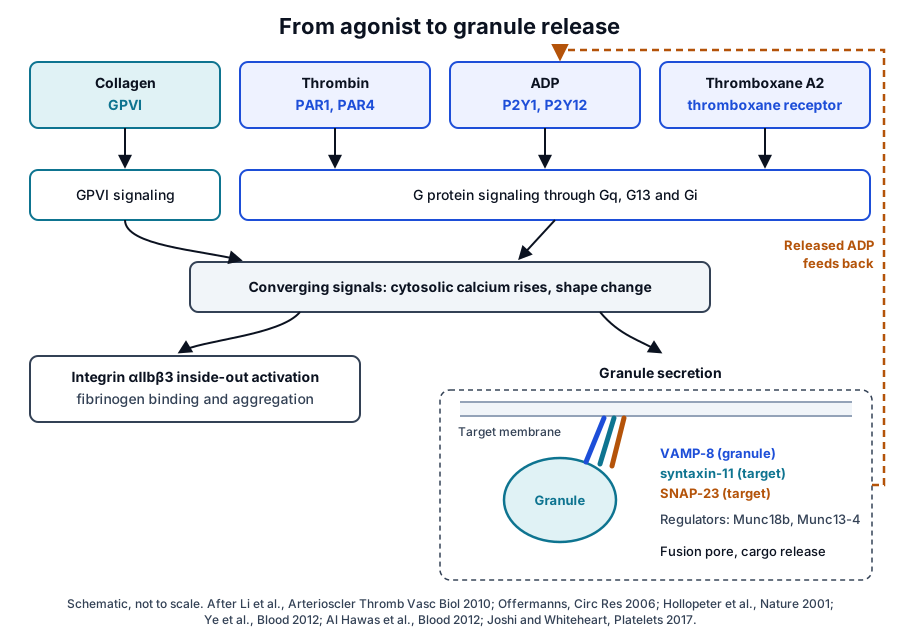
Nieswandt and Watson place the immunoglobulin superfamily receptor GPVI at the center of platelet responses to collagen, with integrin α2β1 as a second collagen receptor. Firm adhesion to collagen requires integrin activation by GPVI signals, reinforced by released ADP and thromboxane A2.[18]
Thrombin acts through the protease-activated receptors, which Coughlin describes as a link between the coagulation cascade and cellular responses.[19] Offermanns traces ADP, thromboxane A2 and thrombin signaling through three G protein pathways, initiated by Gq, G13 and Gi.[20]
Hollopeter and colleagues cloned P2Y12, the Gi-coupled ADP receptor; the second ADP receptor, P2Y1, couples to Gq and mobilizes calcium for shape change and aggregation.[16] Ligand binding to αIIbβ3 then triggers outside-in signaling that drives spreading, further granule secretion and clot retraction.[17]
Versteeg and colleagues frame hemostasis as two linked systems, coagulation and platelet activation. Activated platelets build the hemostatic plug and accelerate coagulation, and blood-borne inhibitors and proteolytic feedback loops later switch both off.[21]
SNARE proteins and their regulators fuse granules with the platelet membrane
Granule release depends on SNARE proteins: a v-SNARE on the granule pairs with t-SNAREs on the target membrane. Joshi and Whiteheart describe a coiled coil of four SNARE domains, one from a VAMP, one from a syntaxin and two from SNAP-23, that draws the two membranes together for fusion.[3]
Ren and colleagues found VAMP-8 required for release from dense granules, alpha granules and lysosomes. Mouse platelets lacking VAMP-8 had a significant secretion defect with normal signaling, morphology and cargo levels, and tetanus toxin pointed to a secondary role for VAMP-2 or VAMP-3.[22]
Ye and colleagues showed that platelets from syntaxin-2 and syntaxin-4 knockout mice secreted normally, while human platelets lacking syntaxin-11 had a robust secretion defect. Syntaxin-11 was the more abundant platelet syntaxin and formed SNARE complexes with VAMP-8 and SNAP-23.[23]
Munc18b and Munc13-4 set the pace of SNARE assembly
Al Hawas and colleagues identified Munc18b as the major platelet Munc18 isoform and found it required for secretion. In human platelets it formed complexes with syntaxin-11, SNAP-23 and VAMP-8, and Munc13-4 and Rab27 associated with these complexes too.[24]
Mouse platelets lacking Munc13-4 showed completely ablated dense granule release and severely reduced alpha granule and lysosome release. Ren and colleagues found Munc13-4 levels lower than SNARE levels and in step with the extent of release, marking it as a limiting factor.[25]
Most granules fuse with the open canalicular system, which raises the platelet surface area by at least two to three times. At higher agonist concentrations, alpha granules can also fuse with each other before fusing with the membrane (Fig. 3).[1] Alpha granule fusion releases cargo and adds membrane to the platelet surface.[7]
Activated platelets release extracellular vesicles alongside granule cargo
Heijnen and colleagues found two vesicle types after stimulation with thrombin or a thrombin receptor agonist peptide: microvesicles of 100 nm to 1 micrometer shed from the surface, and exosomes of 40 to 100 nm released from multivesicular bodies and alpha granules.[26]
The exosomes were selectively enriched in the tetraspanin CD63, while integrin αIIbβ3, GPIbα and P-selectin sat mainly on the microvesicles. Annexin V, factor X and prothrombin bound mainly to the microvesicles as well.[26]
Puhm and colleagues review platelet extracellular vesicles as carriers of lipids, proteins, nucleic acids and organelles. These vesicles can reach lymph, bone marrow and synovial fluid, compartments that platelets themselves cannot enter.[27]
Parsons and colleagues profiled the thrombin-induced releasate of 32 healthy adults by label-free quantitative proteomics. Of 894 proteins identified, 277 were quantified in every donor as a core releasate, and 84% of those overlapped with the proteome of platelet-derived exosomes.[28]
Proteomics, flow cytometry and immunoassays measure platelet granule release
Maynard and colleagues isolated an alpha granule-enriched fraction by sucrose gradient ultracentrifugation and identified 284 proteins by mass spectrometry, 44 of them newly assigned to alpha granules. Their set shared 65% of the proteins detected in an earlier releasate study.[29]
Burkhart and colleagues quantified the whole human platelet proteome at almost 4,000 proteins, with copy number estimates for about 3,700 of them. About 85% of the proteome was constant between healthy donors.[30] Parsons and colleagues likewise found reproducible releasate secretion levels across their 32 donors.[28]
Stenberg and colleagues localized P-selectin, then called GMP-140, to the alpha granule membrane and measured at least a 50-fold rise in surface label after thrombin stimulation.[31] This translocation makes surface P-selectin the main flow cytometry readout of alpha granule release.[1]
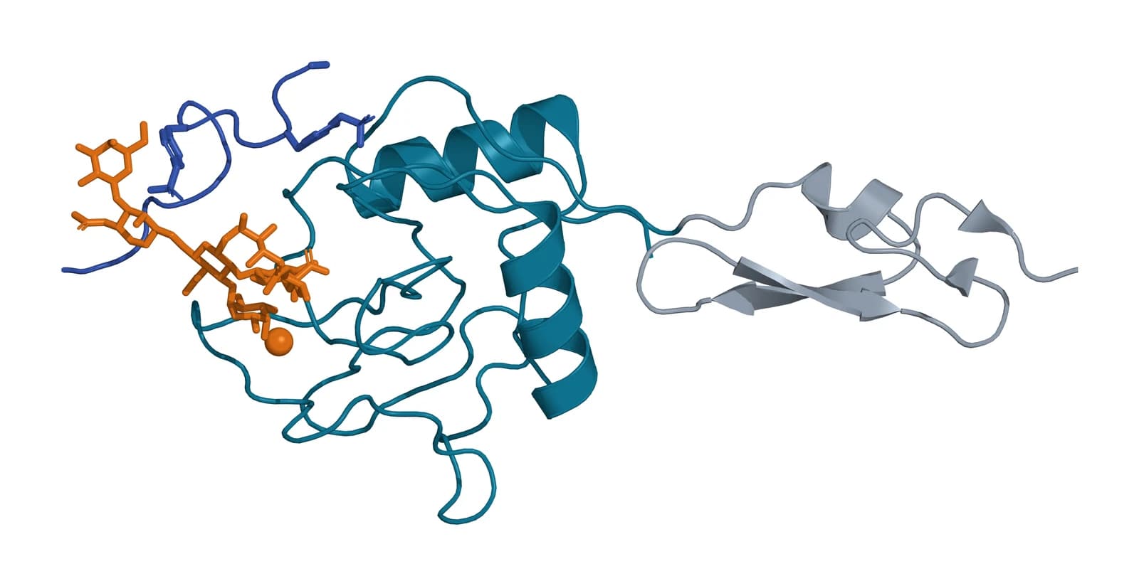
P-selectin is encoded by the SELP gene on chromosome 1q24.2.[32] Somers and colleagues solved its lectin and EGF domains bound to an N-terminal PSGL-1 peptide carrying tyrosine sulfation and sialyl Lewis x, which revealed the basis of the high-affinity P-selectin and PSGL-1 interaction (Fig. 4).[33]
Michelson and colleagues describe whole blood flow cytometry for platelet reactivity, platelet aggregates with one another and with leukocytes, platelet-derived microparticles and calcium flux.[4] Dense granule release is commonly read by ADP and ATP luminescence or serotonin release, and lysosome release by beta-hexosaminidase activity.[1][3]
Immunoassays quantify soluble cargo. Weibrich and colleagues measured growth factors in 115 platelet-rich plasma specimens, reporting PDGF-AB, TGF-beta1 and IGF-I in large amounts and PDGF-BB and TGF-beta2 in small amounts.[34]
In that series, growth factor content correlated only weakly with platelet count in whole blood or in the platelet-rich plasma (r = 0.35). Donor age and sex showed no influence on growth factor concentrations, with IGF-I the exception.[34]
Where to go from here
The PRP preparation variables reference explains how spin settings, platelet concentration and leukocyte content shape a preparation, and the growth factor receptors reference follows PDGF, TGF-beta, VEGF, EGF and IGF-1 to the cells that read them. The platelet-rich fibrin reference describes the fibrin matrix that holds released proteins.
The marrow and adipose composition reference covers the cells in those preparations, and the platelet study reporting reference lists what a complete methods section records. The exosome biogenesis and vesicle protein marker references extend the vesicle biology above. Our team is glad to discuss the methods described on this page.
Annotated References
Numbered in the order they are cited in the text. Each source is annotated with what it supports on this page; peer-reviewed articles are listed with journal, volume, issue and pages, and primary sources are regulations, agency documents and reference databases.
- 1.Peer-reviewedSharda A, Flaumenhaft R. The life cycle of platelet granules. F1000Research. 2018. https://pubmed.ncbi.nlm.nih.gov/29560259/Annotation: Review of platelet granule biogenesis and exocytosis. Cited for granule numbers and sizes, alpha and dense granule cargo, biogenesis machinery, release kinetics by granule type, open canalicular system fusion and the standard release readouts.
- 2.Peer-reviewedHeijnen H, van der Sluijs P. Platelet secretory behaviour: as diverse as the granules … or not? Journal of Thrombosis and Haemostasis. 2015. https://pubmed.ncbi.nlm.nih.gov/26391322/Annotation: Review of platelet secretory behavior. Cited for the three granule types, the summary of alpha granule cargo and the debate over differential secretion of alpha granule subtypes.
- 3.Peer-reviewedJoshi S, Whiteheart SW. The nuts and bolts of the platelet release reaction. Platelets. 2017. https://pubmed.ncbi.nlm.nih.gov/27848265/Annotation: Review of the platelet secretory machinery. Cited for the four-helix SNARE complex of VAMP, syntaxin and SNAP-23 and for the cargo readouts used to measure secretion.
- 4.Peer-reviewedMichelson AD, Barnard MR, Krueger LA, et al. Evaluation of platelet function by flow cytometry. Methods. 2000. https://pubmed.ncbi.nlm.nih.gov/10873480/Annotation: Methods review of whole blood platelet flow cytometry. Cited for the platelet measurements flow cytometry supports, including reactivity, aggregates, microparticles and calcium flux.
- 5.Peer-reviewedThon JN, Italiano JE. Platelets: production, morphology and ultrastructure. Handbook of Experimental Pharmacology. 2012. https://pubmed.ncbi.nlm.nih.gov/22918725/Annotation: Review of platelet production and ultrastructure. Cited for platelet and megakaryocyte size, platelet number and the structural elements of the platelet, including the open canalicular and dense tubular systems.
- 6.Peer-reviewedMachlus KR, Italiano JE Jr. The incredible journey: From megakaryocyte development to platelet formation. The Journal of Cell Biology. 2013. https://pubmed.ncbi.nlm.nih.gov/23751492/Annotation: Review of megakaryocyte maturation and platelet formation. Cited for polyploidization, proplatelet extension into sinusoidal vessels and platelet release by fission.
- 7.Peer-reviewedBlair P, Flaumenhaft R. Platelet alpha-granules: basic biology and clinical correlates. Blood Reviews. 2009. https://pubmed.ncbi.nlm.nih.gov/19450911/Annotation: Landmark review of alpha granule biology. Cited for the two cargo routes in megakaryocytes, granule numbers and volume, serglycin-dependent storage and the membrane added to the surface on fusion.
- 8.Peer-reviewedPokrovskaya ID, Yadav S, Rao A, et al. 3D ultrastructural analysis of α-granule, dense granule, mitochondria, and canalicular system arrangement in resting human platelets. Research and Practice in Thrombosis and Haemostasis. 2020. https://pubmed.ncbi.nlm.nih.gov/31989087/Annotation: Serial block face electron microscopy of 30 resting platelets from three donors. Cited for organelle number against platelet size, alpha granule proximity and the shared alpha granule protein set.
- 9.DatabaseNCBI Gene 5196, PF4 platelet factor 4 (Homo sapiens). Accessed 21 October 2026. https://www.ncbi.nlm.nih.gov/gene/5196
- 10.Peer-reviewedZhang X, Chen L, Bancroft DP, et al. Crystal structure of recombinant human platelet factor 4. Biochemistry. 1994. https://pubmed.ncbi.nlm.nih.gov/8031770/Annotation: X-ray crystal structure of human PF4 at 2.4 Å (PDB 1RHP). Cited for the tetramer and its ring of lysine and arginine side chains with several heparin-binding sites.
- 11.Peer-reviewedItaliano JE Jr, Richardson JL, Patel-Hett S, et al. Angiogenesis is regulated by a novel mechanism: pro- and antiangiogenic proteins are organized into separate platelet alpha granules and differentially released. Blood. 2008. https://pubmed.ncbi.nlm.nih.gov/17962514/Annotation: Immunofluorescence and immunoelectron microscopy of platelets and megakaryocytes. Cited for separate VEGF and endostatin granules and their selective release by PAR1 and PAR4 agonists.
- 12.Peer-reviewedKamykowski J, Carlton P, Sehgal S, et al. Quantitative immunofluorescence mapping reveals little functional coclustering of proteins within platelet α-granules. Blood. 2011. https://pubmed.ncbi.nlm.nih.gov/21622648/Annotation: Quantitative immunofluorescence of 15 alpha granule proteins. Cited for little functional coclustering and a distribution consistent with stochastic packaging.
- 13.Peer-reviewedJonnalagadda D, Izu LT, Whiteheart SW. Platelet secretion is kinetically heterogeneous in an agonist-responsive manner. Blood. 2012. https://pubmed.ncbi.nlm.nih.gov/23086755/Annotation: Time-course release of 28 alpha granule cargo molecules measured by micro-ELISA arrays. Cited for agonist-dependent speed and extent of release, shared release profiles and three classes of release events.
- 14.Peer-reviewedRuiz FA, Lea CR, Oldfield E, et al. Human platelet dense granules contain polyphosphate and are similar to acidocalcisomes of bacteria and unicellular eukaryotes. The Journal of Biological Chemistry. 2004. https://pubmed.ncbi.nlm.nih.gov/15308650/Annotation: Biochemistry of purified human dense granules. Cited for polyphosphate chain length and location, its release with pyrophosphate and serotonin, and the acidocalcisome comparison.
- 15.Peer-reviewedMorrissey JH, Choi SH, Smith SA. Polyphosphate: an ancient molecule that links platelets, coagulation, and inflammation. Blood. 2012. https://pubmed.ncbi.nlm.nih.gov/22517894/Annotation: Review of polyphosphate in blood clotting. Cited for the effects of platelet-sized polyphosphate on factor V, tissue factor pathway inhibitor, fibrin clot structure and factor XI.
- 16.Peer-reviewedHollopeter G, Jantzen HM, Vincent D, et al. Identification of the platelet ADP receptor targeted by antithrombotic drugs. Nature. 2001. https://pubmed.ncbi.nlm.nih.gov/11196645/Annotation: Cloning of the Gi-coupled platelet ADP receptor P2Y12. Cited for P2Y12 and P2Y1 coupling and for secreted ADP as positive feedback on other activators.
- 17.Peer-reviewedLi Z, Delaney MK, O’Brien KA, et al. Signaling during platelet adhesion and activation. Arteriosclerosis, Thrombosis, and Vascular Biology. 2010. https://pubmed.ncbi.nlm.nih.gov/21071698/Annotation: Review of platelet activation signaling. Cited for receptor pathways converging on shape change, secretion and inside-out integrin activation, and for outside-in signaling.
- 18.Peer-reviewedNieswandt B, Watson SP. Platelet-collagen interaction: is GPVI the central receptor? Blood. 2003. https://pubmed.ncbi.nlm.nih.gov/12649139/Annotation: Review of platelet collagen receptors. Cited for GPVI as the central collagen receptor, integrin alpha2 beta1, and reinforcement by released ADP and thromboxane A2.
- 19.Peer-reviewedCoughlin SR. Thrombin signalling and protease-activated receptors. Nature. 2000. https://pubmed.ncbi.nlm.nih.gov/11001069/Annotation: Landmark review of protease-activated receptors. Cited for the PARs as the link between thrombin and cellular responses.
- 20.Peer-reviewedOffermanns S. Activation of platelet function through G protein-coupled receptors. Circulation Research. 2006. https://pubmed.ncbi.nlm.nih.gov/17158345/Annotation: Review of platelet G protein signaling. Cited for ADP, thromboxane A2 and thrombin acting through Gq, G13 and Gi pathways.
- 21.Peer-reviewedVersteeg HH, Heemskerk JW, Levi M, et al. New fundamentals in hemostasis. Physiological Reviews. 2013. https://pubmed.ncbi.nlm.nih.gov/23303912/Annotation: Review of hemostasis. Cited for coagulation and platelet activation as linked components that are later switched off by inhibitors and feedback loops.
- 22.Peer-reviewedRen Q, Barber HK, Crawford GL, et al. Endobrevin/VAMP-8 is the primary v-SNARE for the platelet release reaction. Molecular Biology of the Cell. 2007. https://pubmed.ncbi.nlm.nih.gov/17065550/Annotation: Knockout mouse and permeabilized human platelets. Cited for VAMP-8 as the v-SNARE required for release from all three granule types and a secondary role for VAMP-2 or VAMP-3.
- 23.Peer-reviewedYe S, Karim ZA, Al Hawas R, et al. Syntaxin-11, but not syntaxin-2 or syntaxin-4, is required for platelet secretion. Blood. 2012. https://pubmed.ncbi.nlm.nih.gov/22767500/Annotation: Knockout mouse platelets and syntaxin-11-deficient human platelets. Cited for syntaxin-11 as the required platelet syntaxin and its SNARE complexes with VAMP-8 and SNAP-23.
- 24.Peer-reviewedAl Hawas R, Ren Q, Ye S, et al. Munc18b/STXBP2 is required for platelet secretion. Blood. 2012. https://pubmed.ncbi.nlm.nih.gov/22791290/Annotation: Biochemistry of human platelet Munc18 isoforms. Cited for Munc18b as the major isoform required for secretion and its complexes with syntaxin-11, SNAP-23, VAMP-8, Munc13-4 and Rab27.
- 25.Peer-reviewedRen Q, Wimmer C, Chicka MC, et al. Munc13-4 is a limiting factor in the pathway required for platelet granule release and hemostasis. Blood. 2010. https://pubmed.ncbi.nlm.nih.gov/20435885/Annotation: Platelets from Munc13-4-deficient mice. Cited for the loss of dense granule release, reduced alpha granule and lysosome release, and Munc13-4 as a limiting factor.
- 26.Peer-reviewedHeijnen HF, Schiel AE, Fijnheer R, et al. Activated platelets release two types of membrane vesicles: microvesicles by surface shedding and exosomes derived from exocytosis of multivesicular bodies and alpha-granules. Blood. 1999. https://pubmed.ncbi.nlm.nih.gov/10572093/Annotation: Electron microscopy and Western blot of vesicles from stimulated platelets. Cited for microvesicle and exosome sizes, CD63 enrichment on exosomes and the membrane proteins and coagulation factors bound to microvesicles.
- 27.Peer-reviewedPuhm F, Boilard E, Machlus KR. Platelet Extracellular Vesicles: Beyond the Blood. Arteriosclerosis, Thrombosis, and Vascular Biology. 2021. https://pubmed.ncbi.nlm.nih.gov/33028092/Annotation: Review of platelet extracellular vesicles. Cited for their cargo and their access to lymph, bone marrow and synovial fluid.
- 28.Peer-reviewedParsons MEM, Szklanna PB, Guerrero JA, et al. Platelet Releasate Proteome Profiling Reveals a Core Set of Proteins with Low Variance between Healthy Adults. Proteomics. 2018. https://pubmed.ncbi.nlm.nih.gov/29932309/Annotation: Label-free proteomics of thrombin-induced releasate from 32 healthy adult donors. Cited for the 894 identified and 277 core proteins, their overlap with the platelet exosome proteome and low between-donor variance.
- 29.Peer-reviewedMaynard DM, Heijnen HF, Horne MK, et al. Proteomic analysis of platelet alpha-granules using mass spectrometry. Journal of Thrombosis and Haemostasis. 2007. https://pubmed.ncbi.nlm.nih.gov/17723134/Annotation: First mass spectrometry proteome of an alpha granule-enriched fraction. Cited for 284 proteins, 44 newly assigned, and 65% overlap with an earlier releasate study.
- 30.Peer-reviewedBurkhart JM, Vaudel M, Gambaryan S, et al. The first comprehensive and quantitative analysis of human platelet protein composition allows the comparative analysis of structural and functional pathways. Blood. 2012. https://pubmed.ncbi.nlm.nih.gov/22869793/Annotation: Quantitative mass spectrometry of highly purified human platelets. Cited for the size of the platelet proteome, copy number estimates and the share of proteins that stay constant between healthy donors.
- 31.Peer-reviewedStenberg PE, McEver RP, Shuman MA, et al. A platelet alpha-granule membrane protein (GMP-140) is expressed on the plasma membrane after activation. The Journal of Cell Biology. 1985. https://pubmed.ncbi.nlm.nih.gov/2411738/Annotation: Immunogold electron microscopy of resting and thrombin-stimulated platelets. Cited for P-selectin (GMP-140) in the alpha granule membrane and its at least 50-fold rise on the surface after activation.
- 32.DatabaseNCBI Gene 6403, SELP selectin P (Homo sapiens). Accessed 21 October 2026. https://www.ncbi.nlm.nih.gov/gene/6403
- 33.Peer-reviewedSomers WS, Tang J, Shaw GD, et al. Insights into the molecular basis of leukocyte tethering and rolling revealed by structures of P- and E-selectin bound to SLe(X) and PSGL-1. Cell. 2000. https://pubmed.ncbi.nlm.nih.gov/11081633/Annotation: X-ray crystal structures of P- and E-selectin lectin and EGF domains, PDB 1G1S among them. Cited for P-selectin bound to a sulfated, sialyl Lewis x-modified PSGL-1 peptide and the basis of their high-affinity interaction.
- 34.Peer-reviewedWeibrich G, Kleis WK, Hafner G, et al. Growth factor levels in platelet-rich plasma and correlations with donor age, sex, and platelet count. Journal of Cranio-Maxillo-Facial Surgery. 2002. https://pubmed.ncbi.nlm.nih.gov/12069512/Annotation: Growth factor measurements in 115 platelet-rich plasma specimens from healthy donors. Cited for the relative amounts of PDGF-AB, PDGF-BB, TGF-beta1, TGF-beta2 and IGF-I and their weak correlation with platelet count.
Cite this page
ExaVeyra Sciences Editorial Team. Platelet Granules: Alpha Granules, Dense Granules and Release. ExaVeyra Sciences; 21 October 2026. Available from: https://exaveyra.com/science/platelet-biology/platelet-granule-biology. Accessed [date accessed].
Figure sources
- 35.PDB 1RHP, Crystal structure of recombinant human platelet factor 4. ExaVeyra Sciences, rendered in PyMOL from PDB 1RHP. Structure data: RCSB Protein Data Bank (rcsb.org), CC0. Structure publication: Zhang X, Chen L, Bancroft DP, Lai CK, Maione TE. Crystal structure of recombinant human platelet factor 4. Biochemistry. 1994;33(27):8361-8366.
- 36.PDB 1G1S, P-selectin lectin/EGF domains complexed with PSGL-1 peptide. ExaVeyra Sciences, rendered in PyMOL from PDB 1G1S. Structure data: RCSB Protein Data Bank (rcsb.org), CC0. Structure publication: Somers WS, Tang J, Shaw GD, Camphausen RT. Insights into the molecular basis of leukocyte tethering and rolling revealed by structures of P- and E-selectin bound to SLe(X) and PSGL-1. Cell. 2000;103(3):467-479.