EV Potency Assays: Measuring Biological Activity In Vitro
By Benn Bluestein-Veyra, M.Sc. Organic Chemistry · Updated 20 October 2026
ExaVeyra Sciences is a Miami-based B2B supplier of exosomes for topical aesthetic use to licensed clinics, and it refers licensed prescribers to a 503A compounding pharmacy that prepares formulations against individual prescriptions.
| Question | Answer |
|---|---|
| What this page covers | How EV preparations are characterized for biological activity: in vitro potency assay formats, input metrics, reference standards and assay validation. |
| Who can order | Licensed clinics and practitioners whose active professional license and NPI ExaVeyra has verified. Exosome orders also require a signed professional-use acknowledgment. |
| What ships, and how | Exosome material for topical aesthetic use, characterized by lot, ships overnight in a cold chain. Delivery can take 1 to 3 business days. A certificate of analysis is issued for every lot and provided on request. |
An extracellular vesicle potency assay measures the biological activity of a vesicle preparation as a number, using a readout linked to a proposed mechanism.[1] Published formats for stromal cell vesicles range from T cell proliferation and macrophage polarization to endothelial tube formation and reporter cell lines.[2]
This page covers how regulators define potency, how the vesicle societies apply that definition, the in vitro readouts in current use, how dose is expressed, and how an assay is validated. Each section describes published characterization methods.
A potency assay quantifies biological activity linked to a mechanism
ICH Q6B, issued by FDA as guidance in August 1999, defines biological activity as the specific ability of a product to achieve a defined biological effect. Potency is the quantitative measure of that activity, expressed in units and based on the attribute linked to the relevant biological properties.[3]
The FDA guidance Potency Tests for Cellular and Gene Therapy Products, finalized in January 2011, states that a potency assay ideally represents the mechanism of action. It also recognizes that many complex biologics act through several activities, which makes the most relevant attribute hard to choose.[4]
The same guidance lists what a potency measurement provides: quantitative data, appropriate reference materials and controls, and documented accuracy, sensitivity, specificity and reproducibility.[4] Those requirements frame every assay format described below.
The guidance also values assays evaluated during development that are impractical for lot release. Such assays provide information about product attributes related to biological activity, which is the role most published vesicle assays play today.[4]
In December 2023, FDA issued a draft guidance, Potency Assurance for Cellular and Gene Therapy Products, that describes a science- and risk-based potency assurance strategy. The strategy combines manufacturing process design and control, material control, in-process testing and potency lot release assays.[5]
ISEV and ISCT working groups define potency for stromal cell vesicles
In 2019, Witwer and colleagues wrote on behalf of members of four societies, SOCRATES, ISEV, ISCT and ISBT, that stromal cell small vesicles should be defined by quantifiable metrics. They named cellular origin, the presence of lipid membrane vesicles, and physical and biochemical integrity.[6]
The same group proposed that new preparations be measured against a well-characterized biological reference. A shared reference lets laboratories express results relative to the same material. Their stated aim was to map vesicle biology and activity onto quantifiable features of each preparation.[6]
Gimona and colleagues, twenty-one researchers in the field, followed in 2021 with a framework for potency tests consistent with ICH guidelines. They proposed identifying the vesicle attribute most relevant to the intended biological activity and quantifying that attribute directly.[1]
Nguyen and colleagues reviewed the functional assays in use and argued for well-standardized quantitative formats. Such assays allow comparison of preparations from different batches, manufacturing platforms and cell sources.[2] MISEV2023 sets the general reporting framework for functional studies of vesicles.[7]
Rohde and colleagues described quality testing for umbilical cord stromal cell vesicles under the principle that the process is the product.[8] Willis and colleagues identified defining potency metrics as one of the central steps in standardizing the field.[9]
T cell proliferation assays measure immunomodulatory activity
Pachler and colleagues evaluated an in vitro immunomodulation assay for stromal cell vesicles in 2017. Vesicles inhibited mitogen-stimulated T cell proliferation in a dose-dependent manner comparable to the parent cells, and the authors proposed validating the format toward release criteria.[10]
The same study ran an alloantigen-driven mixed leukocyte reaction, where inhibition was reported for the parent cells alone.[10] The choice of stimulus therefore shapes what a T cell assay reports, which is why the stimulus is part of the method description.
Monguió-Tortajada and colleagues separated umbilical cord stromal cell vesicles by size-exclusion chromatography. The vesicle fractions inhibited polyclonal T cell proliferation like the parent cells, while non-vesicle fractions and whole conditioned medium raised pro-inflammatory cytokine secretion.[11]
The same authors characterized the vesicle fractions as positive for CD9, CD63, CD73 and CD90 and sized them by nanoparticle tracking and cryo-electron microscopy.[11] Identity data reported beside the functional readout make such a result interpretable.
Wong and colleagues measured T cell proliferation by dye dilution and flow cytometry in stimulated peripheral blood mononuclear cells. Inhibition tracked secretome components larger than 100 kDa in a dose-dependent manner.[12]
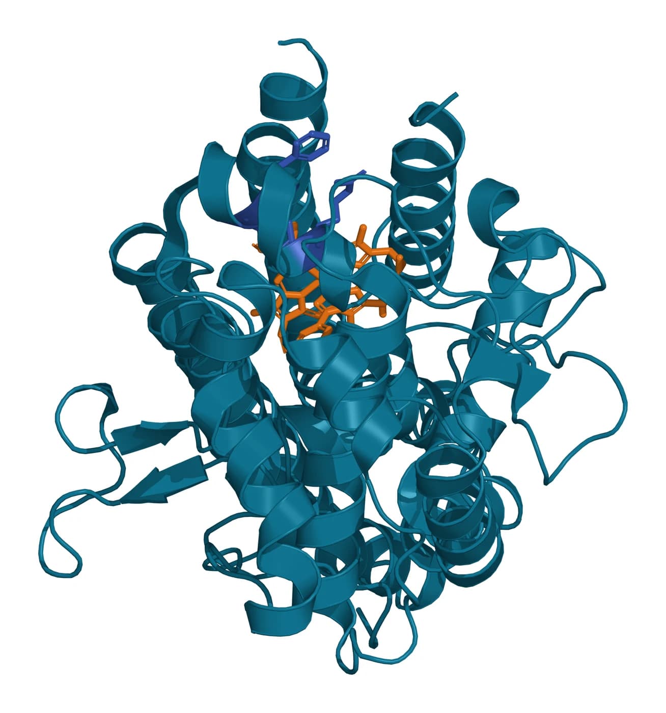
Molecular readouts can sit beside the cellular one. In stromal cell potency work, Niekamp and colleagues traced part of T cell suppression to tryptophan depletion by indoleamine 2,3-dioxygenase 1, IDO1 (Fig. 1).[13] IDO1 is a heme enzyme that catalyzes the first, rate-limiting step of tryptophan catabolism.[14]
Macrophage polarization and reporter cells capture myeloid signaling
Lo Sicco and colleagues found that vesicles from adipose stromal cells were taken up by bone marrow-derived macrophages and shifted them from an M1 to an M2 phenotype. Vesicles from normoxic and hypoxic cultures both produced the shift.[15]
Wang and colleagues co-cultured RAW264.7 macrophages with stromal cell vesicles and scored polarization by flow cytometry. IL-10 rose by ELISA while IL-1β, TNF-α and IL-6 fell, and the authors linked the shift to TGF-β1 carried by the vesicles.[16]
Polarization readouts report on the whole preparation. In the study by Monguió-Tortajada and colleagues, CD163 and CD206 induction on monocytes came from the non-vesicle fraction.[11] Purification method is therefore part of the readout.
Reporter cell lines turn signaling into a single measurable output. Zhang and colleagues showed that stromal cell exosomes induced MyD88-dependent secreted alkaline phosphatase in THP1-Xblue cells, a THP-1 line carrying an NF-κB reporter, with IL-10 and TGF-β1 transcripts induced alongside.[17]
Wong and colleagues used a THP-1 dual reporter line to follow NF-κB and IRF pathways in parallel. Reporter inhibition tracked soluble factors below 5 kDa, partly through prostaglandin E2, while T cell proliferation tracked larger components.[12] Two readouts from one preparation can measure different active fractions.
Endothelial and fibroblast assays quantify network formation and migration
Endothelial cells plated on reconstituted basement membrane rapidly form capillary-like structures. The process involves adhesion, migration, alignment, protease secretion and tubule formation, and it is the most widely used in vitro angiogenesis assay.[18]
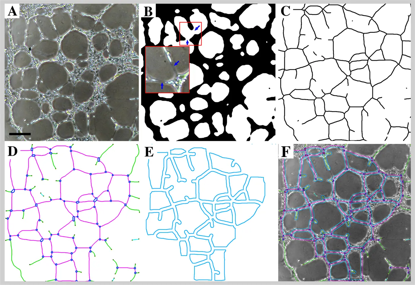
Image analysis turns the network into numbers. Carpentier and colleagues described the Angiogenesis Analyzer for ImageJ, which segments and skeletonizes HUVEC networks and counts junctions, segments, branches and meshes (Fig. 2).[19]
Anderson and colleagues identified 1,927 proteins in stromal cell exosomes, including PDGF, EGF, FGF and NF-κB pathway proteins. Blocking NF-κB signaling with a specific inhibitor showed that the angiogenic response of endothelial cells to the exosomes depended on that pathway.[20]
Fibroblast migration is read in a scratch or transwell format. A scratch assay images a gap in a cell monolayer at set intervals to quantify the migration rate, and a transwell assay counts the cells that cross a porous membrane toward a chemoattractant gradient.[21]
Dose metrics determine how potency results compare
A potency result is as comparable as its dose metric. Willis and colleagues reviewed how the field expresses vesicle dose, and each common metric measures a different property of the preparation, so two studies using different metrics can report the same material differently.[9]
- Particle number, from nanoparticle tracking or tunable resistive pulse sensing, which counts vesicles and any non-vesicle material in the same size range.[9]
- Total protein, which is fast and inexpensive and can include protein not associated with vesicles.[9]
- Cell equivalents, the number of producing cells a dose came from, which ties dose to the culture that made it.[9]
Gupta and colleagues examined 64 preclinical vesicle studies and found wide variation in dosing strategies, independent of purification method and cell source. They proposed anchoring dose to a qualitative measure such as the activity of the relevant cargo.[22]
Hagey and colleagues exposed fibroblasts to vesicles from twelve cell sources at doses from 20 to 200,000 per cell. Dose had a larger effect on the transcriptional response than cell source, and source-specific responses appeared at the lower doses.[23]
At high doses the same study recorded reduced exocytosis and increased lysosomal activity in recipient cells.[23] A potency assay therefore reports most clearly when it covers a defined dose range and states the metric used.
Reference materials and validation make an extracellular vesicle potency assay reproducible
ICH Q6B asks that biological assay results be calibrated against an international or national reference standard where one exists. Where none exists, a characterized in-house reference material is established and results are expressed as in-house units.[3]
For vesicles, Witwer and colleagues proposed a well-characterized stromal cell vesicle preparation as the biological reference.[6] Geeurickx and colleagues built trackable recombinant vesicles as a reference material and used them to measure recovery, intra-method variability and inter-user variability.[24]
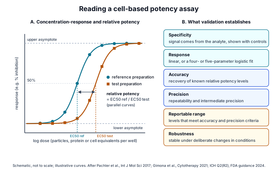
Cell-based assays often give an S-shaped response bounded by upper and lower asymptotes. ICH Q2(R2) describes fitting such curves with four- or five-parameter logistic models and evaluates linearity separately from curve shape (Fig. 3).[25]
Q2(R2) organizes validation around a small set of performance characteristics. For a relative potency assay it describes testing several potency levels across the reportable range, and it places robustness, such as tolerance to changes in plate type, reagent lots and incubation conditions, in assay development.[25]
- Specificity: the signal comes from the analyte, shown with reference materials or other characterized controls.[25]
- Accuracy: agreement between measured and known values, for example by comparison with a reference material.[25]
- Precision: repeatability within a run and intermediate precision across days, analysts and equipment.[25]
- Response and range: the model that relates dose to signal, and the lowest to highest levels that meet the accuracy and precision criteria.[25]
Gimona and colleagues noted that potency assays for stromal cell vesicles should be developed to comply with regulatory guidance for assay validation.[1] Pachler and colleagues described validation of their T cell format as the step toward reliable release criteria.[10]
An assay matrix captures several mechanisms at once
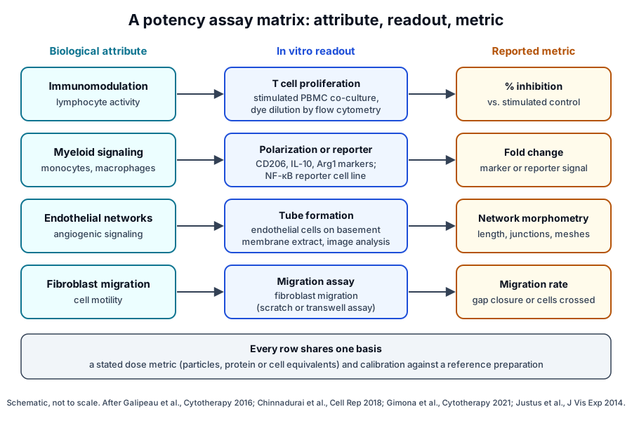
The FDA guidance notes that a product with a complex mechanism or several biological activities may need more than one assay to measure potency. For that case it describes an assay matrix, a set of complementary biological and analytical assays.[4]
An ISCT workshop in May 2015 applied the matrix idea to stromal cells. Galipeau and colleagues named three preferred methods: quantitative RNA analysis of selected genes, flow cytometry of functional surface markers, and protein-based secretome assays.[26]
Chinnadurai and colleagues built such a matrix from secretome analysis and an RNA-based gene array. A cytokine signature correlated with T cell suppression, and interferon-γ served as a surrogate for activated blood cells.[27]
Niekamp and colleagues validated a matrix covering T cell suppression, regulatory T cell induction and macrophage polarization across ten donors. They also reported that the cells secrete vesicles that shifted T cells and monocytes toward regulatory phenotypes in vitro.[13]
Where to go from here
The reference materials page covers how vesicle standards are built and characterized, and the isolation methods page explains how separation shapes what an assay measures. The protein markers page covers the identity markers that accompany a potency result.
The quality references on how exosomes are tested and on reading a certificate of analysis show how these measurements are documented. Verified practices can request lot documentation, and our team is glad to discuss the methods described on this page.
Annotated References
Numbered in the order they are cited in the text. Each source is annotated with what it supports on this page; peer-reviewed articles are listed with journal, volume, issue and pages, and primary sources are regulations, agency documents and reference databases.
- 1.Peer-reviewedGimona M, Brizzi MF, Choo ABH, et al. Critical considerations for the development of potency tests for therapeutic applications of mesenchymal stromal cell-derived small extracellular vesicles. Cytotherapy. 2021;23(5):373-380. https://pubmed.ncbi.nlm.nih.gov/33934807/Annotation: Expert perspective on potency test development for stromal cell vesicles. Cited for identifying and quantifying the vesicle attribute most relevant to the intended activity, and for developing assays in line with ICH validation guidance.
- 2.Peer-reviewedNguyen VVT, Witwer KW, Verhaar MC, et al. Functional assays to assess the therapeutic potential of extracellular vesicles. Journal of Extracellular Vesicles. 2020;10(1):e12033. https://pubmed.ncbi.nlm.nih.gov/33708360/Annotation: Review of in vitro and in vivo functional assays for vesicles. Cited for the range of formats in use and for standardized quantitative assays that compare batches, platforms and cell sources.
- 3.RegulatoryFDA, Q6B Specifications: Test Procedures and Acceptance Criteria for Biotechnological/Biological Products (August 1999). Accessed 20 October 2026. https://www.fda.gov/regulatory-information/search-fda-guidance-documents/q6b-specifications-test-procedures-and-acceptance-criteria-biotechnologicalbiological-products
- 4.RegulatoryFDA, Potency Tests for Cellular and Gene Therapy Products, Final Guidance for Industry (January 2011). Accessed 20 October 2026. https://www.fda.gov/regulatory-information/search-fda-guidance-documents/potency-tests-cellular-and-gene-therapy-products
- 5.RegulatoryFDA, Potency Assurance for Cellular and Gene Therapy Products, Draft Guidance for Industry (December 2023). Accessed 20 October 2026. https://www.fda.gov/regulatory-information/search-fda-guidance-documents/potency-assurance-cellular-and-gene-therapy-products
- 6.Peer-reviewedWitwer KW, Van Balkom BWM, Bruno S, et al. Defining mesenchymal stromal cell (MSC)-derived small extracellular vesicles for therapeutic applications. Journal of Extracellular Vesicles. 2019;8(1):1609206. https://pubmed.ncbi.nlm.nih.gov/31069028/Annotation: Joint statement by members of SOCRATES, ISEV, ISCT and ISBT. Cited for defining stromal cell vesicles by quantifiable metrics and for measuring new preparations against a characterized biological reference.
- 7.Peer-reviewedWelsh JA, Goberdhan DCI, O’Driscoll L, et al. Minimal information for studies of extracellular vesicles (MISEV2023): From basic to advanced approaches. Journal of Extracellular Vesicles. 2024;13(2):e12404. https://pubmed.ncbi.nlm.nih.gov/38326288/Annotation: ISEV minimal information guidelines. Cited as the general reporting framework for functional studies of extracellular vesicles.
- 8.Peer-reviewedRohde E, Pachler K, Gimona M. Manufacturing and characterization of extracellular vesicles from umbilical cord-derived mesenchymal stromal cells for clinical testing. Cytotherapy. 2019;21(6):581-592. https://pubmed.ncbi.nlm.nih.gov/30979664/Annotation: Perspective on manufacturing and quality testing of umbilical cord stromal cell vesicles. Cited for the principle that the process is the product.
- 9.Peer-reviewedWillis GR, Kourembanas S, Mitsialis SA. Toward Exosome-Based Therapeutics: Isolation, Heterogeneity, and Fit-for-Purpose Potency. Frontiers in Cardiovascular Medicine. 2017;4:63. https://pubmed.ncbi.nlm.nih.gov/29062835/Annotation: Review of culture conditions, harvesting, dosage and potency of stromal cell exosomes. Cited for defining potency metrics as a central step in standardizing the field, and (full text, Dose Evaluation section and Table 3) for cell equivalents, protein and particle counting as the common dose metrics with their limitations.
- 10.Peer-reviewedPachler K, Ketterl N, Desgeorges A, et al. An In Vitro Potency Assay for Monitoring the Immunomodulatory Potential of Stromal Cell-Derived Extracellular Vesicles. International Journal of Molecular Sciences. 2017;18(7):1413. https://pubmed.ncbi.nlm.nih.gov/28671586/Annotation: In vitro T cell proliferation and mixed leukocyte reaction assays. Cited for dose-dependent inhibition of mitogen-stimulated proliferation by vesicles, the stimulus dependence of the result, and validation toward release criteria.
- 11.Peer-reviewedMonguió-Tortajada M, Roura S, Gálvez-Montón C, et al. Nanosized UCMSC-derived extracellular vesicles but not conditioned medium exclusively inhibit the inflammatory response of stimulated T cells: implications for nanomedicine. Theranostics. 2017;7(2):270-284. https://pubmed.ncbi.nlm.nih.gov/28042333/Annotation: Size-exclusion fractions of umbilical cord stromal cell secretome tested on T cells and monocytes. Cited for vesicle fractions inhibiting T cell proliferation and for monocyte CD163 and CD206 induction by the non-vesicle fraction.
- 12.Peer-reviewedWong C, Rous I, Lemieux S, et al. Size-dependent immunomodulation by MSC secretome: soluble factors target innate pathways, components larger than 100 kDa regulate T cell proliferation. Stem Cell Research & Therapy. 2025;16(1):556. https://pubmed.ncbi.nlm.nih.gov/41074052/Annotation: THP-1 dual reporter and dye dilution assays on fractionated secretome. Cited for reporter inhibition by small soluble factors partly through prostaglandin E2, and T cell inhibition by components larger than 100 kDa.
- 13.Peer-reviewedNiekamp P, Gu D, Jiang J, et al. Development and validation of a potency assay matrix for optimized and consistent manufacture of clinical mesenchymal stem/stromal cells. Frontiers in Immunology. 2026;17:1725191. https://pubmed.ncbi.nlm.nih.gov/41798920/Annotation: Validated stromal cell potency matrix across ten donors. Cited for T cell, regulatory T cell and macrophage readouts, IDO1-mediated tryptophan depletion, and vesicle effects on T cells and monocytes.
- 14.DatabaseNCBI Gene 3620, IDO1 indoleamine 2,3-dioxygenase 1 (Homo sapiens). Accessed 20 October 2026. https://www.ncbi.nlm.nih.gov/gene/3620
- 15.Peer-reviewedLo Sicco C, Reverberi D, Balbi C, et al. Mesenchymal Stem Cell-Derived Extracellular Vesicles as Mediators of Anti-Inflammatory Effects: Endorsement of Macrophage Polarization. Stem Cells Translational Medicine. 2017;6(3):1018-1028. https://pubmed.ncbi.nlm.nih.gov/28186708/Annotation: Characterization of adipose stromal cell vesicles and their uptake by macrophages. Cited only for the in vitro shift of bone marrow-derived macrophages from an M1 to an M2 phenotype.
- 16.Peer-reviewedWang Y, Han B, Wang Y, et al. Mesenchymal stem cell-secreted extracellular vesicles carrying TGF-β1 up-regulate miR-132 and promote mouse M2 macrophage polarization. Journal of Cellular and Molecular Medicine. 2020;24(21):12750-12764. https://pubmed.ncbi.nlm.nih.gov/32965772/Annotation: Co-culture of RAW264.7 macrophages with stromal cell vesicles. Cited for flow cytometry of M2 polarization, ELISA of IL-10 and inflammatory cytokines, and the link to vesicular TGF-β1.
- 17.Peer-reviewedZhang B, Yin Y, Lai RC, et al. Mesenchymal stem cells secrete immunologically active exosomes. Stem Cells and Development. 2014;23(11):1233-1244. https://pubmed.ncbi.nlm.nih.gov/24367916/Annotation: Reporter cell and monocyte studies of stromal cell exosomes. Cited only for MyD88-dependent NF-κB reporter activity in THP1-Xblue cells and induction of IL-10 and TGF-β1 transcripts.
- 18.Peer-reviewedArnaoutova I, George J, Kleinman HK, et al. The endothelial cell tube formation assay on basement membrane turns 20: state of the science and the art. Angiogenesis. 2009;12(3):267-274. https://pubmed.ncbi.nlm.nih.gov/19399631/Annotation: Review of the endothelial tube formation assay. Cited for the steps the assay captures and its status as the most widely used in vitro angiogenesis assay.
- 19.Peer-reviewedCarpentier G, Berndt S, Ferratge S, et al. Angiogenesis Analyzer for ImageJ - A comparative morphometric analysis of "Endothelial Tube Formation Assay" and "Fibrin Bead Assay". Scientific Reports. 2020;10(1):11568. https://pubmed.ncbi.nlm.nih.gov/32665552/Annotation: Image analysis method for endothelial networks formed by HUVEC. Cited for automated morphometry of junctions, segments, branches and meshes, and as the source of Fig. 2.
- 20.Peer-reviewedAnderson JD, Johansson HJ, Graham CS, et al. Comprehensive Proteomic Analysis of Mesenchymal Stem Cell Exosomes Reveals Modulation of Angiogenesis via Nuclear Factor-KappaB Signaling. Stem Cells. 2016;34(3):601-613. https://pubmed.ncbi.nlm.nih.gov/26782178/Annotation: Proteomics of stromal cell exosomes with functional validation in endothelial cells. Cited for the angiogenic protein profile and the dependence of the endothelial response on NF-κB signaling.
- 21.Peer-reviewedJustus CR, Leffler N, Ruiz-Echevarria M, Yang LV. In vitro cell migration and invasion assays. Journal of Visualized Experiments. 2014. https://pubmed.ncbi.nlm.nih.gov/24962652/Annotation: Methods paper on two cell migration formats. Cited for the scratch assay, imaged at set intervals to quantify migration rate, and the transwell assay, which counts cells crossing a porous membrane toward a chemoattractant gradient.
- 22.Peer-reviewedGupta D, Zickler AM, El Andaloussi S. Dosing extracellular vesicles. Advanced Drug Delivery Reviews. 2021;178:113961. https://pubmed.ncbi.nlm.nih.gov/34481030/Annotation: Review of dosing strategies across 64 preclinical vesicle studies. Cited for the variation in dosing strategies and the proposal to anchor dose to the activity of the relevant cargo.
- 23.Peer-reviewedHagey DW, Ojansivu M, Bostancioglu BR, et al. The cellular response to extracellular vesicles is dependent on their cell source and dose. Science Advances. 2023;9(35):eadh1168. https://pubmed.ncbi.nlm.nih.gov/37656796/Annotation: Transcriptional profiling of fibroblasts exposed to vesicles from twelve cell sources across a wide dose range. Cited for dose outweighing cell source, and for source-specific responses at lower doses.
- 24.Peer-reviewedGeeurickx E, Tulkens J, Dhondt B, et al. The generation and use of recombinant extracellular vesicles as biological reference material. Nature Communications. 2019;10(1):3288. https://pubmed.ncbi.nlm.nih.gov/31337761/Annotation: Development of trackable recombinant vesicles as reference material. Cited for using a reference to measure recovery and intra-method and inter-user variability.
- 25.RegulatoryFDA, Q2(R2) Validation of Analytical Procedures (March 2024). Accessed 20 October 2026. https://www.fda.gov/regulatory-information/search-fda-guidance-documents/q2r2-validation-analytical-procedures
- 26.Peer-reviewedGalipeau J, Krampera M, Barrett J, et al. International Society for Cellular Therapy perspective on immune functional assays for mesenchymal stromal cells as potency release criterion for advanced phase clinical trials. Cytotherapy. 2016;18(2):151-159. https://pubmed.ncbi.nlm.nih.gov/26724220/Annotation: ISCT workshop consensus on immune functional assays for stromal cells. Cited for the matrix approach and its three preferred analytic methods.
- 27.Peer-reviewedChinnadurai R, Rajan D, Qayed M, et al. Potency Analysis of Mesenchymal Stromal Cells Using a Combinatorial Assay Matrix Approach. Cell Reports. 2018;22(9):2504-2517. https://pubmed.ncbi.nlm.nih.gov/29490284/Annotation: Secretome and RNA array matrix for stromal cell potency. Cited for a cytokine signature correlated with T cell suppression and interferon-γ as a surrogate for activated blood cells.
Cite this page
ExaVeyra Sciences Editorial Team. EV Potency Assays: Measuring Biological Activity In Vitro. ExaVeyra Sciences; 20 October 2026. Available from: https://exaveyra.com/science/exosome-biochemistry/extracellular-vesicle-potency-assays. Accessed [date accessed].
Figure sources
- 28.PDB 2D0T, Human indoleamine 2,3-dioxygenase 1 (IDO1). ExaVeyra Sciences, rendered in PyMOL from PDB 2D0T. Structure data: RCSB Protein Data Bank (rcsb.org), CC0. Structure publication: Sugimoto H, Oda S, Otsuki T, et al. Crystal structure of human indoleamine 2,3-dioxygenase: catalytic mechanism of O2 incorporation by a heme-containing dioxygenase. Proc Natl Acad Sci U S A. 2006;103(8):2611-2616.