Extracellular Vesicle Protein Markers: MISEV2023 Categories
By Benn Bluestein-Veyra, M.Sc. Organic Chemistry · Updated 20 October 2026
ExaVeyra Sciences is a Miami-based B2B supplier of exosomes for topical aesthetic use to licensed clinics, and it refers licensed prescribers to a 503A compounding pharmacy that prepares formulations against individual prescriptions.
| Question | Answer |
|---|---|
| What this page covers | The protein categories MISEV2023 uses to characterize EV preparations: transmembrane and cytosolic EV proteins, co-isolated non-EV components and their biology. |
| Who can order | Licensed clinics and practitioners whose active professional license and NPI ExaVeyra has verified. Exosome orders also require a signed professional-use acknowledgment. |
| What ships, and how | Exosome material for topical aesthetic use, characterized by lot, ships overnight in a cold chain. Delivery can take 1 to 3 business days. A certificate of analysis is issued for every lot and provided on request. |
Extracellular vesicle markers are proteins whose presence or depletion shows that a preparation contains lipid-bilayer vesicles and how free it is of other particles. The MISEV2023 guidelines from the International Society for Extracellular Vesicles group them into five categories and ask for at least one protein from each of the first three.[1]
This page covers the biology behind each category, the proteomic studies that refined the classic exosome markers, why tetraspanin levels differ between cell types, and the methods that read these proteins.
MISEV2023 sorts extracellular vesicle markers into five protein categories
The category framework was introduced in MISEV2018, and MISEV2023 recommends the same five-component structure.[1][2] It built on the 2014 position statement, which first set out minimal biochemical standards for attributing cargo or functions to vesicles.[3]
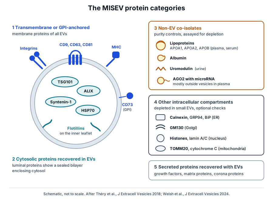
Categories 1 and 2 establish that vesicles are present, and category 3 assesses purity from common co-isolates. Category 4 adds information on possible intracellular origins, and category 5 covers secreted proteins that associate with vesicles after release (Fig. 1).[1]
- Category 1: transmembrane or GPI-anchored proteins of the plasma membrane and endosomes, such as tetraspanins, integrins and MHC molecules.[1]
- Category 2: cytosolic proteins recovered in vesicles, such as TSG101, ALIX, syntenin-1 and flotillins.[1]
- Category 3: major components of co-isolated non-vesicular particles, such as apolipoproteins, albumin and uromodulin.[1][2]
- Category 4: proteins of other compartments, such as calnexin of the endoplasmic reticulum and histones of the nucleus.[1]
- Category 5: secreted proteins recovered with vesicles, including growth factors, matrix proteins and corona proteins.[1]
MISEV2023 names only a few examples per category to keep the choice of proteins open. Other proteins can be assigned by their subcellular location and membrane topology, and the guideline recommends showing enrichment or depletion relative to the source material.[1]
Category 5 proteins such as fibronectin, galectin-3-binding protein and fetuin-A can bind vesicles after release, and MISEV2023 relates the blood-derived ones to the vesicle corona.[1] MISEV2018 asks that their detection be accompanied by study of the vesicle receptor that binds them.[2]
Transmembrane proteins demonstrate a vesicle membrane
MISEV2018 describes category 1 proteins as hallmarks of any vesicle type, because their presence shows the lipid bilayer specific to vesicles.[2] MISEV2023 groups them as multi-pass transmembrane proteins such as tetraspanins, cell- or tissue-specific proteins, and GPI- or lipid-anchored proteins such as CD73.[1]
The category has deep roots. Raposo and colleagues showed in 1996 that B lymphocytes release the internal vesicles of their MHC class II compartments, which they named exosomes, and that the surface protein composition of these exosomes differed from that of the plasma membrane.[4]
Escola and colleagues then found these exosomes enriched in the tetraspanins CD37, CD53, CD63, CD81 and CD82. The tetraspanins concentrated on the internal membranes of the compartment, while LAMP-1 and LAMP-2 stayed on its limiting membrane, which pointed to active protein sorting.[5]
Single-pass category 1 proteins include MHC class I and II, integrins, the transferrin receptor, LAMP1 and LAMP2, syndecans, EMMPRIN and ADAM10. GPI- or lipid-anchored examples include glypican 1, CD73 and the complement regulator CD59.[1]
Tetraspanins are regarded as among the most abundant membrane proteins of extracellular vesicles.[6] Their four-helix fold is known from crystal structures of CD81, which revealed an intramembrane cholesterol-binding pocket (Fig. 2a),[7] and of CD9, which adopts a reversed cone shape (Fig. 2b).[8]
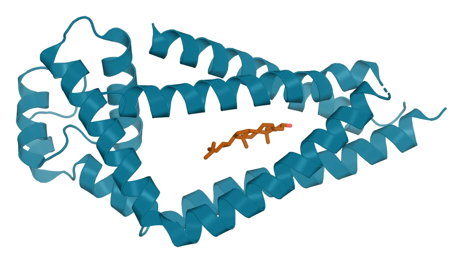
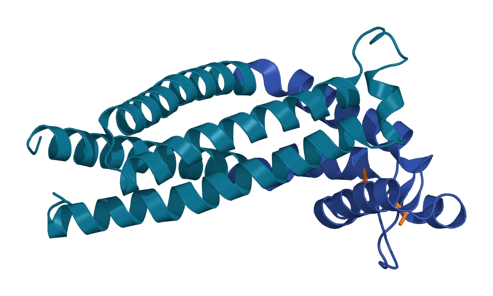
Fig. 2 | Tetraspanin CD81 with cholesterol and Tetraspanin CD9.
Cytosolic proteins show that a bilayer encloses cell contents
Category 2 proteins show that a preparation contains bilayers enclosing intracellular material. MISEV2018 separates proteins with lipid or membrane protein binding ability, which are presumably incorporated actively, from promiscuous cytosolic components such as cytoskeletal proteins and metabolic enzymes.[2]
The first group includes the ESCRT protein TSG101, the ESCRT accessory protein ALIX, syntenin-1, flotillins and caveolins.[1] ALIX, encoded by PDCD6IP, functions in the ESCRT pathway during intraluminal vesicle formation in endosomes (Fig. 3a).[9]
Baietti and colleagues linked several of these proteins in one pathway. Syntenin binds ALIX directly through LYPX(n)L motifs, and syntenin exosomes depended on heparan sulfate, syndecans, ALIX and the ESCRT machinery.[10]
Syntenin-1, encoded by SDCBP, carries two tandem PDZ domains that bind the cytoplasmic C-terminal tails of transmembrane proteins.[11] Kang and colleagues solved this PDZ tandem and showed that each domain binds peptides independently, with PDZ2 binding syndecan-4 (Fig. 3b).[12]
Raft-associated and chaperone proteins complete the picture. de Gassart and colleagues detected raft-associated proteins in exosomes from several cell types,[13] and MISEV2018 lists HSP70 among cytosolic proteins incorporated promiscuously into vesicles.[2] MISEV2023 places HSC70, HSP84, actin, tubulin and GAPDH in the same group and notes that both heat shock proteins are also abundant in non-vesicular particles.[1]
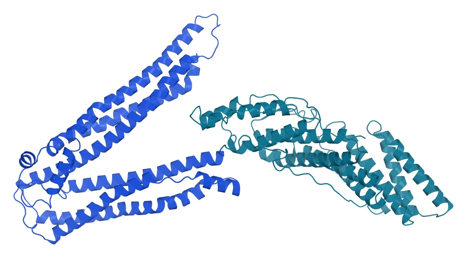
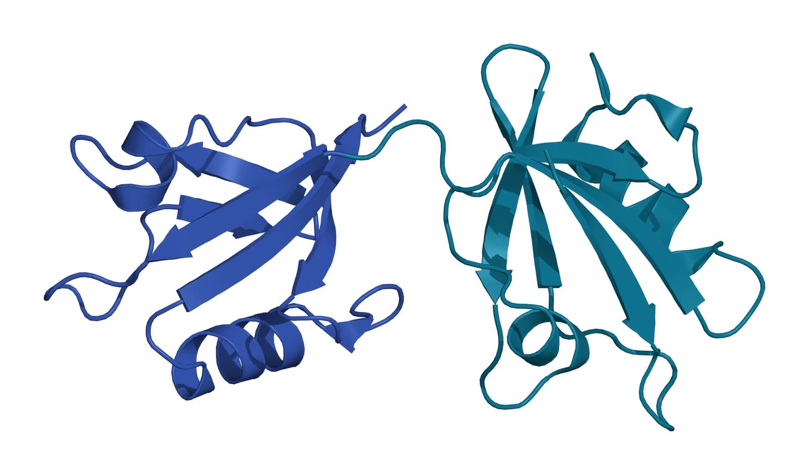
Fig. 3 | ALIX (Bro1 and V domains) and Syntenin-1 PDZ tandem.
Proteomic comparisons refined the classic exosome markers
Kowal and colleagues compared vesicle populations from human dendritic cells separated by sedimentation speed, density flotation and immuno-isolation. Several classic exosome markers, including MHC, flotillin and HSP70, were present similarly in all vesicle types.[14]
The same study defined five protein categories with different relative abundance across vesicle populations. It showed exosomal and non-exosomal subpopulations within small vesicles, and proposed separating them by immuno-isolation with CD63, CD81 or CD9.[14]
Jeppesen and colleagues combined high-resolution density gradients with direct immunoaffinity capture on tetraspanins. Argonaute 1 to 4, glycolytic enzymes and cytoskeletal proteins were undetectable in the captured exosomes, and annexin A1 marked microvesicles shed from the plasma membrane.[15]
In that dataset, ALIX and syntenin-1 associated strongly with CD63, CD81 and CD9 positive vesicles. TSG101 was weak on CD63 and CD81 positive vesicles and stronger on CD9 positive vesicles, and the most abundant non-vesicular proteins were metabolic enzymes such as GAPDH.[15]
Syntenin-1 is the most consistent exosome protein across cell types
Kugeratski and colleagues used super-SILAC quantitative proteomics on exosomes from 14 human cell lines of epithelial, mesenchymal and blood origin. They quantified 1,212 exosome proteins and identified 22 that were universally enriched.[16]
Syntenin-1 was the most consistently abundant of these proteins. It was also detected in exosomes from other species and from human plasma and urine, which led the authors to propose it as a putative universal exosome marker.[16]
Six of the 22 enriched proteins were membrane proteins, including CD47 and integrin beta 1, fifteen were intraluminal and one was an extracellular matrix protein. The enriched set also included biogenesis-related proteins and GTPases.[16]
Among the conventional markers, ALIX and TSG101 were the two found ubiquitous and abundant in all 14 cell lines. Flotillins and heat shock proteins varied, and the 15 consistently depleted proteins were mainly nuclear, with calnexin among the validated depleted set.[16]
Co-isolates and organelle proteins measure purity
MISEV2018 proposes apolipoproteins A1, A2 and B and albumin as negative markers for plasma and serum vesicles, and recommends reporting their depletion as a measured quantity.[2] Apolipoprotein A-I, the main protein of high-density lipoprotein, forms a horseshoe-shaped amphipathic helix (Fig. 4).[17]
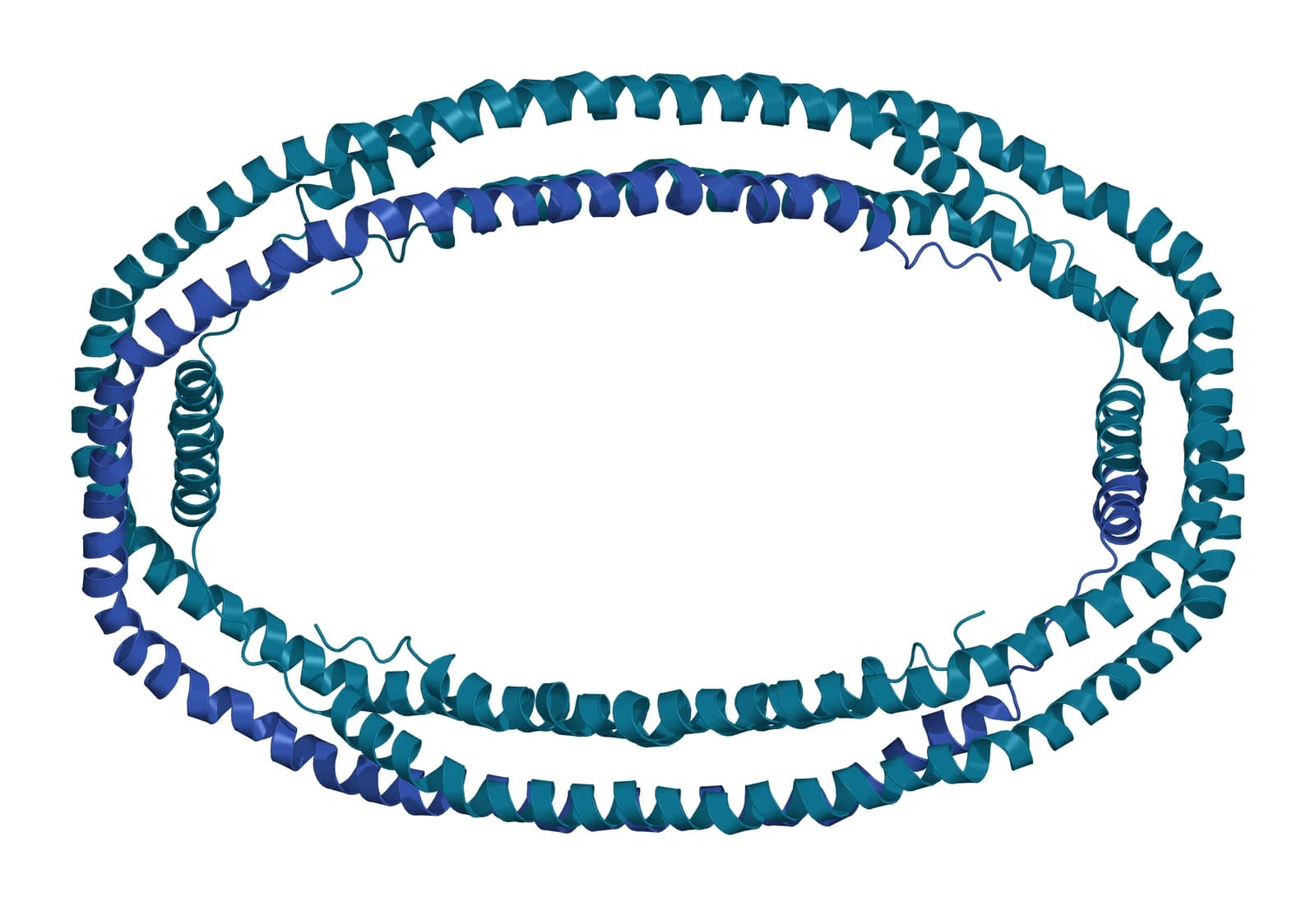
Lipoprotein particles overlap vesicles in plasma. Sódar and colleagues found that most circulating particles in the vesicle size range lacked vesicle markers and were mainly low-density lipoprotein, which co-purified with vesicles and associated with them on mixing.[18]
Karimi and colleagues showed that size-exclusion chromatography alone left lipoproteins in the plasma vesicle fractions. Adding a density cushion reduced lipoprotein contamination 100-fold and allowed 1,187 vesicle proteins to be identified.[19]
MISEV2023 widened category 3 to include immunoglobulins in blood, 14-3-3 proteins, and components enriched in exomeres and supermeres such as HSP90, TGFBI and lactate dehydrogenase.[1]
Uromodulin and Argonaute complexes
In urine, polymeric uromodulin, also called Tamm-Horsfall protein, entraps vesicles in the low-speed pellet. Fernández-Llama and colleagues showed that dithiothreitol disrupts this polymer network and shifts vesicle proteins to the high-speed pellet.[20]
Most circulating microRNA in plasma co-fractionated with protein complexes containing Argonaute 2, as Arroyo and colleagues and Turchinovich and colleagues reported in 2011.[21][22] MISEV2018 notes that Argonaute also occurs inside vesicles, so its abundance is read relative to the vesicle fraction.[2]
Calnexin, GM130 and histones mark other compartments
Category 4 proteins belong to the endoplasmic reticulum, Golgi, mitochondria, nucleus or autophagosomes. MISEV2018 describes them as present in some vesicle types and expected to be depleted in small vesicles of plasma membrane or endosomal origin.[2]
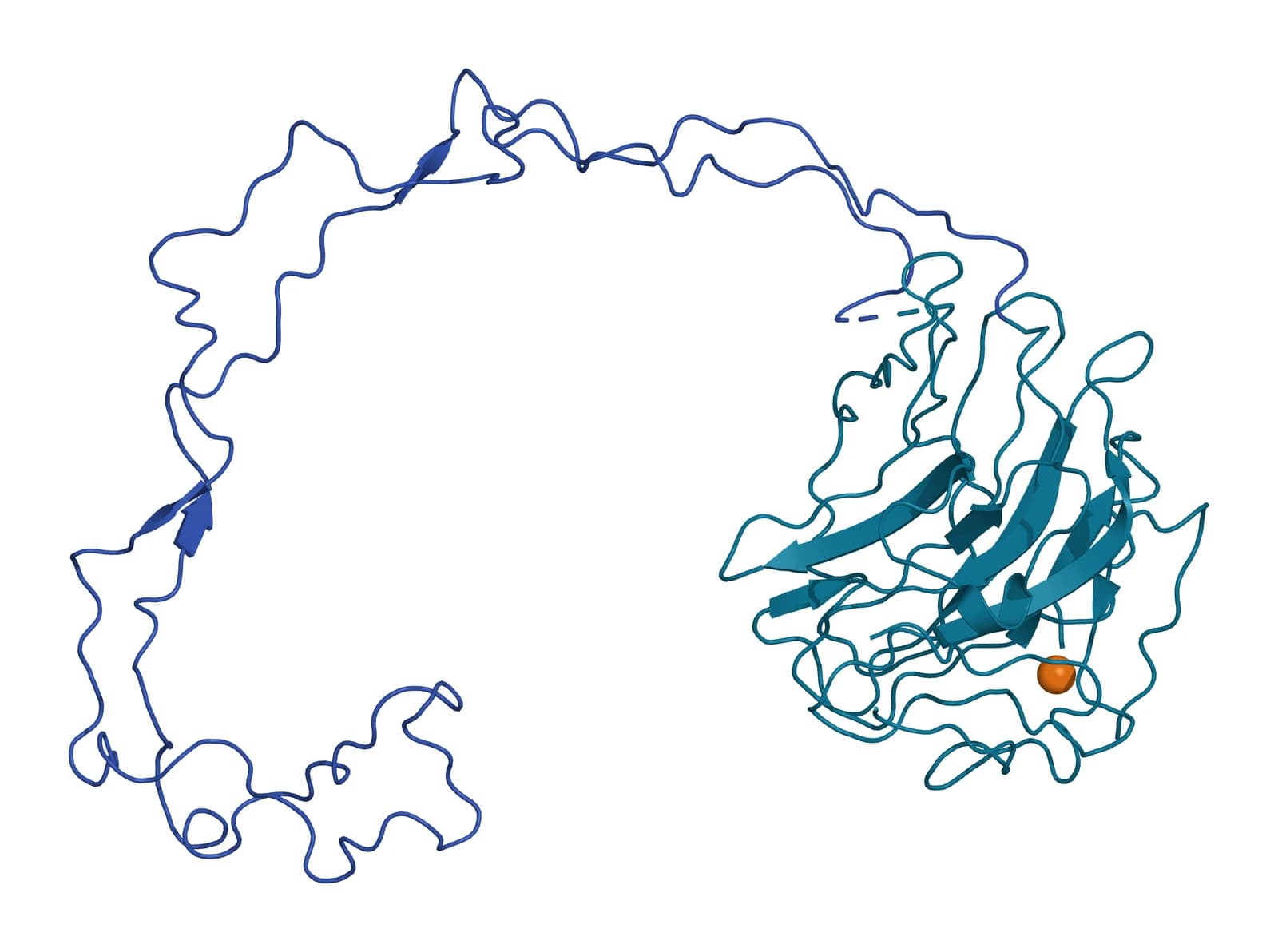
Calnexin is the most widely used of these proteins. It is a lectin-like chaperone of the endoplasmic reticulum whose lumenal domain pairs a beta sandwich, like those of legume lectins, with an extended proline-rich arm about 140 Å long (Fig. 5).[23]
MISEV2023 treats category 4 analysis as optional, since these proteins may appear in some vesicle subtypes or conditions. Its examples include GM130 for the Golgi, histones and lamin A/C for the nucleus, and TOMM20 for mitochondria.[1]
Tetraspanin levels vary with the cell of origin
MISEV2018 placed CD9 among cell-specific markers, noting its absence from NK cells, B cells and some mesenchymal stromal cells,[2] and MISEV2023 lists it with CD63, CD81 and CD82 as a multi-pass tetraspanin.[1] Kugeratski and colleagues found that low tetraspanin levels on exosomes from some cell lines mirrored low expression in the parent cells.[16]
Route of release matters as well. Mathieu and colleagues showed that CD9 resides mainly at the plasma membrane and CD63 in late endosomes, with CD9 released in vesicles more abundantly.[24] Ai and colleagues found that endocytosis reduces vesicular secretion of CD81, CD9 and CD63, and that syntenin promotes CD63 release by blocking its endocytosis.[25]
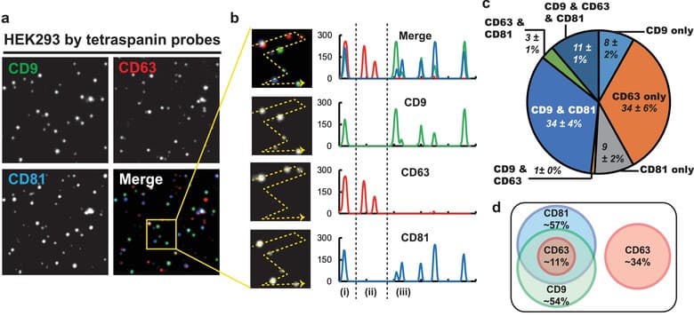
Single-vesicle methods show this variation directly. Han and colleagues imaged individual vesicles from HEK293 cells by total internal reflection microscopy and found that about half of the tetraspanin signals were single-positive, most often CD63 alone (Fig. 6).[26]
Karimi and colleagues reported a similar pattern in human blood. Very few plasma or serum vesicles carried both CD63 and CD81, and CD9 positive vesicles were more numerous in EDTA plasma and carried the platelet protein CD41a, which points to a platelet origin.[27] CD63 positive vesicles were enriched in serum, and CD81 positive vesicles were the rarest subpopulation in both fluids.[27]
Western blot, flow cytometry and single-vesicle imaging read the categories
Western blotting reports category proteins for a bulk preparation, ideally alongside the source cells or fluid so enrichment and depletion can be compared.[1] Kugeratski and colleagues used it to detect syntenin-1 in exosomes from 100 human plasma samples and 5 urine samples.[16] Mass spectrometry extends the same comparison to the whole proteome.[14][16]
Flow cytometry measures proteins on individual particles, and vesicle signals partly overlap instrument background noise.[28] The MIFlowCyt-EV framework sets out what such experiments report, including calibration, controls and staining.[29]
Single-vesicle imaging and bead capture reveal co-expression patterns that bulk assays average out.[26][27] MISEV2023 notes that some category markers are harder to use in single-vesicle techniques, which may need their own controls.[1] The tetraspanin marker panel reference covers these measurements in detail.
Where to go from here
The tetraspanin marker panel reference explains how CD9, CD63 and CD81 are measured, and the MISEV2023 compliance reference sets out how category results are documented. The biogenesis reference follows TSG101, ALIX and syntenin-1 into the endosome where exosomes form.
The tetraspanin family reference covers the structures behind category 1. Verified practices can request the certificate of analysis for a lot, and our team can explain how its marker data were obtained.
Annotated References
Numbered in the order they are cited in the text. Each source is annotated with what it supports on this page; peer-reviewed articles are listed with journal, volume, issue and pages, and primary sources are regulations, agency documents and reference databases.
- 1.Peer-reviewedWelsh JA, Goberdhan DCI, O'Driscoll L, et al. Minimal information for studies of extracellular vesicles (MISEV2023): From basic to advanced approaches. Journal of Extracellular Vesicles. 2024;13(2):e12404. https://pubmed.ncbi.nlm.nih.gov/38326288/Annotation: ISEV reporting guideline. Cited for the five-category protein framework, the request for at least one protein from categories 1 to 3, the optional status of category 4 and the examples in each category.
- 2.Peer-reviewedThéry C, Witwer KW, Aikawa E, et al. Minimal information for studies of extracellular vesicles 2018 (MISEV2018): a position statement of the International Society for Extracellular Vesicles and update of the MISEV2014 guidelines. Journal of Extracellular Vesicles. 2018;7(1):1535750. https://pubmed.ncbi.nlm.nih.gov/30637094/Annotation: ISEV reporting guideline that introduced the protein categories. Cited for the rationale of each category, the apolipoprotein and albumin negative markers, CD9 as a cell-specific tetraspanin and Argonaute inside vesicles.
- 3.Peer-reviewedLötvall J, Hill AF, Hochberg F, et al. Minimal experimental requirements for definition of extracellular vesicles and their functions: a position statement from the International Society for Extracellular Vesicles. Journal of Extracellular Vesicles. 2014;3:26913. https://pubmed.ncbi.nlm.nih.gov/25536934/Annotation: The first ISEV position statement. Cited for the minimal biochemical standards that preceded the MISEV2018 categories.
- 4.Peer-reviewedRaposo G, Nijman HW, Stoorvogel W, et al. B lymphocytes secrete antigen-presenting vesicles. Journal of Experimental Medicine. 1996;183(3):1161-1172. https://pubmed.ncbi.nlm.nih.gov/8642258/Annotation: Immunoelectron microscopy of B lymphoblastoid cells. Cited for the release of MHC class II carrying exosomes whose surface composition differed from the plasma membrane.
- 5.Peer-reviewedEscola JM, Kleijmeer MJ, Stoorvogel W, et al. Selective enrichment of tetraspan proteins on the internal vesicles of multivesicular endosomes and on exosomes secreted by human B-lymphocytes. Journal of Biological Chemistry. 1998;273(32):20121-20127. https://pubmed.ncbi.nlm.nih.gov/9685355/Annotation: Subcellular localization in B lymphocytes. Cited for the enrichment of CD37, CD53, CD63, CD81 and CD82 on exosomes and internal endosomal membranes, with LAMP-1 and LAMP-2 on the limiting membrane.
- 6.Peer-reviewedAndreu Z, Yáñez-Mó M. Tetraspanins in extracellular vesicle formation and function. Frontiers in Immunology. 2014;5:442. https://pubmed.ncbi.nlm.nih.gov/25278937/Annotation: Review of tetraspanins in vesicle biology. Cited for tetraspanins as among the most abundant membrane proteins of extracellular vesicles.
- 7.Peer-reviewedZimmerman B, Kelly B, McMillan BJ, et al. Crystal Structure of a Full-Length Human Tetraspanin Reveals a Cholesterol-Binding Pocket. Cell. 2016;167(4):1041-1051.e11. https://pubmed.ncbi.nlm.nih.gov/27881302/Annotation: Crystal structure of full-length human CD81. Cited for the tetraspanin fold and its intramembrane cholesterol-binding pocket.
- 8.Peer-reviewedUmeda R, Satouh Y, Takemoto M, et al. Structural insights into tetraspanin CD9 function. Nature Communications. 2020;11(1):1606. https://pubmed.ncbi.nlm.nih.gov/32231207/Annotation: Crystal structure of full-length CD9. Cited for the reversed cone shape of the CD9 fold.
- 9.DatabaseNCBI Gene 10015, PDCD6IP (ALIX) (Homo sapiens). Accessed 20 October 2026. https://www.ncbi.nlm.nih.gov/gene/10015
- 10.Peer-reviewedBaietti MF, Zhang Z, Mortier E, et al. Syndecan-syntenin-ALIX regulates the biogenesis of exosomes. Nature Cell Biology. 2012;14(7):677-685. https://pubmed.ncbi.nlm.nih.gov/22660413/Annotation: Cell biology of exosome formation. Cited for syntenin binding ALIX through LYPX(n)L motifs and for syntenin exosomes depending on syndecans, heparan sulfate, ALIX and ESCRTs.
- 11.DatabaseNCBI Gene 6386, SDCBP syndecan binding protein (syntenin-1) (Homo sapiens). Accessed 20 October 2026. https://www.ncbi.nlm.nih.gov/gene/6386
- 12.Peer-reviewedKang BS, Cooper DR, Jelen F, et al. PDZ tandem of human syntenin: crystal structure and functional properties. Structure. 2003;11(4):459-468. https://pubmed.ncbi.nlm.nih.gov/12679023/Annotation: Crystal structure of the syntenin PDZ tandem with binding studies. Cited for independent peptide binding by each PDZ domain and syndecan-4 binding by PDZ2.
- 13.Peer-reviewedde Gassart A, Geminard C, Fevrier B, et al. Lipid raft-associated protein sorting in exosomes. Blood. 2003;102(13):4336-4344. https://pubmed.ncbi.nlm.nih.gov/12881314/Annotation: Immunoblot and detergent fractionation of exosomes. Cited for raft-associated proteins in exosomes from several cell types.
- 14.Peer-reviewedKowal J, Arras G, Colombo M, et al. Proteomic comparison defines novel markers to characterize heterogeneous populations of extracellular vesicle subtypes. Proceedings of the National Academy of Sciences of the United States of America. 2016;113(8):E968-E977. https://pubmed.ncbi.nlm.nih.gov/26858453/Annotation: Quantitative proteomics of separated dendritic cell vesicle populations. Cited for MHC, flotillin and HSP70 being present in all vesicle types and for tetraspanin immuno-isolation of subpopulations.
- 15.Peer-reviewedJeppesen DK, Fenix AM, Franklin JL, et al. Reassessment of Exosome Composition. Cell. 2019;177(2):428-445.e18. https://pubmed.ncbi.nlm.nih.gov/30951670/Annotation: Density gradient and direct immunoaffinity capture of exosomes. Cited for Argonaute and glycolytic enzymes being undetectable in exosomes, annexin A1 on microvesicles, and ALIX and syntenin-1 on tetraspanin-positive vesicles.
- 16.Peer-reviewedKugeratski FG, Hodge K, Lilla S, et al. Quantitative proteomics identifies the core proteome of exosomes with syntenin-1 as the highest abundant protein and a putative universal biomarker. Nature Cell Biology. 2021;23(6):631-641. https://pubmed.ncbi.nlm.nih.gov/34108659/Annotation: Super-SILAC proteomics of exosomes from 14 human cell lines. Cited for the 22 enriched core proteins, syntenin-1 as the most consistent, ALIX and TSG101 as ubiquitous, and tetraspanin levels tracking the parent cell.
- 17.Peer-reviewedBorhani DW, Rogers DP, Engler JA, et al. Crystal structure of truncated human apolipoprotein A-I suggests a lipid-bound conformation. Proceedings of the National Academy of Sciences of the United States of America. 1997;94(23):12291-12296. https://pubmed.ncbi.nlm.nih.gov/9356442/Annotation: Crystal structure of apolipoprotein A-I residues 44 to 243. Cited for its horseshoe-shaped amphipathic helix and its place as the main protein of high-density lipoprotein.
- 18.Peer-reviewedSódar BW, Kittel Á, Pálóczi K, et al. Low-density lipoprotein mimics blood plasma-derived exosomes and microvesicles during isolation and detection. Scientific Reports. 2016;6:24316. https://pubmed.ncbi.nlm.nih.gov/27087061/Annotation: Flow cytometry and electron microscopy of plasma particles. Cited for low-density lipoprotein making up most vesicle-sized particles, co-purifying with vesicles and associating with them.
- 19.Peer-reviewedKarimi N, Cvjetkovic A, Jang SC, et al. Detailed analysis of the plasma extracellular vesicle proteome after separation from lipoproteins. Cellular and Molecular Life Sciences. 2018;75(15):2873-2886. https://pubmed.ncbi.nlm.nih.gov/29441425/Annotation: Separation and proteomics of plasma vesicles. Cited for lipoproteins co-eluting with vesicles in size-exclusion chromatography and the 100-fold reduction with a density cushion.
- 20.Peer-reviewedFernández-Llama P, Khositseth S, Gonzales PA, et al. Tamm-Horsfall protein and urinary exosome isolation. Kidney International. 2010;77(8):736-742. https://pubmed.ncbi.nlm.nih.gov/20130532/Annotation: Western blot and electron microscopy of urinary vesicle isolation. Cited for polymeric uromodulin entrapping vesicles and dithiothreitol releasing them.
- 21.Peer-reviewedArroyo JD, Chevillet JR, Kroh EM, et al. Argonaute2 complexes carry a population of circulating microRNAs independent of vesicles in human plasma. Proceedings of the National Academy of Sciences of the United States of America. 2011;108(12):5003-5008. https://pubmed.ncbi.nlm.nih.gov/21383194/Annotation: Fractionation of human plasma and serum. Cited for most circulating microRNAs co-fractionating with Argonaute 2 protein complexes.
- 22.Peer-reviewedTurchinovich A, Weiz L, Langheinz A, et al. Characterization of extracellular circulating microRNA. Nucleic Acids Research. 2011;39(16):7223-7233. https://pubmed.ncbi.nlm.nih.gov/21609964/Annotation: Filtration, ultracentrifugation and immunoprecipitation of extracellular microRNA. Cited for most plasma microRNA being associated with Argonaute 2 outside vesicles.
- 23.Peer-reviewedSchrag JD, Bergeron JJ, Li Y, et al. The Structure of calnexin, an ER chaperone involved in quality control of protein folding. Molecular Cell. 2001;8(3):633-644. https://pubmed.ncbi.nlm.nih.gov/11583625/Annotation: Crystal structure of the calnexin lumenal domain. Cited for its legume lectin-like beta sandwich and the extended 140 Å proline-rich arm.
- 24.Peer-reviewedMathieu M, Névo N, Jouve M, et al. Specificities of exosome versus small ectosome secretion revealed by live intracellular tracking of CD63 and CD9. Nature Communications. 2021;12(1):4389. https://pubmed.ncbi.nlm.nih.gov/34282141/Annotation: Live tracking of CD9 and CD63. Cited for CD9 residing at the plasma membrane and CD63 in late endosomes, with CD9 released in vesicles more abundantly.
- 25.Peer-reviewedAi Y, Guo C, Garcia-Contreras M, et al. Endocytosis blocks the vesicular secretion of exosome marker proteins. Science Advances. 2024;10(19):eadi9156. https://pubmed.ncbi.nlm.nih.gov/38718108/Annotation: Cell biology of tetraspanin marker secretion. Cited for endocytosis reducing vesicular secretion of CD81, CD9 and CD63, and syntenin promoting CD63 release by blocking its endocytosis.
- 26.Peer-reviewedHan C, Kang H, Yi J, et al. Single-vesicle imaging and co-localization analysis for tetraspanin profiling of individual extracellular vesicles. Journal of Extracellular Vesicles. 2021;10(3):e12047. https://pubmed.ncbi.nlm.nih.gov/33456726/Annotation: Total internal reflection imaging of single vesicles. Cited for distinct tetraspanin co-expression patterns, with about half of HEK293 vesicle signals single-positive and CD63 alone the most common.
- 27.Peer-reviewedKarimi N, Dalirfardouei R, Dias T, et al. Tetraspanins distinguish separate extracellular vesicle subpopulations in human serum and plasma - Contributions of platelet extracellular vesicles in plasma samples. Journal of Extracellular Vesicles. 2022;11(5):e12213. https://pubmed.ncbi.nlm.nih.gov/35524458/Annotation: Bead capture, single-particle imaging and flow cytometry of blood vesicles from healthy donors. Cited for rare CD63 and CD81 double-positive vesicles and platelet CD41a on CD9 positive vesicles.
- 28.Peer-reviewedWelsh JA, Arkesteijn GJA, Bremer M, et al. A compendium of single extracellular vesicle flow cytometry. Journal of Extracellular Vesicles. 2023;12(2):e12299. https://pubmed.ncbi.nlm.nih.gov/36759917/Annotation: ISEV, ISAC and ISTH working group compendium. Cited for vesicle signals overlapping flow cytometer background noise.
- 29.Peer-reviewedWelsh JA, Van Der Pol E, Arkesteijn GJA, et al. MIFlowCyt-EV: a framework for standardized reporting of extracellular vesicle flow cytometry experiments. Journal of Extracellular Vesicles. 2020;9(1):1713526. https://pubmed.ncbi.nlm.nih.gov/32128070/Annotation: Working group reporting framework. Cited for the reporting of calibration, controls and staining in vesicle flow cytometry.
Cite this page
ExaVeyra Sciences Editorial Team. Extracellular Vesicle Protein Markers: MISEV2023 Categories. ExaVeyra Sciences; 20 October 2026. Available from: https://exaveyra.com/science/exosome-biochemistry/extracellular-vesicle-protein-markers. Accessed [date accessed].
Figure sources
- 30.PDB 5TCX, Tetraspanin CD81 with cholesterol. ExaVeyra Sciences, rendered in PyMOL from PDB 5TCX. Structure data: RCSB Protein Data Bank (rcsb.org), CC0. Structure publication: Zimmerman B, Kelly B, McMillan BJ, et al. Crystal structure of a full-length human tetraspanin reveals a cholesterol-binding pocket. Cell. 2016;167(4):1041-1051.e11.
- 31.PDB 6K4J, Tetraspanin CD9. ExaVeyra Sciences, rendered in PyMOL from PDB 6K4J. Structure data: RCSB Protein Data Bank (rcsb.org), CC0. Structure publication: Umeda R, Satouh Y, Takemoto M, et al. Structural insights into tetraspanin CD9 function. Nat Commun. 2020;11(1):1606.
- 32.PDB 2OEV, ALIX (Bro1 and V domains). ExaVeyra Sciences, rendered in PyMOL from PDB 2OEV. Structure data: RCSB Protein Data Bank (rcsb.org), CC0. Structure publication: Fisher RD, Chung HY, Zhai Q, et al. Structural and biochemical studies of ALIX/AIP1 and its role in retrovirus budding. Cell. 2007;128(5):841-852.
- 33.PDB 1N99, Syntenin-1 PDZ tandem. ExaVeyra Sciences, rendered in PyMOL from PDB 1N99. Structure data: RCSB Protein Data Bank (rcsb.org), CC0. Structure publication: Kang BS, Cooper DR, Jelen F, et al. PDZ tandem of human syntenin: crystal structure and functional properties. Structure. 2003;11:459-468.
- 34.PDB 1AV1, Apolipoprotein A-I (residues 44 to 243). ExaVeyra Sciences, rendered in PyMOL from PDB 1AV1. Structure data: RCSB Protein Data Bank (rcsb.org), CC0. Structure publication: Borhani DW, Rogers DP, Engler JA, Brouillette CG. Crystal structure of truncated human apolipoprotein A-I suggests a lipid-bound conformation. Proc Natl Acad Sci U S A. 1997;94(23):12291-12296.
- 35.PDB 1JHN, Calnexin lumenal domain. ExaVeyra Sciences, rendered in PyMOL from PDB 1JHN. Structure data: RCSB Protein Data Bank (rcsb.org), CC0. Structure publication: Schrag JD, Bergeron JJ, Li Y, et al. The structure of calnexin, an ER chaperone involved in quality control of protein folding. Mol Cell. 2001;8:633-644.