Extracellular Vesicle Reference Materials and Standardization
By Benn Bluestein-Veyra, M.Sc. Organic Chemistry · Updated 20 October 2026
ExaVeyra Sciences is a Miami-based B2B supplier of exosomes for topical aesthetic use to licensed clinics, and it refers licensed prescribers to a 503A compounding pharmacy that prepares formulations against individual prescriptions.
| Question | Answer |
|---|---|
| What this page covers | Reference materials for EV measurement: recombinant and biological reference vesicles, NIST and ISEV initiatives, and why counts and sizes vary between labs. |
| Who can order | Licensed clinics and practitioners whose active professional license and NPI ExaVeyra has verified. Exosome orders also require a signed professional-use acknowledgment. |
| What ships, and how | Exosome material for topical aesthetic use, characterized by lot, ships overnight in a cold chain. Delivery can take 1 to 3 business days. A certificate of analysis is issued for every lot and provided on request. |
An extracellular vesicle reference material is a sample with stable, well-characterized properties that laboratories measure alongside their own vesicles, so that sizes, concentrations and marker signals can be compared.[1] The need is measurable: the same vesicle sample gives a different size distribution and concentration on each detection technique.[2]
This page covers why EV measurements vary between laboratories, the metrology terms that define a reference material, and the synthetic, recombinant and biological reference materials now in use. It closes with the reporting frameworks and ISEV initiatives that coordinate standardization across the field.
Instrument sensitivity and settings drive variation between laboratories
Van der Pol and colleagues measured one vesicle sample with electron microscopy, two flow cytometers, nanoparticle tracking analysis and resistive pulse sensing in 2014. Each technique reported its own size distribution and concentration, and the differences traced mainly to the smallest vesicle each method could detect (Fig. 1).[2]
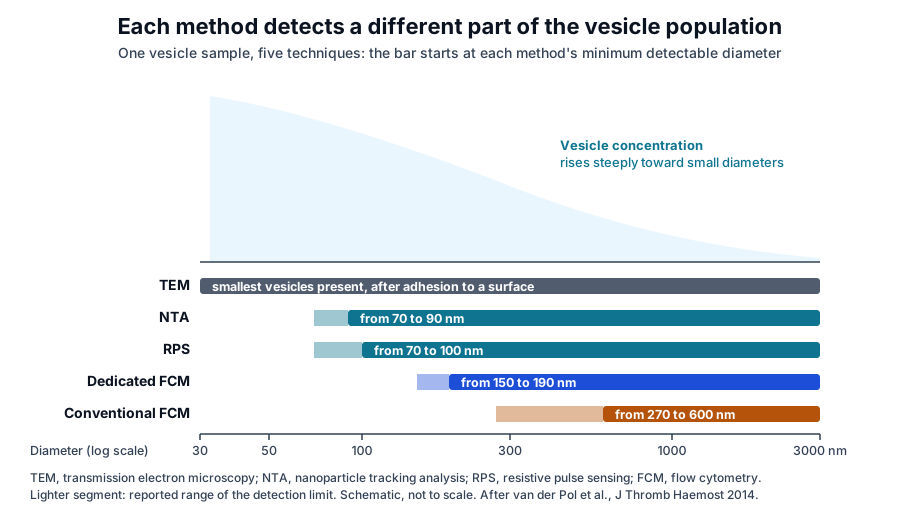
They reported minimum detectable diameters of 70 to 90 nm for nanoparticle tracking analysis, 70 to 100 nm for resistive pulse sensing, 150 to 190 nm for a dedicated flow cytometer and 270 to 600 nm for a conventional flow cytometer. Electron microscopy resolved the smallest vesicles present, after adhesion to a surface.[2]
Nanoparticle tracking analysis depends on settings and hardware
Vestad and colleagues measured vesicles and synthetic particles on two NanoSight NS500 instruments in different laboratories. Within-day coefficients of variation were 1 to 12 percent, and identical software settings gave significantly different results between the two instruments, a gap that instrument-optimized settings largely closed.[3]
Bachurski and colleagues compared the NanoSight NS300 and the ZetaView against electron microscopy and single-particle interferometric imaging. The NS300 reported about twice the concentration of polystyrene and silica nanospheres; the ZetaView gave more accurate concentrations, and the NS300 gave sizes of higher resolution.[4]
Flow cytometers differ in sensitivity and flow rate
In a study run through the ISTH Scientific and Standardization Committee, van der Pol and colleagues sent calibration beads and platelet vesicle samples to 33 participants operating 46 flow cytometers. Instruments set to the same nominal flow rate differed six-fold in measured flow rate, which made flow rate calibration essential.[5]
A 2025 NIST study by Poudel and colleagues characterized commercially available vesicles from two human cell sources by cryo-electron microscopy, particle tracking, field-flow fractionation and resistive pulse sensing. Size distributions from every method skewed larger than cryo-electron microscopy, and concentrations differed by up to two orders of magnitude.[6]
Metrology defines a reference material by its specified properties
NIST defines a reference material as one sufficiently homogeneous and stable in one or more specified properties to be fit for its intended use in a measurement process. Its uses include calibrating a measurement system, assessing a measurement procedure, assigning values to other materials and quality control.[7]
A certified reference material adds a certificate that states the property value, its uncertainty and a statement of metrological traceability. NIST Standard Reference Materials are certified reference materials whose certified values are traceable to an internationally recognized higher-order reference system, such as the SI.[7]
Traceability connects a reported number to the SI
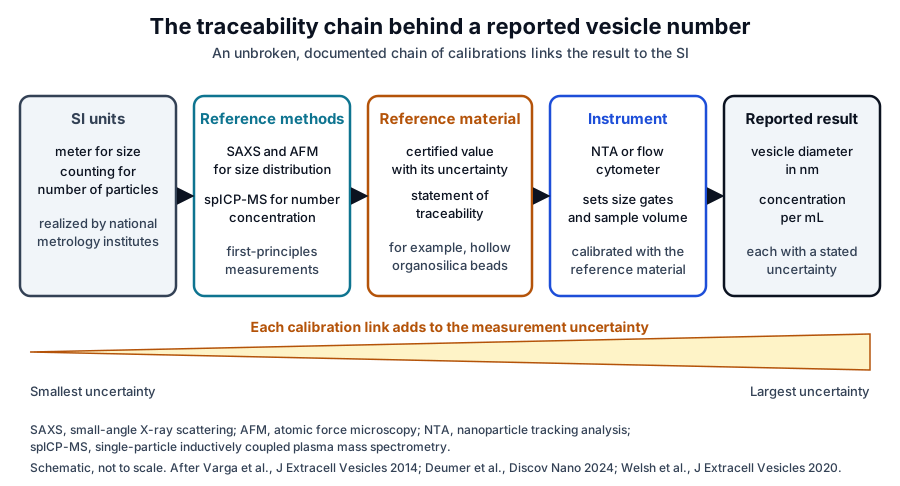
NIST adopts the international vocabulary definition of metrological traceability: a result is related to a reference through a documented, unbroken chain of calibrations, each of which contributes to the measurement uncertainty (Fig. 2). Under the same policy, the provider of a result supports its traceability claim and the user assesses it.[8]
For vesicles, the chain begins with methods that measure size and number from first principles. Varga and colleagues sized erythrocyte vesicles by tracking analysis, resistive pulse sensing, electron microscopy and small-angle X-ray scattering, found modal sizes near 130 nm, and identified X-ray scattering as a promising traceable method.[9]
Deumer and colleagues applied that approach to candidate calibrants. They measured the size distribution of hollow organosilica beads of 200 to 500 nm by small-angle X-ray scattering and atomic force microscopy, and their number concentration by single-particle inductively coupled plasma mass spectrometry.[10]
National metrology programs are building vesicle reference materials
The NIST Quantitative Flow Cytometry Measurements program develops reference materials and procedures for quantitative measurement of cells, extracellular vesicles and other bioparticles. It includes a consensus-based protocol for assigning certified number concentrations to sub-micrometer particle suspensions.[11]
Refractive index links light scatter to vesicle size
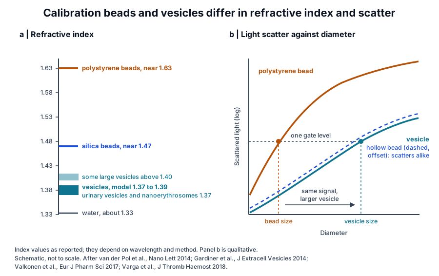
Optical instruments detect a vesicle by the light it scatters, which depends on both its diameter and its refractive index. The flow cytometry compendium from Welsh and colleagues explains this interaction and why vesicle signals overlap with background noise on instruments designed for cells.[12]
Van der Pol and colleagues determined the refractive index of single particles in suspension with nanoparticle tracking analysis in 2014, separating silica from polystyrene beads by index alone. They measured a mean refractive index of 1.37 for vesicles from human urine.[13]
Gardiner and colleagues combined tracking data with Mie scattering theory. Vesicles from several cell and fluid sources had modal indices of 1.37 to 1.39, some large vesicles above 200 nm exceeded 1.40, and the silica and polystyrene calibration microspheres sat near 1.47 and 1.63 (Fig. 3).[14]
Polystyrene gates select different vesicle sizes on each instrument
Varga and colleagues pointed out that the refractive index mismatch between polystyrene and vesicles makes a bead-based scatter gate select larger vesicles, and a different vesicle size on each flow cytometer.[15] Van der Pol and colleagues therefore set gates by estimated vesicle diameter, using beads together with a light scattering model.[5]
In that study, 31 instruments detected the 1,200 to 3,000 nm vesicle diameter gate. Inter-laboratory variability in platelet vesicle concentration was 81 percent with the diameter gate, compared with 139 percent with the bead gate.[5]
Synthetic particles calibrate size, scatter and concentration
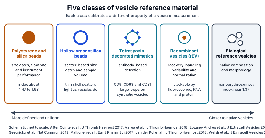
Polystyrene and silica beads remain the most accessible calibrants. Cointe and colleagues tested a bead-based strategy in 44 laboratories running 52 cytometers of 14 types; it suited 85 percent of the instruments, and all of them correctly ranked platelet vesicle levels in two plasma samples.[16]
Inter-laboratory variability of platelet vesicle counts in that workshop was 37 and 28 percent for the two samples. Instruments that used forward scatter and side scatter as the sizing parameter gave equivalent results.[16]
Hollow organosilica beads scatter light as vesicles do
Varga and colleagues prepared monodisperse hollow organosilica beads with a homogeneous shell. Two-angle light scattering showed that the beads scatter light much as platelet vesicles do, so a hollow bead of a given size gates vesicles of the same size.[15]
Deumer and colleagues assigned traceable size and concentration values to four hollow bead types. Concentrations measured by particle tracking and two independent flow cytometric methods agreed with the reference values within the calculated uncertainty.[10]
Fluorescence calibration expresses signals in standard units
Cook and colleagues described quantitative flow cytometry tools that optimize detector settings for small particles and calibrate their signals. They demonstrated cross-platform comparison of vesicle measurements across instrument configurations and sensitivities, using a commercially available vesicle reference material.[17]
Recombinant extracellular vesicles provide a trackable biological reference
Geeurickx and colleagues introduced recombinant extracellular vesicles, called rEV, as a biological reference material in 2019. Characterized with complementary methods, rEV were stable and carried physical and biochemical traits characteristic of sample vesicles.[18]
Because rEV are trackable, they can be quantified with fluorescence, RNA and protein methods available in routine laboratories. Spiked into biofluids, they reported the recovery of common separation methods and the variability that sample handling introduced within a method and between users.[18]
The authors also used rEV to normalize vesicle counts and improve the sensitivity of enumeration.[18] The ISEV Rigor and Standardization editorial lists recombinant vesicles and synthetic nanoparticles among the dedicated reference materials in development.[19]
Biological reference vesicles and mimetics approach native composition
Valkonen and colleagues surveyed vesicle researchers and found a clear, recognized need for reference materials optimized for calibrating vesicle measurements. Candidate materials from the literature, such as liposomes, each resembled vesicles in some properties, and the authors set out to match size and refractive index more closely.[20]
They evaluated nanoerythrosomes, vesicles produced from erythrocytes. Compared with erythrocyte vesicles, nanoerythrosomes had similar morphology and a similar refractive index of 1.37, larger diameters with 70 percent above 200 nm, and stronger staining for CD235a and membrane lipid (Fig. 4).[20]
Tetraspanin-decorated mimetics present defined epitopes
Lozano-Andrés and colleagues described a vesicle mimetic that presents the large extracellular loops of CD9, CD63 and CD81 on synthetic nanovesicles. The mimetic shared biophysical properties with natural vesicles and was recognized by antibodies against these common markers, which suits it to antibody-based detection methods.[21]
Cell-derived materials are characterized across methods
In the NIST intermethod study, mass spectrometry identified syntenin-1 and the tetraspanins CD9, CD63 and CD81 in commercially available vesicles. Small RNA sequencing at two independent laboratories showed notable inter-laboratory variation, a measured baseline for developing cell-derived reference materials.[6]
Reporting frameworks make vesicle measurements comparable
Welsh and colleagues, working from ISEV workshops in Birmingham in 2017 and Ghent in 2019, set out which reference materials are required and available for refractive index, epitope abundance, size and concentration. Their paper focuses on nanoparticle tracking analysis and flow cytometry, the most widely used optical methods.[1]
MIFlowCyt-EV defines what to report for flow cytometry
MIFlowCyt-EV, developed by a working group from ISEV, ISAC and ISTH, sets the minimum information to report for vesicle flow cytometry. It covers sample staining, vesicle detection and measurement, calibration and experimental design, and it leaves the choice of protocol open as instruments evolve.[22]
EV-TRACK and MISEV2023 centralize methods reporting
The EV-TRACK consortium launched a crowdsourced knowledgebase at evtrack.org in 2017 to centralize vesicle biology and methodology. Its stated goal is to help authors, reviewers, editors and funders put experimental guidelines into practice.[23]
MISEV2023, compiled with ISEV task forces and more than 1,000 researchers, summarizes current approaches for producing, separating and characterizing vesicles from cell culture, body fluids and tissues. It reports advances in vesicle metrology alongside the strengths and limitations of each method.[24]
The ISEV Rigor and Standardization Subcommittee coordinates the field
ISEV formally announced its Rigor and Standardization Subcommittee at ISEV2019 in Kyoto, co-chaired by Juan Manuel Falcón-Pérez and Rienk Nieuwland. Topic-specific task forces, which community members can propose, produce position papers, interlaboratory comparisons, protocols and educational material.[19]
Early products included a 2019 Ghent workshop on standardization and reference materials and the reference material paper from Welsh and colleagues. The editorial notes that the metrologists among its authors brought traceability of vesicle measurements within reach.[19]
The subcommittee’s 2019 worldwide survey received more than 600 full or partial responses. Ultracentrifugation and density gradients were still the most used separation methods, size exclusion chromatography had grown, and most respondents asked for more attention to separation and characterization.[25]
Where to go from here
The single-vesicle analysis and isolation methods references cover the instruments and separation steps that reference materials calibrate. The quality references on particle counting, MISEV2023 documentation and certificate interpretation explain how these measurements appear in lot records.
Lot records list the characterization methods behind each particle count and size. Verified practices can request lot documentation, and our team is glad to discuss the methods described on this page.
Annotated References
Numbered in the order they are cited in the text. Each source is annotated with what it supports on this page; peer-reviewed articles are listed with journal, volume, issue and pages, and primary sources are regulations, agency documents and reference databases.
- 1.Peer-reviewedWelsh JA, van der Pol E, Bettin BA, et al. Towards defining reference materials for measuring extracellular vesicle refractive index, epitope abundance, size and concentration. Journal of Extracellular Vesicles. 2020;9(1):1816641. https://pubmed.ncbi.nlm.nih.gov/33062218/Annotation: ISEV workshop paper on reference materials for optical vesicle measurement. Cited for the properties to be standardized: refractive index, epitope abundance, size and concentration.
- 2.Peer-reviewedvan der Pol E, Coumans FA, Grootemaat AE, et al. Particle size distribution of exosomes and microvesicles determined by transmission electron microscopy, flow cytometry, nanoparticle tracking analysis, and resistive pulse sensing. Journal of Thrombosis and Haemostasis. 2014;12(7):1182-1192. https://pubmed.ncbi.nlm.nih.gov/24818656/Annotation: Comparison of five techniques on one vesicle sample. Cited for method-specific size distributions and concentrations and the minimum detectable diameter of each method.
- 3.Peer-reviewedVestad B, Llorente A, Neurauter A, et al. Size and concentration analyses of extracellular vesicles by nanoparticle tracking analysis: a variation study. Journal of Extracellular Vesicles. 2017;6(1):1344087. https://pubmed.ncbi.nlm.nih.gov/28804597/Annotation: Variation study on two NanoSight NS500 instruments. Cited for intra-assay coefficients of variation and for the effect of identical versus instrument-optimized settings.
- 4.Peer-reviewedBachurski D, Schuldner M, Nguyen PH, et al. Extracellular vesicle measurements with nanoparticle tracking analysis - An accuracy and repeatability comparison between NanoSight NS300 and ZetaView. Journal of Extracellular Vesicles. 2019;8(1):1596016. https://pubmed.ncbi.nlm.nih.gov/30988894/Annotation: Instrument comparison against electron microscopy and interferometric imaging. Cited for the nanosphere concentration overestimate on one instrument and the accuracy and resolution profile of each.
- 5.Peer-reviewedvan der Pol E, Sturk A, van Leeuwen T, et al. Standardization of extracellular vesicle measurements by flow cytometry through vesicle diameter approximation. Journal of Thrombosis and Haemostasis. 2018;16(6):1236-1245. https://pubmed.ncbi.nlm.nih.gov/29575716/Annotation: ISTH interlaboratory study across 46 flow cytometers. Cited for six-fold flow rate differences and for diameter gates lowering inter-laboratory variability from 139 to 81 percent.
- 6.Peer-reviewedPoudel S, Nelson DL, Yen JH, et al. Intermethod Characterization of Commercially Available Extracellular Vesicles as Reference Materials. Biomolecules. 2025;16(1):66. https://pubmed.ncbi.nlm.nih.gov/41594606/Annotation: NIST intermethod characterization of commercial vesicles. Cited for size distributions skewing larger than cryo-EM, concentration differences of up to two orders of magnitude, and the proteomic and RNA findings.
- 7.StandardNIST, Standard Reference Materials: SRM Definitions. Accessed 20 October 2026. https://www.nist.gov/srm/srm-definitions
- 8.StandardNIST Policy on Metrological Traceability. Accessed 20 October 2026. https://www.nist.gov/calibrations/traceability
- 9.Peer-reviewedVarga Z, Yuana Y, Grootemaat AE, et al. Towards traceable size determination of extracellular vesicles. Journal of Extracellular Vesicles. 2014;3:23298. https://pubmed.ncbi.nlm.nih.gov/24511372/Annotation: Multi-method sizing of erythrocyte vesicles including small-angle X-ray scattering. Cited for modal sizes near 130 nm and X-ray scattering as a route to traceable sizing.
- 10.Peer-reviewedDeumer J, Schürmann R, Gaál A, et al. Traceable characterization of hollow organosilica beads as potential reference materials for extracellular vesicle measurements with optical techniques. Discover Nano. 2024;19(1):14. https://pubmed.ncbi.nlm.nih.gov/38252361/Annotation: Traceable size and number concentration measurements of hollow organosilica beads. Cited for the SAXS, AFM and spICP-MS reference values and the agreement of optical methods within uncertainty.
- 11.StandardNIST, Quantitative Flow Cytometry Measurements (programs and projects). Accessed 20 October 2026. https://www.nist.gov/programs-projects/quantitative-flow-cytometry-measurements
- 12.Peer-reviewedWelsh JA, Arkesteijn GJA, Bremer M, et al. A compendium of single extracellular vesicle flow cytometry. Journal of Extracellular Vesicles. 2023;12(2):e12299. https://pubmed.ncbi.nlm.nih.gov/36759917/Annotation: ISEV, ISAC and ISTH background compendium on vesicle flow cytometry. Cited for the interaction of light with vesicles and the overlap of vesicle signals with background noise.
- 13.Peer-reviewedvan der Pol E, Coumans FA, Sturk A, et al. Refractive index determination of nanoparticles in suspension using nanoparticle tracking analysis. Nano Letters. 2014;14(11):6195-6201. https://pubmed.ncbi.nlm.nih.gov/25256919/Annotation: Refractive index measurement of single particles by tracking analysis. Cited for separating silica from polystyrene by index and the mean index of 1.37 for urinary vesicles.
- 14.Peer-reviewedGardiner C, Shaw M, Hole P, et al. Measurement of refractive index by nanoparticle tracking analysis reveals heterogeneity in extracellular vesicles. Journal of Extracellular Vesicles. 2014;3:25361. https://pubmed.ncbi.nlm.nih.gov/25425324/Annotation: Tracking analysis with Mie scattering modeling of vesicle refractive index. Cited for modal indices of 1.37 to 1.39, values above 1.40 for some large vesicles, and the calibration microsphere indices.
- 15.Peer-reviewedVarga Z, van der Pol E, Pálmai M, et al. Hollow organosilica beads as reference particles for optical detection of extracellular vesicles. Journal of Thrombosis and Haemostasis. 2018;16:1646-1655. https://pubmed.ncbi.nlm.nih.gov/29877049/Annotation: Preparation and light scattering characterization of hollow organosilica beads. Cited for the refractive index mismatch of polystyrene gates and for hollow beads scattering light as platelet vesicles do.
- 16.Peer-reviewedCointe S, Judicone C, Robert S, et al. Standardization of microparticle enumeration across different flow cytometry platforms: results of a multicenter collaborative workshop. Journal of Thrombosis and Haemostasis. 2017;15(1):187-193. https://pubmed.ncbi.nlm.nih.gov/27662257/Annotation: Multicenter workshop of a bead-based standardization strategy on 52 cytometers. Cited for instrument compatibility, correct ranking of samples and inter-laboratory variability of 37 and 28 percent.
- 17.Peer-reviewedCook S, Tang VA, Lannigan J, et al. Quantitative flow cytometry enables end-to-end optimization of cross-platform extracellular vesicle studies. Cell Reports Methods. 2023;3(12):100664. https://pubmed.ncbi.nlm.nih.gov/38113854/Annotation: Methods and software for quantitative flow cytometry of small particles. Cited for detector optimization and cross-platform comparison with a commercial vesicle reference material.
- 18.Peer-reviewedGeeurickx E, Tulkens J, Dhondt B, et al. The generation and use of recombinant extracellular vesicles as biological reference material. Nature Communications. 2019;10(1):3288. https://pubmed.ncbi.nlm.nih.gov/31337761/Annotation: Characterization and use of recombinant vesicles (rEV) as a biological reference material. Cited for rEV stability, vesicle-like traits, multi-platform quantification, recovery and handling assessment, and normalization.
- 19.Peer-reviewedNieuwland R, Falcón-Pérez JM, Théry C, et al. Rigor and standardization of extracellular vesicle research: Paving the road towards robustness. Journal of Extracellular Vesicles. 2020;10(2):e12037. https://pubmed.ncbi.nlm.nih.gov/33343835/Annotation: ISEV editorial on rigor and standardization, read in full text. Cited for the formation, leadership and task forces of the Rigor and Standardization Subcommittee and the reference materials in development.
- 20.Peer-reviewedValkonen S, van der Pol E, Böing A, et al. Biological reference materials for extracellular vesicle studies. European Journal of Pharmaceutical Sciences. 2017;98:4-16. https://pubmed.ncbi.nlm.nih.gov/27622921/Annotation: Researcher survey and evaluation of nanoerythrosomes as a biological reference material. Cited for the recognized need for reference materials and the morphology, refractive index and staining comparison.
- 21.Peer-reviewedLozano-Andrés E, Libregts SF, Toribio V, et al. Tetraspanin-decorated extracellular vesicle-mimetics as a novel adaptable reference material. Journal of Extracellular Vesicles. 2019;8(1):1573052. https://pubmed.ncbi.nlm.nih.gov/30863514/Annotation: Characterization of synthetic nanovesicles presenting tetraspanin large extracellular loops. Cited for their vesicle-like biophysical properties and antibody recognition as a reference material.
- 22.Peer-reviewedWelsh JA, Van Der Pol E, Arkesteijn GJA, et al. MIFlowCyt-EV: a framework for standardized reporting of extracellular vesicle flow cytometry experiments. Journal of Extracellular Vesicles. 2020;9(1):1713526. https://pubmed.ncbi.nlm.nih.gov/32128070/Annotation: ISEV, ISAC and ISTH consensus reporting framework for vesicle flow cytometry. Cited for its scope of staining, detection, calibration and experimental design.
- 23.Peer-reviewedEV-TRACK Consortium, Van Deun J, Mestdagh P, et al. EV-TRACK: transparent reporting and centralizing knowledge in extracellular vesicle research. Nature Methods. 2017;14(3):228-232. https://pubmed.ncbi.nlm.nih.gov/28245209/Annotation: Description of the EV-TRACK crowdsourced knowledgebase. Cited for centralizing vesicle methodology and helping put experimental guidelines into practice.
- 24.Peer-reviewedWelsh JA, Goberdhan DCI, O’Driscoll L, et al. Minimal information for studies of extracellular vesicles (MISEV2023): From basic to advanced approaches. Journal of Extracellular Vesicles. 2024;13(2):e12404. https://pubmed.ncbi.nlm.nih.gov/38326288/Annotation: ISEV position statement on vesicle production, separation and characterization. Cited for its community input and its summary of methods and vesicle metrology.
- 25.Peer-reviewedRoyo F, Théry C, Falcón-Pérez JM, et al. Methods for Separation and Characterization of Extracellular Vesicles: Results of a Worldwide Survey Performed by the ISEV Rigor and Standardization Subcommittee. Cells. 2020;9(9):1955. https://pubmed.ncbi.nlm.nih.gov/32854228/Annotation: Community survey of separation and characterization practice by the ISEV subcommittee. Cited for the response count and the trends in separation methods.
Cite this page
ExaVeyra Sciences Editorial Team. Extracellular Vesicle Reference Materials and Standardization. ExaVeyra Sciences; 20 October 2026. Available from: https://exaveyra.com/science/exosome-biochemistry/extracellular-vesicle-reference-materials. Accessed [date accessed].