Reading an Exosome Certificate of Analysis
Reviewed by ExaVeyra Sciences editorial team · Updated 1 October 2026
ExaVeyra Sciences is a Miami-based B2B supplier of exosomes for topical aesthetic use to licensed clinics, and it refers licensed prescribers to a 503A compounding pharmacy that prepares formulations against individual prescriptions.
| Question | Answer |
|---|---|
| What this is | How to read a certificate structurally in three passes, what each section can and cannot establish, and where to go for the assay behind each line. |
| Who can buy | Licensed clinics and practitioners, after ExaVeyra verifies an active professional license and NPI and, for exosome material, a signed professional-use acknowledgment. |
| What ships or is provided | Exosome formats for topical aesthetic use, characterized per lot, ship cold chain by overnight shipping, and arrival can take 1 to 3 business days. A certificate of analysis is issued for every lot and provided on request. |
A certificate of analysis is the densest document a supplier will send you and the one most often skimmed for the word "pass". Read structurally rather than line by line it takes about three minutes, and most of what it can tell you comes from three passes that have nothing to do with the values.
Each assay also has its own reference page, linked in the table below. The sections after the table cover the biology behind each result and a line-by-line checklist for the values themselves.
First pass: does this document describe your material
Before any result matters, establish that the certificate belongs to the vial in front of you. A lot number on the document that matches the lot on the container is the whole of this pass.
A certificate with no lot number is a specification sheet describing what the product is supposed to be. That is a useful document and a different one, and the two get sent interchangeably.
Second pass: start with the test list
The most common misreading is treating the absence of a result as a pass. A certificate reports the assays that were run, so an attribute nobody measured simply does not appear, and its absence looks identical to a clean result.
Read the left-hand column first and ask what is missing from it. That question is answerable in seconds and it is where most of the information is.
Third pass: check that each result names its method
Particle counts and marker percentages are not portable between instruments. The same preparation measured two ways produces two different numbers, and neither is wrong.
So a result without a named method cannot be set against a competing quote. Method first, then numbers, is the order that makes two certificates comparable at all.
Where each line is covered properly
| Section of the certificate | What it establishes | Covered in |
|---|---|---|
| Identity and marker panel | That the material is vesicular in origin, to the extent the fraction reported supports it. | The tetraspanin marker panel reference. |
| Particle concentration and size | How many detectable particles were present, under one method with one detection floor. | The particle counting reference. |
| Sterility, endotoxin, mycoplasma | Three separate contamination questions, each with its own blind spot. | The contamination testing reference. |
| Storage and handling conditions | The condition the material was validated to hold, which the label governs. | The cold chain reference. |
| Overall documentation completeness | Whether the package reports what a reporting standard says a study should. | The MISEV2023 reference. |
Those references go deep on one assay each. The sections that follow cover the biology a certificate is measuring, then a checklist for reading the values line by line.
Exosome identity starts with how the vesicle forms
An exosome is a small extracellular vesicle, 30 to 150 nanometers across, formed inside the producing cell through the endosomal pathway. Late endosomes mature into multivesicular bodies, which hold intraluminal vesicles. When a multivesicular body fuses with the plasma membrane, those vesicles are released as exosomes.
That route of formation separates exosomes from microvesicles, which bud directly from the plasma membrane, and from apoptotic bodies released during cell death. The marker section of a certificate is built around it, because the markers trace that endosomal origin.
Each tetraspanin points to a different part of the cell
CD9, CD63 and CD81 belong to the tetraspanin family, proteins that cross the membrane four times and organize it into microdomains. Each sits in a different compartment of the producing cell, so each says something slightly different about where a vesicle came from.
CD63 is the tetraspanin most closely tied to the endosomal compartment. In producing cells it is mainly confined to multivesicular bodies, the compartment where exosomes form, which makes it the marker most directly linked to exosome origin.
CD9 sits mainly on the plasma membrane and appears on the surface of secreted vesicles. It associates with partner proteins such as CD9P-1 and EWI-2, which travel into vesicles with it, and it is one of the most reliably detected positive markers.
CD81 is distributed across both the plasma membrane and the endosomal compartments. In isolated small vesicle preparations it is frequently the most enriched of the three, and it forms a scaffolding complex with the same partner proteins as CD9.
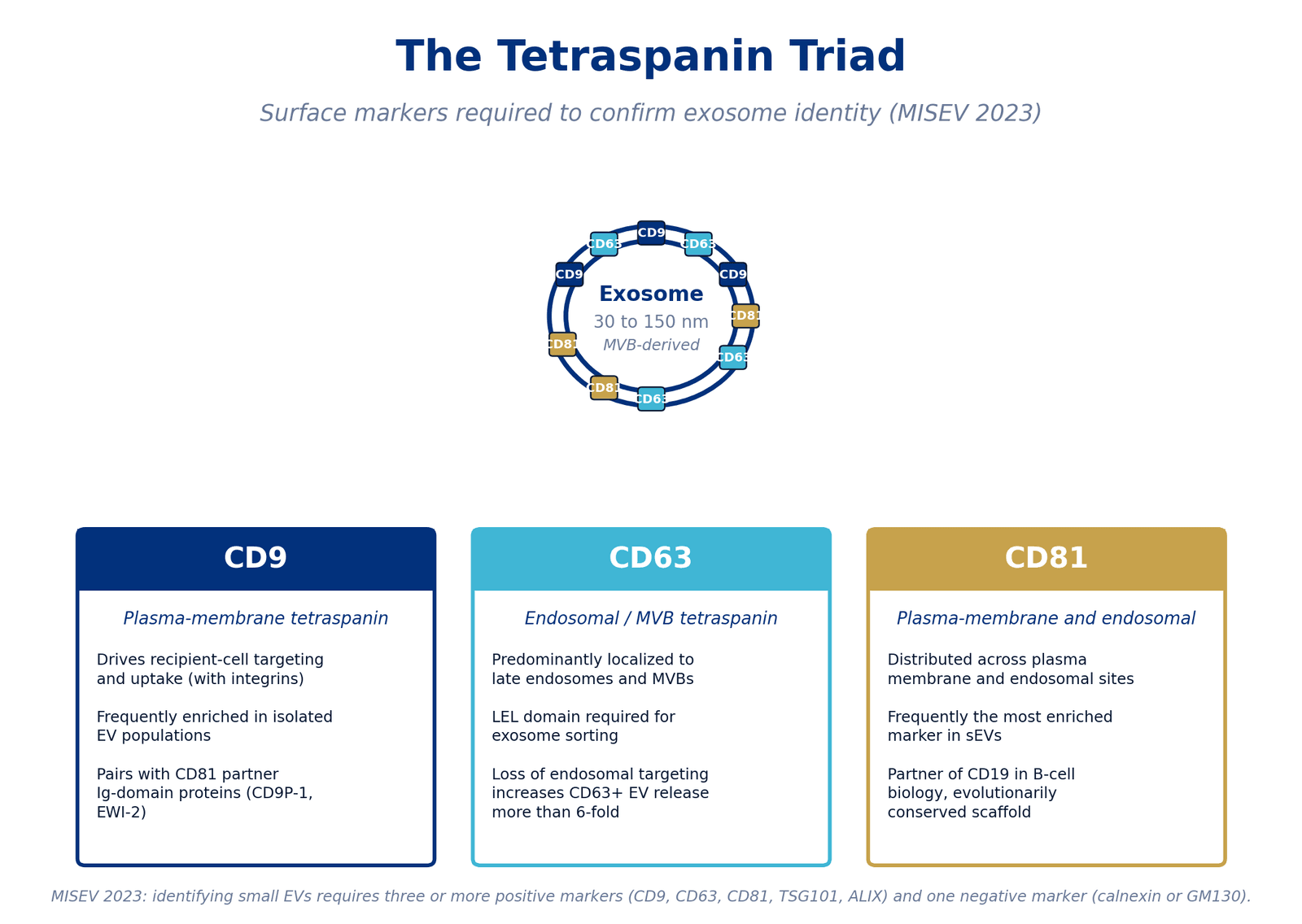
MISEV guidance pairs several positive vesicle markers with at least one negative marker. The tetraspanins are commonly reported alongside cytosolic proteins such as TSG101 or ALIX. Calnexin and GM130 are the negative markers most often reported, and their absence indicates low contamination from cell debris.
Cargo carries the signature of the producing cell
Each vesicle carries proteins, lipids, messenger RNA and microRNA drawn from the cell that made it. Surface markers establish what a vesicle is, while the cargo reflects the biology of its parent cell.
Published work on vesicles from mesenchymal stromal cells finds their microRNA cargo clustering around a small number of signaling pathways. PI3K/Akt, NF-κB, TGF-β and VEGF signaling recur across that literature.
| microRNA | Reported direct targets | Pathway |
|---|---|---|
| miR-21 | TGFBR2 | TGF-β receptor signaling |
| miR-125b | TGFBR1 | TGF-β receptor signaling |
| miR-146a | IRAK1, TRAF6 | NF-κB signaling |
| miR-126 | SPRED1, PIK3R2 | VEGF and PI3K signaling |
| miR-181 | TLR4 axis | Innate immune signaling |
The table records molecular targets reported in the published literature. It describes biology a supplier can disclose about a preparation and makes no statement about what any product does in use.
Cargo varies with tissue source, donor, passage number and culture conditions. A supplier who can name the tissue source and describe the dominant microRNA profile has given you something to compare between lots and between sources.
Source disclosure lets you compare cargo across suppliers
Vesicles from different tissue sources differ measurably. A 2021 proteomic comparison of umbilical cord and adipose tissue preparations found 198 differentially expressed proteins, with immune and complement pathways more strongly enriched in the umbilical cord material.
A 2020 comparison under serum-free and xeno-free conditions found bone marrow preparations most active on dermal fibroblast proliferation in vitro. Umbilical cord preparations in the same study carried TGF-β alongside VEGF-A, FGF-2, HGF and PDGF-BB.
So a generic "MSC exosome" description leaves a first-order variable unstated. Ask for the producing cell type, the tissue of origin, the donor screening criteria and the passage number at harvest.
Twelve checks cover nearly every line on a certificate
Read each line for three things: the method, the result with its unit, and whether it is lot specific. The twelve checks below follow the order most certificates use.
- Cell source: the producing cell type, tissue of origin, donor screening criteria, passage number at harvest and culture media, with xeno-free conditions noted.
- Particle concentration: particles per mL by a named method, with the raw histogram or tracking video available on request.
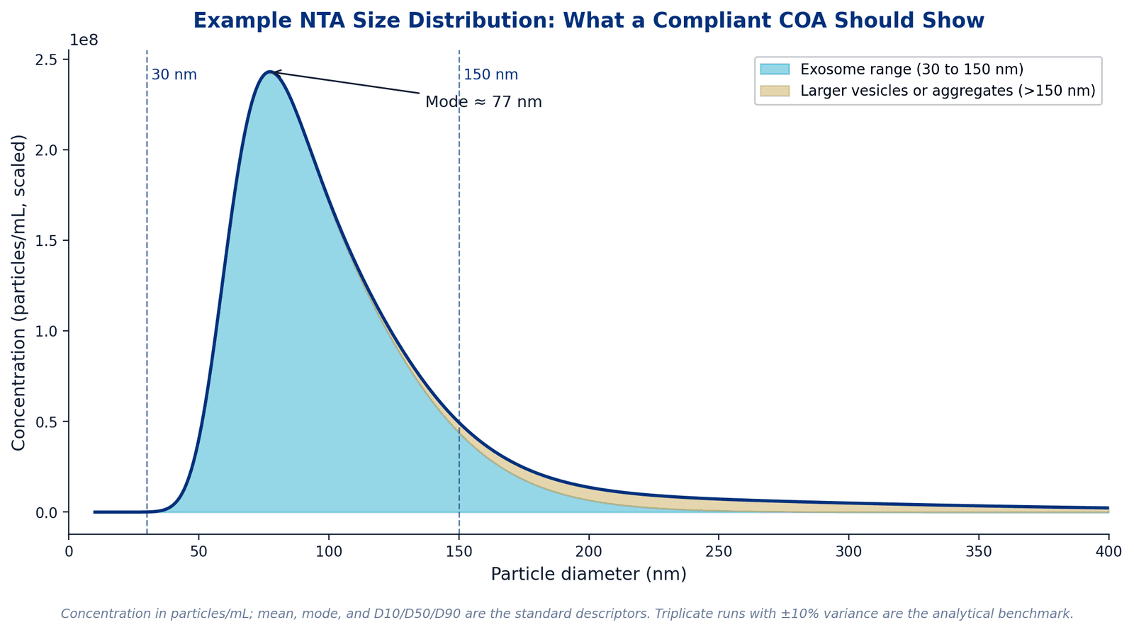
- Size distribution: mean, mode and the D10, D50 and D90 percentiles, with the bulk of particles between 30 and 150 nm.
- Positive markers: several vesicle markers, such as CD9, CD63 and CD81 with TSG101 or ALIX, each with its detection method.
- Negative markers: calnexin or GM130 at minimum, reported alongside the positive panel.
- Sterility: USP <71>, reported as no growth for the specific lot.
- Endotoxin: USP <85> or USP <86>, as a measured value in EU/mL against a stated limit.
- Mycoplasma: EP 2.6.7 by PCR or culture, negative for the specific lot.
- Residual host DNA: a quantified value with its method named.
- Total protein: by BCA or an equivalent assay, which makes the particle-to-protein ratio calculable.
- Storage and stability: the manufacturer’s storage condition, -20°C for long-term storage and 4°C after thawing per its handling instructions, with the stability period validated for it.
- Manufacturing site: named on the document, so its registration status can be checked independently.
The particle-to-protein ratio is the quickest purity signal on the page. A high ratio suggests a clean preparation, while a low one suggests protein carried over from culture media or cell lysate.
A line that misses any of these checks is a reason to ask a question before the material is accepted. Most gaps close with a single request, because the supplier usually holds the data already.
A full size distribution shows what a single number hides
Nanoparticle tracking analysis reports both concentration and size. The distribution is most informative with mean, mode and percentiles, and with any particles above 150 nm quantified rather than left out.
Raw data is a normal request, whether as the histogram or the tracking video. Triplicate runs agreeing within about 10 percent are the usual benchmark for comparing one lot with the next.
What sits alongside the certificate
A complete certificate tells you the material was measured and how. It does not establish potency, it does not establish suitability for any patient or procedure, and it says nothing about a container opened outside the supplier’s control.
That is the correct division. Judging suitability is a prescriber determination on a named patient, and no document substitutes for it.
How to get the documentation
Every document described on this page is one a supplier can send on request, and the request is routine rather than adversarial. Ask during pricing, when it is a normal part of qualifying a source, and a supplier who holds the package will send it within a business day.
ExaVeyra releases lot-specific certificates, characterization data and the pharmacy formulary to practices once NPI verification is complete. Verification is what separates a public catalog from wholesale access, and it is the same step that makes pricing visible.
Sources
- 21 CFR Part 1271, human cells, tissues, and cellular and tissue-based products (accessed 29 August 2026)
- 21 CFR Part 610, General Biological Products Standards (accessed 29 August 2026)
Scientific literature
Each source carries the kind of study it was and, where the study enrolled people, how many. Study design decides what a result can establish, so it is stated rather than left to be inferred. Each line also says what that source is carrying on this page.
- Welsh JA, Goberdhan DCI, O’Driscoll L, et al. Minimal information for studies of extracellular vesicles (MISEV2023): from basic to advanced approaches. Journal of Extracellular Vesicles, 2024. doi:10.1002/jev2.12404 PMID:38326288Consensus statementLandmarkSource 1 supports: The reporting standard a characterization package is usually assembled against. Cited here for the structural point that completeness of reporting is a separate question from the values reported, for its guidance on reporting positive and negative markers together, and for the biogenesis distinction between exosomes and other vesicles.
- Fan Y, et al. Differential proteomics argues against a general role for CD9, CD81 or CD63 in the sorting of proteins into extracellular vesicles. Journal of Extracellular Vesicles, 2023. doi:10.1002/jev2.12352In vitroSource 2 supports: Cited for the partner Ig-domain proteins that travel into vesicles with CD9 and CD81, and for CD81 as frequently the most enriched tetraspanin in small vesicle preparations.
- Jankovičová J, et al. Tetraspanins, More than Markers of Extracellular Vesicles in Reproduction. International Journal of Molecular Sciences, 2020. doi:10.3390/ijms21207568Narrative reviewSource 3 supports: Review of tetraspanin biology. Cited for CD9, CD63 and CD81 as canonical vesicle markers, their membrane microdomains, and the confinement of CD63 to multivesicular bodies.
- Zhao X, et al. Application of mesenchymal stem cell exosomes in the treatment of skin wounds. Smart Materials in Medicine, 2023. doi:10.1016/j.smaim.2023.04.006Narrative reviewSource 4 supports: Review of microRNA cargo in mesenchymal stromal cell vesicles. Cited only for miR-146a and its reported targets IRAK1 and TRAF6 in NF-κB signaling.
- Liu B, et al. Proteomics Analyses Reveal Functional Differences between Exosomes of Mesenchymal Stem Cells Derived from The Umbilical Cord and Those Derived from The Adipose Tissue. Cell Journal, 2021. doi:10.22074/cellj.2021.6969In vitroSource 5 supports: Proteomic comparison of umbilical cord and adipose tissue vesicles. Cited for the 198 differentially expressed proteins that make tissue source a first-order variable.
- Zhang Y, et al. Exosomes derived from human umbilical cord blood mesenchymal stem cells stimulate regenerative wound healing via transforming growth factor-β receptor inhibition. Stem Cell Research & Therapy, 2021. doi:10.1186/s13287-021-02517-0PreclinicalSource 6 supports: Cited only for miR-21-5p and miR-125b-5p as vesicle cargo and their reported targets in TGF-β receptor signaling, TGFBR2 and TGFBR1.
- Hoang DH, et al. Differential Wound Healing Capacity of Mesenchymal Stem Cell-Derived Exosomes Originated From Bone Marrow, Adipose Tissue and Umbilical Cord Under Serum- and Xeno-Free Condition. Frontiers in Molecular Biosciences, 2020. doi:10.3389/fmolb.2020.00119In vitroSource 7 supports: Side-by-side comparison of three tissue sources under serum-free and xeno-free conditions. Cited for source-dependent growth factor content and in vitro fibroblast activity.
- Ti D, et al. Mesenchymal stem cells-derived exosomal microRNAs contribute to wound inflammation. Science China Life Sciences, 2016. doi:10.1007/s11427-016-0240-4Narrative reviewSource 8 supports: Review of microRNA cargo in mesenchymal stromal cell vesicles. Cited for miR-181 and its association with TLR4 signaling.
- Fish JE, et al. miR-126 Regulates Angiogenic Signaling and Vascular Integrity. Developmental Cell, 2008. doi:10.1016/j.devcel.2008.07.008PreclinicalLandmarkSource 9 supports: Foundational study of miR-126 in VEGF signaling. Cited for its reported direct targets SPRED1 and PIK3R2.