CD81: Tetraspanin Structure and the CD19 Co-Receptor
Reviewed by ExaVeyra Sciences editorial team · Updated 19 October 2026
ExaVeyra Sciences is a Miami-based B2B supplier of exosomes for topical aesthetic use to licensed clinics, and it refers licensed prescribers to a 503A compounding pharmacy that prepares formulations against individual prescriptions.
| Question | Answer |
|---|---|
| What this page covers | The first full-length tetraspanin structure, the cholesterol pocket, how CD81 chaperones CD19 in the B cell co-receptor, its T cell synapse role and vesicles. |
| Who can order | Licensed clinics and practitioners whose active professional license and NPI ExaVeyra has verified. Exosome orders also require a signed professional-use acknowledgment. |
| What ships, and how | Exosome material for topical aesthetic use, characterized by lot, ships overnight in a cold chain. Delivery can take 1 to 3 business days. A certificate of analysis is issued for every lot and provided on request. |
CD81 is the tetraspanin whose structure taught the field what this family of proteins looks like.1 It was the first tetraspanin crystallized in full, the first seen with a lipid bound inside it, and the first captured in complex with the partner receptor it escorts to the cell surface.2,3
This page covers those structures, the partnership between CD81 and CD19 in B cells, its part in organizing the T cell synapse and in cell fusion, and its place as one of the three conventional markers on extracellular vesicles.
Understanding the structure of CD81
CD81 is a tetraspanin of 236 amino acids.4 Its large extracellular loop spans residues 113 to 201 and is held by two disulfide bonds, and in 2001 Kitadokoro and colleagues showed that this loop folds into a mushroom-like domain of five helices.5
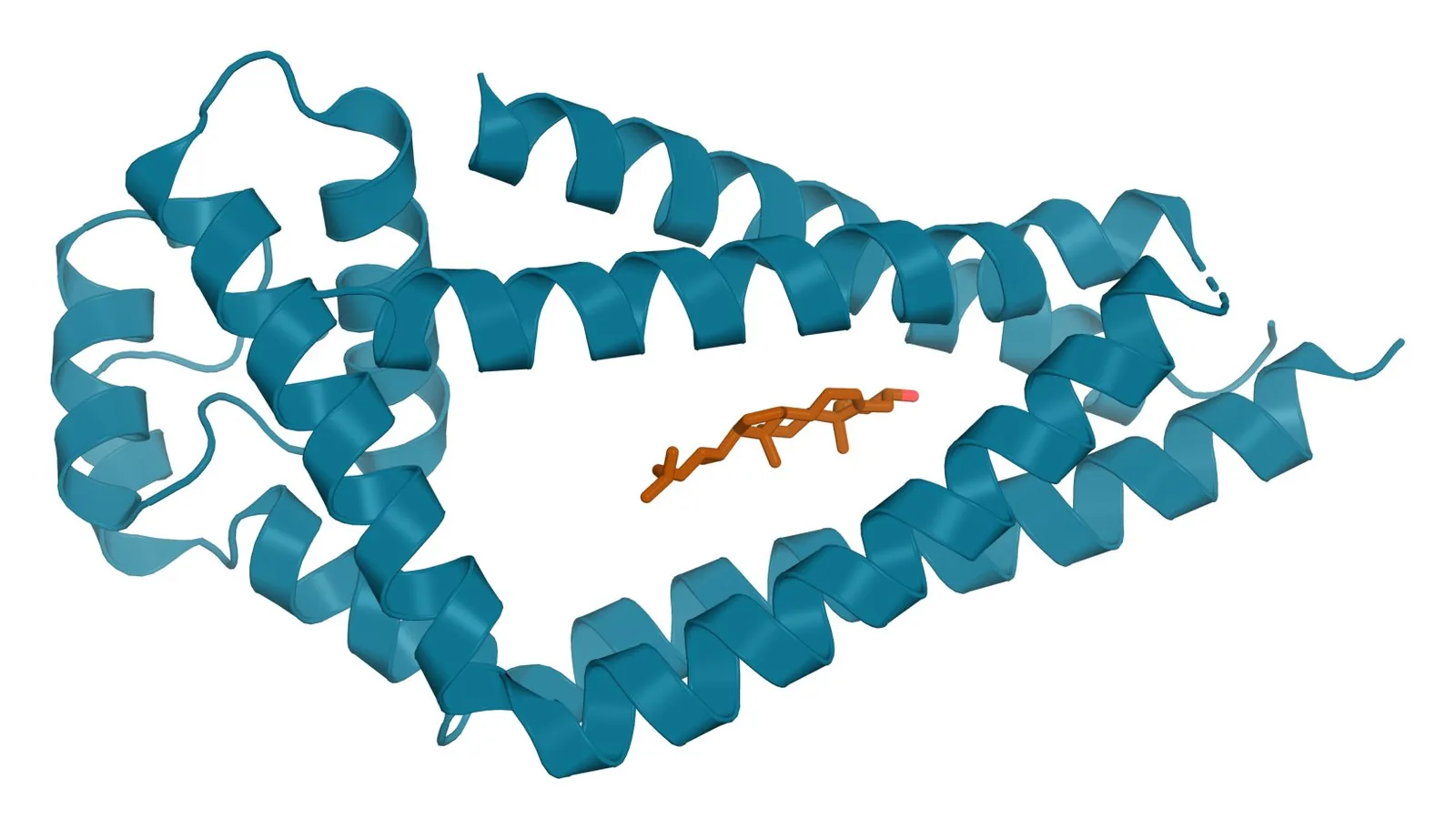
Zimmerman and colleagues reported the full-length crystal structure in 2016 (Fig. 1). The four membrane helices pack as two largely separated pairs, capped at the outer leaflet by the large loop, and they converge at the inner leaflet around a pocket that held a cholesterol molecule.2
Their simulations found a second conformation in which the large loop swings away from the membrane, and cholesterol binding appeared to modulate CD81 activity in cells.2 The structure therefore suggested a switch between closed and open states.
CD81 opens to bind its partners
Yang and colleagues compared CD81 with the tetraspanin CD53, which crystallized in an open state. In the closed conformation of CD81 the large loop rotates toward the membrane, and contacts between the two extracellular loops support the open state needed for partner binding.6
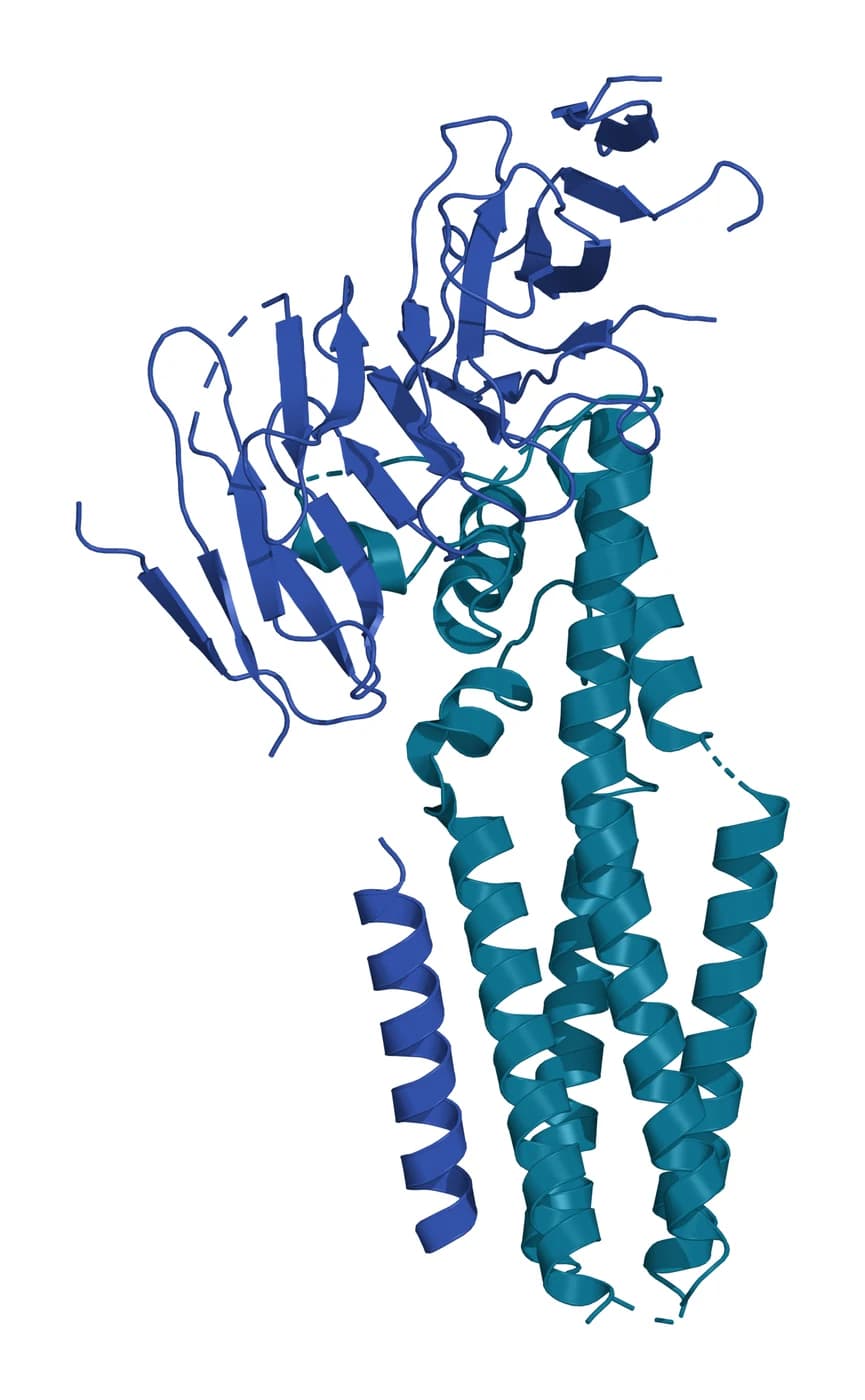
The CD19 complex confirmed the switch directly (Fig. 2). In the cryo-EM structure from Susa and colleagues, CD81 opens its large loop to expose a hydrophobic surface for CD19 and rearranges its membrane helices so that the cholesterol pocket closes (Fig. 3).3
CD81 escorts CD19 in the B cell co-receptor
On B cells, CD81 and CD19 are core subunits of the co-receptor complex that works alongside the B cell receptor (Fig. 4).7 Signaling through this complex is a critical determinant of B cell development and activation.3
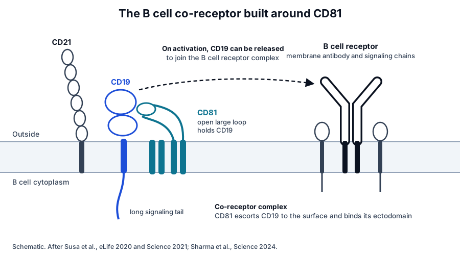
Susa and colleagues showed in 2020 that CD81 uses its ectodomain to traffic CD19 to the cell surface. An antibody that binds only activated B cells recognized an epitope on CD81 that is hidden when CD19 is bound, indicating that the partnership is regulated when the cell is activated.7
Sharma and colleagues identified one regulator. The glycolipid globotriaosylceramide on germinal center B cells binds CD19 and releases it from CD81, allowing CD19 to join the B cell receptor complex and supporting the production of high-affinity antibodies.8
The importance of this partnership was clear before the structures. Maecker and colleagues found in 1998 that mice lacking CD81 on B cells had reduced interleukin 4 and antibody production in T helper type 2 responses.10
CD81 organizes the T cell immune synapse
When a T cell recognizes an antigen-presenting cell, the two form an organized contact called the immune synapse. Rocha-Perugini and colleagues found that CD81 interacts with the adhesion molecule ICAM-1 and with CD3 of the T cell receptor at that contact.11
Those interactions increased as the synapse matured and defined its sequential stages. CD81 controlled how long CD3 stayed in the contact area, sustaining the activation signal.11
Super-resolution imaging adds the spatial picture. Zuidscherwoude and colleagues found that CD81 forms its own nanoclusters on B cells and dendritic cells, sitting closer to its partner CD19 than to other tetraspanins.12
CD81 and CD9 share roles in cell fusion
CD81 works alongside CD9 at the oocyte surface. Rubinstein and colleagues found that female mice lacking CD81 had about 40 percent lower fertility because their oocytes fused poorly with sperm, and mice lacking both CD81 and CD9 were completely infertile.13
In muscle, Charrin and colleagues found that CD9 and CD81 associate with their shared partner EWI-F and keep myotube fusion in check during regeneration.14 Stipp and colleagues had earlier shown that EWI-2 binds CD81 and CD9 directly and in high proportion.15
Macrophages follow the same pattern. Takeda and colleagues found that macrophages lacking both CD81 and CD9 formed multinucleated cells more readily, indicating that the two tetraspanins normally restrain macrophage fusion.16
CD81 on extracellular vesicles
CD81 is one of the three tetraspanins most enriched on small extracellular vesicles.17 Kowal and colleagues proposed CD81-based immuno-isolation as one way to separate vesicle subpopulations,18 and single-vesicle platforms resolve how many vesicles carry it.
Those platforms can follow normal physiology. McIlvenna and colleagues imaged single vesicles in plasma from healthy volunteers and found more CD9, CD63 and CD81 positive vesicles in circulation after high-intensity interval exercise.19
Cell biology studies explain part of the variation. Ai and colleagues showed that endocytosis reduces vesicular release of CD81 and directs it toward degradation,20 and Breitwieser and colleagues found that tetraspanin profiles of stromal cell vesicles shift with culture conditions.21
Where to go from here
The CD9 reference covers the closest relative of CD81, and the tetraspanin family page explains the shared fold and the tetraspanin web. The quality reference on marker panels explains how the share of vesicles carrying CD81 is measured.
Verified practices can request lot-specific characterization data with the certificate of analysis. Our team can explain what a CD81 result on a panel represents and how it was measured.
References
Numbered in the order they are cited in the text. Each study carries its design and, where people were enrolled, how many, with a line saying what it supports on this page. Primary sources are regulations, agency documents and reference databases.
- 1.Susa KJ, Kruse AC, Blacklow SC. Tetraspanins: structure, dynamics, and principles of partner-protein recognition. Trends in Cell Biology. 2024. doi:10.1016/j.tcb.2023.09.003. PMID: 37783654.Narrative reviewReference 1 supports: Review of tetraspanin structures and partner recognition. Cited for the place of the CD81 structures in the wider family.
- 2.Zimmerman B, Kelly B, McMillan BJ, et al. Crystal Structure of a Full-Length Human Tetraspanin Reveals a Cholesterol-Binding Pocket. Cell. 2016. doi:10.1016/j.cell.2016.09.056. PMID: 27881302.In vitroReference 2 supports: The full-length CD81 crystal structure. Cited for the two helix pairs, the cholesterol pocket and the open conformation seen in simulations.
- 3.Susa KJ, Rawson S, Kruse AC, et al. Cryo-EM structure of the B cell co-receptor CD19 bound to the tetraspanin CD81. Science. 2021. doi:10.1126/science.abd9836. PMID: 33446559.In vitroReference 3 supports: Cryo-EM structure of the CD19 and CD81 complex. Cited for the opening of the large loop and the closure of the cholesterol pocket on CD19 binding.
- 4.NCBI Gene 975, CD81 molecule (Homo sapiens). Accessed 19 October 2026.Primary source
- 5.Kitadokoro K, Bordo D, Galli G, et al. CD81 extracellular domain 3D structure: insight into the tetraspanin superfamily structural motifs. EMBO Journal. 2001. doi:10.1093/emboj/20.1.12. PMID: 11226150.In vitroLandmarkReference 5 supports: The first structure of a tetraspanin large loop. Cited for the five-helix mushroom-like fold of the CD81 large extracellular loop.
- 6.Yang Y, Liu XR, Greenberg ZJ, et al. Open conformation of tetraspanins shapes interaction partner networks on cell membranes. EMBO Journal. 2020. doi:10.15252/embj.2020105246. PMID: 32974937.In vitroReference 6 supports: Structural comparison of CD53 and CD81. Cited for the closed conformation of CD81 and the loop contacts that support the open state.
- 7.Susa KJ, Seegar TC, Blacklow SC, et al. A dynamic interaction between CD19 and the tetraspanin CD81 controls B cell co-receptor trafficking. eLife. 2020. doi:10.7554/eLife.52337. PMID: 32338599.In vitroReference 7 supports: Trafficking and antibody epitope studies. Cited for CD81 using its ectodomain to carry CD19 to the surface and for the activation-regulated interaction.
- 8.Sharma P, Zhang X, Ly K, et al. The lipid globotriaosylceramide promotes germinal center B cell responses and antiviral immunity. Science. 2024. doi:10.1126/science.adg0564. PMID: 38359115.PreclinicalReference 8 supports: Germinal center B cell studies. Cited for globotriaosylceramide releasing CD19 from CD81 so that CD19 can join the B cell receptor complex.
- 9.Termini CM, Gillette JM. Tetraspanins Function as Regulators of Cellular Signaling. Frontiers in Cell and Developmental Biology. 2017. doi:10.3389/fcell.2017.00034. PMID: 28428953.Narrative reviewReference 9 supports: Review of tetraspanin signaling scaffolds. Cited for the general role of tetraspanins such as CD81 in organizing signaling partners.
- 10.Maecker HT, Do MS, Levy S. CD81 on B cells promotes interleukin 4 secretion and antibody production during T helper type 2 immune responses. Proceedings of the National Academy of Sciences of the United States of America. 1998. doi:10.1073/pnas.95.5.2458. PMID: 9482907.PreclinicalLandmarkReference 10 supports: Immune responses in CD81-null mice. Cited for the requirement of B cell CD81 for optimal interleukin 4 and antibody production.
- 11.Rocha-Perugini V, Zamai M, González-Granado JM, et al. CD81 controls sustained T cell activation signaling and defines the maturation stages of cognate immunological synapses. Molecular and Cellular Biology. 2013. doi:10.1128/MCB.00302-13. PMID: 23858057.In vitroReference 11 supports: Quantitative imaging of T cell and antigen-presenting cell contacts. Cited for CD81 interacting with ICAM-1 and CD3 and sustaining CD3 at the synapse.
- 12.Zuidscherwoude M, Göttfert F, Dunlock VM, et al. The tetraspanin web revisited by super-resolution microscopy. Scientific Reports. 2015. doi:10.1038/srep12201. PMID: 26183063.In vitroReference 12 supports: STED imaging of tetraspanin nanoclusters. Cited for CD81 clusters lying closer to CD19 than to other tetraspanins.
- 13.Rubinstein E, Ziyyat A, Prenant M, et al. Reduced fertility of female mice lacking CD81. Developmental Biology. 2006. doi:10.1016/j.ydbio.2005.11.031. PMID: 16380109.PreclinicalLandmarkReference 13 supports: Fertility of CD81-null and CD9 and CD81 double-null mice. Cited for the 40 percent reduction in female fertility and complete infertility without both tetraspanins.
- 14.Charrin S, Latil M, Soave S, et al. Normal muscle regeneration requires tight control of muscle cell fusion by tetraspanins CD9 and CD81. Nature Communications. 2013. doi:10.1038/ncomms2675. PMID: 23575678.PreclinicalReference 14 supports: Mouse regeneration studies and myoblast culture. Cited for CD81 and CD9 associating with EWI-F and keeping myotube fusion in check.
- 15.Stipp CS, Kolesnikova TV, Hemler ME. EWI-2 is a major CD9 and CD81 partner and member of a novel Ig protein subfamily. Journal of Biological Chemistry. 2001. doi:10.1074/jbc.M107338200. PMID: 11504738.In vitroLandmarkReference 15 supports: Identification of EWI-2. Cited for its direct, high-proportion association with CD81 and CD9.
- 16.Takeda Y, Tachibana I, Miyado K, et al. Tetraspanins CD9 and CD81 function to prevent the fusion of mononuclear phagocytes. Journal of Cell Biology. 2003. doi:10.1083/jcb.200212031. PMID: 12796480.PreclinicalLandmarkReference 16 supports: Macrophage fusion in CD9- and CD81-null mice. Cited for the shared role of the two tetraspanins in restraining macrophage fusion.
- 17.Rubinstein E, Théry C, Zimmermann P. Tetraspanins affect membrane structures and the trafficking of molecular partners: what impact on extracellular vesicles? Biochemical Society Transactions. 2025. doi:10.1042/BST20240523. PMID: 40135387.Narrative reviewReference 17 supports: Review of tetraspanins on vesicles. Cited for CD81 as one of the three widely used vesicle markers.
- 18.Kowal J, Arras G, Colombo M, et al. Proteomic comparison defines novel markers to characterize heterogeneous populations of extracellular vesicle subtypes. Proceedings of the National Academy of Sciences of the United States of America. 2016. doi:10.1073/pnas.1521230113. PMID: 26858453.In vitroReference 18 supports: Proteomics of vesicle subtypes. Cited for CD81-based immuno-isolation of vesicle subpopulations.
- 19.McIlvenna LC, Parker HJ, Seabright AP, et al. Single vesicle analysis reveals the release of tetraspanin positive extracellular vesicles into circulation with high intensity intermittent exercise. Journal of Physiology. 2023. doi:10.1113/JP284047. PMID: 36855276.Clinical study7 participantsReference 19 supports: Single-vesicle imaging of plasma from seven healthy volunteers. Cited for the rise in CD9, CD63 and CD81 positive vesicles in circulation after high-intensity exercise.
- 20.Ai Y, Guo C, Garcia-Contreras M, et al. Endocytosis blocks the vesicular secretion of exosome marker proteins. Science Advances. 2024. doi:10.1126/sciadv.adi9156. PMID: 38718108.In vitroReference 20 supports: Cell biology of tetraspanin marker secretion. Cited for endocytosis reducing vesicular release of CD81 and directing it to degradation.
- 21.Breitwieser K, Koch LF, Tertel T, et al. Detailed Characterization of Small Extracellular Vesicles from Different Cell Types Based on Tetraspanin Composition by ExoView R100 Platform. International Journal of Molecular Sciences. 2022. doi:10.3390/ijms23158544. PMID: 35955677.In vitroReference 21 supports: Single-vesicle tetraspanin profiling. Cited for stromal cell vesicle tetraspanin profiles that shift with culture conditions.
Figure sources
- 22.PDB 5TCX, Tetraspanin CD81 with cholesterol. ExaVeyra Sciences, rendered in PyMOL from PDB 5TCX. Structure data: RCSB Protein Data Bank (rcsb.org), CC0. Structure publication: Zimmerman B, Kelly B, McMillan BJ, et al. Crystal structure of a full-length human tetraspanin reveals a cholesterol-binding pocket. Cell. 2016;167(4):1041-1051.e11.
- 23.PDB 7JIC, CD19 bound to tetraspanin CD81. ExaVeyra Sciences, rendered in PyMOL from PDB 7JIC. Structure data: RCSB Protein Data Bank (rcsb.org), CC0. Structure publication: Susa KJ, Rawson S, Kruse AC, Blacklow SC. Cryo-EM structure of the B cell co-receptor CD19 bound to the tetraspanin CD81. Science. 2021;371(6526):300-305.