CD9: Structure, Partners and Biological Functions
Reviewed by ExaVeyra Sciences editorial team · Updated 19 October 2026
ExaVeyra Sciences is a Miami-based B2B supplier of exosomes for topical aesthetic use to licensed clinics, and it refers licensed prescribers to a 503A compounding pharmacy that prepares formulations against individual prescriptions.
| Question | Answer |
|---|---|
| What this page covers | The CD9 crystal structure and its curvature-sensing cone shape, its EWI-2, EWI-F and integrin partners, its role in gamete fusion, and its place on vesicles. |
| Who can order | Licensed clinics and practitioners whose active professional license and NPI ExaVeyra has verified. Exosome orders also require a signed professional-use acknowledgment. |
| What ships, and how | Exosome material for topical aesthetic use, characterized by lot, ships overnight in a cold chain. Delivery can take 1 to 3 business days. A certificate of analysis is issued for every lot and provided on request. |
CD9 is a tetraspanin of 228 amino acids found on the surface of most human cell types, from oocytes and platelets to stromal cells.1 Its crystal structure explains much of what it does: a cone-shaped protein that settles into curved membrane and gathers a defined set of partners around it.2
This page covers that structure, the partner proteins CD9 binds, its essential role in fertilization, how it moderates adhesion and cell fusion, and why it is one of the three conventional markers of extracellular vesicles.
Understanding the structure of CD9
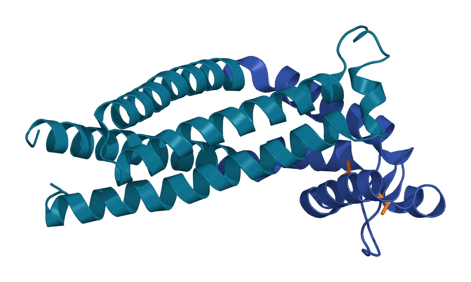
CD9 follows the tetraspanin plan of four membrane helices with a small and a large extracellular loop. Its large loop runs from about residue 112 to 195 and is held together by two disulfide bonds, and the protein carries palmitate on several cysteines next to its membrane helices.1
Umeda and colleagues solved the crystal structure of CD9 in 2020 (Fig. 1). The protein adopts a reversed cone shape that bent the crystalline lipid layers around it, which explains its enrichment in regions of high membrane curvature.2
Dharan and colleagues later measured that preference directly. In membrane tubes pulled from cell-derived vesicles, CD9 partitioned toward positively curved membrane, the geometry of microvilli and small vesicles.3
EWI-2 and EWI-F are the closest partners of CD9
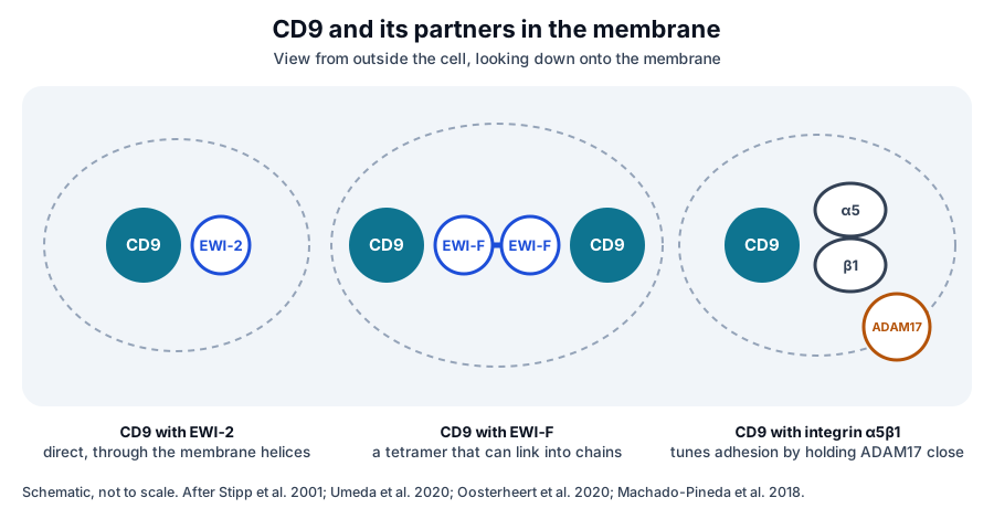
Stipp and colleagues identified EWI-2 as a major partner of CD9 and CD81 in 2001. About 70 percent of cellular CD9 and CD81 was bound to it in the cell line they studied, and the two proteins could be cross-linked directly.4
EWI-2 and its relative EWI-F belong to a small immunoglobulin subfamily that shares a glutamate, tryptophan, isoleucine motif.4 The cryo-EM structure from Umeda and colleagues showed that CD9 binds EWI-2 mainly through small residues in the transmembrane region.2
Oosterheert and colleagues found that CD9 and EWI-F assemble as a tetramer, two EWI-F molecules joined through their ectodomains with one CD9 bound to each (Fig. 2). That arrangement suggests how short chains of tetraspanin complexes could form the larger microdomain network.5
CD9 is essential for sperm and egg fusion
The best-defined physiological role of CD9 is in fertilization.6 Le Naour and colleagues showed in 2000 that oocytes from CD9-deficient female mice were ovulated normally but did not fuse with sperm, and the affected females had severely reduced fertility.7
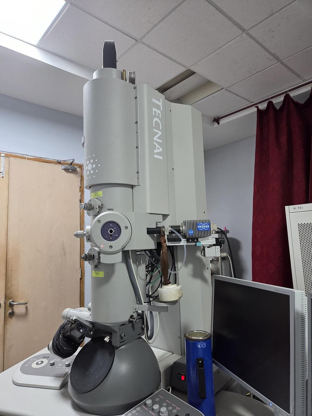
Later studies traced this to the oocyte surface. CD9-deficient oocytes have shorter and sparser microvilli (Fig. 3),8 and Inoue and colleagues found that CD9 also confines GPI-anchored proteins such as the sperm receptor JUNO to the region of the oocyte where fusion occurs.9
CD9 continues to act once sperm binds. Wang and colleagues showed that CD9 concentrates at the binding site and is needed for the kinase activation and actin remodeling that follow,10 and Cohen and colleagues linked the CD9 partners EWI-2, EWI-F and the ERM proteins to the same process.11
CD9 tunes adhesion through integrins
CD9 associates with integrins of the beta 1, beta 2 and beta 3 subfamilies.13 Machado-Pineda and colleagues found that it moderates adhesion through integrin alpha 5 beta 1 by strengthening the integrin’s side-by-side association with the enzyme ADAM17.14
Reyes and colleagues showed that CD9 also binds the leukocyte integrin LFA-1 through its large extracellular loop. Rather than changing the integrin’s affinity, CD9 changed how LFA-1 molecules cluster on the cell surface.13
In blood-forming stem and progenitor cells from cord blood, Leung and colleagues found that CD9 levels rise in response to the chemokine SDF-1. Blocking CD9 reduced migration toward SDF-1, placing CD9 within the signaling that guides these cells to the marrow.15
CD9 and CD81 keep cell fusion in balance
Outside fertilization, CD9 often works to limit fusion rather than promote it. Takeda and colleagues showed that macrophages lacking CD9 and CD81 formed multinucleated cells more readily, indicating that the two tetraspanins normally restrain macrophage fusion.16
Muscle shows the same principle. Charrin and colleagues found that CD9 and CD81 associate with EWI-F in myoblasts and that muscle regeneration in mice depends on these proteins keeping myotube fusion in check.17
CD9 on extracellular vesicles
CD9 is one of the most consistently detected proteins on small extracellular vesicles.18 Kowal and colleagues proposed immuno-isolation with CD9, CD63 or CD81 to separate vesicle subpopulations, after finding that several older markers were present on all vesicle types (Fig. 4).19
Mathieu and colleagues followed CD9 and CD63 inside living cells. CD9 resides mainly at the plasma membrane and CD63 in late endosomes, and CD9 was released in vesicles more abundantly, consistent with budding directly from the cell surface.20
Ai and colleagues added that endocytosis reduces vesicular release of CD9 and directs it toward degradation.21 Together these studies explain why the CD9 content of a vesicle preparation reflects both the cell type and the route by which its vesicles were made.
Where to go from here
The CD81 reference covers the closest relative of CD9, and the CD63 reference covers the endosomal tetraspanin it is most often compared with. The quality reference on marker panels explains how the share of vesicles carrying CD9 is measured.
Verified practices can request lot-specific characterization data with the certificate of analysis. Our team can explain what a CD9 result on a panel represents and how it was measured.
References
Numbered in the order they are cited in the text. Each study carries its design and, where people were enrolled, how many, with a line saying what it supports on this page. Primary sources are regulations, agency documents and reference databases.
- 1.NCBI Gene 928, CD9 molecule (Homo sapiens). Accessed 19 October 2026.Primary source
- 2.Umeda R, Satouh Y, Takemoto M, et al. Structural insights into tetraspanin CD9 function. Nature Communications. 2020. doi:10.1038/s41467-020-15459-7. PMID: 32231207.In vitroReference 2 supports: Crystal structure of CD9 and cryo-EM of CD9 with EWI-2. Cited for the reversed cone shape, the transmembrane binding of EWI-2 and the role of the large loop in gamete fusion.
- 3.Dharan R, Goren S, Cheppali SK, et al. Transmembrane proteins tetraspanin 4 and CD9 sense membrane curvature. Proceedings of the National Academy of Sciences of the United States of America. 2022. doi:10.1073/pnas.2208993119. PMID: 36252000.In vitroReference 3 supports: Membrane tube measurements of curvature sensing. Cited for the preference of CD9 for positively curved membrane.
- 4.Stipp CS, Kolesnikova TV, Hemler ME. EWI-2 is a major CD9 and CD81 partner and member of a novel Ig protein subfamily. Journal of Biological Chemistry. 2001. doi:10.1074/jbc.M107338200. PMID: 11504738.In vitroLandmarkReference 4 supports: Identification of EWI-2. Cited for about 70 percent of CD9 and CD81 bound to it, for direct cross-linking, and for the EWI immunoglobulin subfamily.
- 5.Oosterheert W, Xenaki KT, Neviani V, et al. Implications for tetraspanin-enriched microdomain assembly based on structures of CD9 with EWI-F. Life Science Alliance. 2020. doi:10.26508/lsa.202000883. PMID: 32958604.In vitroReference 5 supports: Structures of CD9 with EWI-F. Cited for the tetrameric complex and the concatenation model of microdomain assembly.
- 6.Jankovičová J, Simon M, Antalíková J, et al. Role of tetraspanin CD9 molecule in fertilization of mammals. Physiological Research. 2015. doi:10.33549/physiolres.932876. PMID: 25536312.Narrative reviewReference 6 supports: Review of CD9 across the steps of fertilization. Cited for its abundance on the oocyte membrane and in perivitelline vesicles.
- 7.Le Naour F, Rubinstein E, Jasmin C, et al. Severely reduced female fertility in CD9-deficient mice. Science. 2000. doi:10.1126/science.287.5451.319. PMID: 10634790.PreclinicalLandmarkReference 7 supports: The knockout study that established the role of CD9 in fertilization. Cited for normal ovulation with failed sperm and oocyte fusion.
- 8.Benammar A, Ziyyat A, Lefèvre B, et al. Tetraspanins and Mouse Oocyte Microvilli Related to Fertilizing Ability. Reproductive Sciences. 2017. doi:10.1177/1933719116678688. PMID: 27879451.PreclinicalReference 8 supports: Electron microscopy of oocyte microvilli in tetraspanin-deleted mice. Cited for reduced microvillus density on CD9-deficient oocytes.
- 9.Inoue N, Saito T, Wada I. Unveiling a novel function of CD9 in surface compartmentalization of oocytes. Development. 2020. doi:10.1242/dev.189985. PMID: 32665248.PreclinicalReference 9 supports: Analysis of CD9- and JUNO-disrupted mouse oocytes. Cited for CD9 confining GPI-anchored proteins, including JUNO, to the fusion-competent oocyte surface.
- 10.Wang H, Hong X, Kinsey WH. Sperm-oocyte signaling: the role of IZUMO1R and CD9 in PTK2B activation and actin remodeling at the sperm binding site. Biology of Reproduction. 2021. doi:10.1093/biolre/ioab048. PMID: 33724343.PreclinicalReference 10 supports: Imaging and knockout oocytes at the sperm binding site. Cited for CD9 concentrating there and driving kinase activation and actin remodeling.
- 11.Cohen J, Wang L, Marques S, et al. Oocyte ERM and EWI Proteins Are Involved in Mouse Fertilization. Frontiers in Cell and Developmental Biology. 2022. doi:10.3389/fcell.2022.863729. PMID: 35359433.PreclinicalReference 11 supports: siRNA inhibition of EWI and ERM proteins in mouse oocytes. Cited for the link between CD9 partners, the cortical cytoskeleton and fertilization.
- 12.Inoue N. Molecular mechanisms leading to gamete fusion. Fukushima Journal of Medical Science. 2025. doi:10.5387/fms.24-00046. PMID: 40058809.Narrative reviewReference 12 supports: Current review of gamete fusion factors. Cited for CD9 as the first oocyte fusion factor identified, followed by IZUMO1 and JUNO.
- 13.Reyes R, Monjas A, Yánez-Mó M, et al. Different states of integrin LFA-1 aggregation are controlled through its association with tetraspanin CD9. Biochimica et Biophysica Acta. 2015. doi:10.1016/j.bbamcr.2015.05.018. PMID: 26003300.In vitroReference 13 supports: Biochemistry and imaging of CD9 with LFA-1 on leukocytes. Cited for the large loop interaction and for CD9 acting on integrin clustering rather than affinity.
- 14.Machado-Pineda Y, Cardeñes B, Reyes R, et al. CD9 Controls Integrin α5β1-Mediated Cell Adhesion by Modulating Its Association With the Metalloproteinase ADAM17. Frontiers in Immunology. 2018. doi:10.3389/fimmu.2018.02474. PMID: 30455686.In vitroReference 14 supports: Adhesion assays on integrin alpha 5 beta 1. Cited for CD9 moderating adhesion by strengthening the cis association of the integrin with ADAM17.
- 15.Leung KT, Chan KY, Ng PC, et al. The tetraspanin CD9 regulates migration, adhesion, and homing of human cord blood CD34+ hematopoietic stem and progenitor cells. Blood. 2011. doi:10.1182/blood-2010-04-281329. PMID: 21063023.In vitroReference 15 supports: Transcriptome and migration studies of cord blood progenitors. Cited for SDF-1 raising CD9 levels and CD9 blockade reducing migration.
- 16.Takeda Y, Tachibana I, Miyado K, et al. Tetraspanins CD9 and CD81 function to prevent the fusion of mononuclear phagocytes. Journal of Cell Biology. 2003. doi:10.1083/jcb.200212031. PMID: 12796480.PreclinicalLandmarkReference 16 supports: Macrophage fusion in CD9- and CD81-null mice. Cited for the two tetraspanins restraining macrophage fusion.
- 17.Charrin S, Latil M, Soave S, et al. Normal muscle regeneration requires tight control of muscle cell fusion by tetraspanins CD9 and CD81. Nature Communications. 2013. doi:10.1038/ncomms2675. PMID: 23575678.PreclinicalReference 17 supports: Mouse regeneration studies and myoblast culture. Cited for CD9 and CD81 associating with EWI-F and keeping myotube fusion in check.
- 18.Andreu Z, Yáñez-Mó M. Tetraspanins in extracellular vesicle formation and function. Frontiers in Immunology. 2014. doi:10.3389/fimmu.2014.00442. PMID: 25278937.Narrative reviewReference 18 supports: Review of tetraspanins in vesicle biology. Cited for tetraspanins as among the most abundant membrane proteins of extracellular vesicles.
- 19.Kowal J, Arras G, Colombo M, et al. Proteomic comparison defines novel markers to characterize heterogeneous populations of extracellular vesicle subtypes. Proceedings of the National Academy of Sciences of the United States of America. 2016. doi:10.1073/pnas.1521230113. PMID: 26858453.In vitroReference 19 supports: Quantitative proteomics of separated vesicle populations. Cited for immuno-isolation with CD9, CD63 or CD81 to resolve vesicle subtypes.
- 20.Mathieu M, Névo N, Jouve M, et al. Specificities of exosome versus small ectosome secretion revealed by live intracellular tracking of CD63 and CD9. Nature Communications. 2021. doi:10.1038/s41467-021-24384-2. PMID: 34282141.In vitroReference 20 supports: Live tracking of CD9 and CD63 from synthesis to residence. Cited for CD9 residing at the plasma membrane and being released in vesicles more abundantly than CD63.
- 21.Ai Y, Guo C, Garcia-Contreras M, et al. Endocytosis blocks the vesicular secretion of exosome marker proteins. Science Advances. 2024. doi:10.1126/sciadv.adi9156. PMID: 38718108.In vitroReference 21 supports: Cell biology of tetraspanin marker secretion. Cited for endocytosis reducing vesicular release of CD9 and directing it to degradation.
Figure sources
- 22.PDB 6K4J, Tetraspanin CD9. ExaVeyra Sciences, rendered in PyMOL from PDB 6K4J. Structure data: RCSB Protein Data Bank (rcsb.org), CC0. Structure publication: Umeda R, Satouh Y, Takemoto M, et al. Structural insights into tetraspanin CD9 function. Nat Commun. 2020;11(1):1606.