Esta referencia está disponible actualmente en inglés. La versión en español está en preparación.
Exosome vs Extracellular Vesicle: MISEV2023 Nomenclature
Por Benn Bluestein-Veyra, M.Sc. en Química Orgánica · Actualizado el 20 de octubre de 2026
ExaVeyra Sciences es un proveedor B2B con sede en Miami que suministra exosomas para uso estético tópico a clínicas con licencia, y remite a los prescriptores con licencia a una farmacia de preparados compuestos 503A que prepara formulaciones según prescripciones individuales.
| Pregunta | Respuesta |
|---|---|
| Qué cubre esta página | How MISEV2023 names extracellular vesicles: exosomes, ectosomes and microvesicles by biogenesis, small and large EVs by size, and when the word exosome applies. |
| Quién puede hacer pedidos | Clínicas y profesionales con licencia profesional activa y NPI verificados por ExaVeyra. Los pedidos de exosomas requieren además un reconocimiento firmado de uso profesional. |
| Qué se envía y cómo | El material de exosomas para uso estético tópico, caracterizado por lote, se envía en cadena de frío por exprés nocturno. La entrega puede tardar 1 a 3 días hábiles. Se emite un certificado de análisis para cada lote y se entrega a solicitud. |
The question of exosome vs extracellular vesicle comes down to evidence. Extracellular vesicle (EV) is the generic term for particles released from cells that are delimited by a lipid bilayer and cannot replicate on their own, and an exosome is the subset of EVs that originates in the multivesicular body.[1]
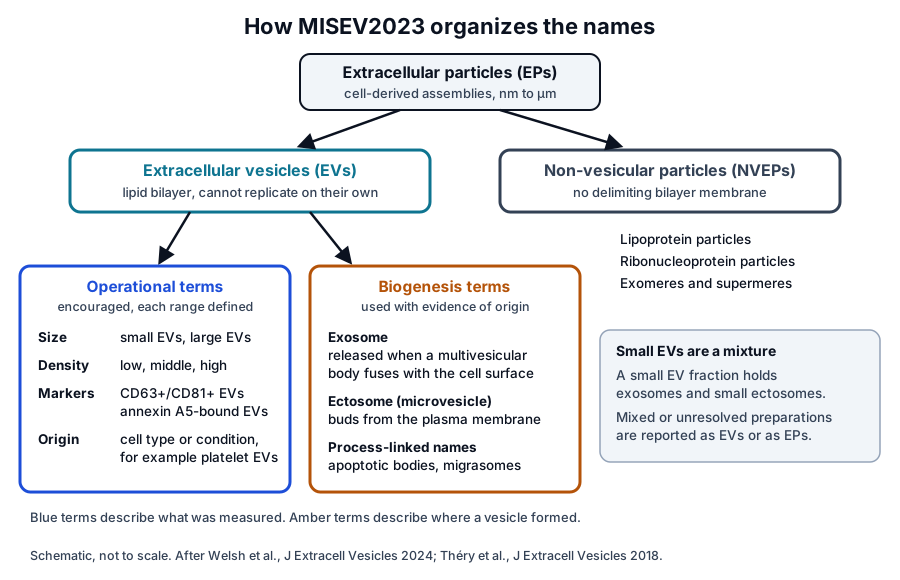
The guidelines of the International Society for Extracellular Vesicles (ISEV) reserve biogenesis names for preparations whose origin has been demonstrated. Other preparations are named with operational terms for size, density, composition or source.[1][2]
Extracellular vesicle is the generic term for bilayer-bounded particles released by cells
MISEV2023 kept the 2018 definition of an EV and removed the word "naturally" from it, so that EVs from engineered cells or varied culture conditions also fit. ISEV recommends the generic term and its operational extensions over terms tied to biogenesis pathways that are difficult to establish.[1]
The guidelines place EVs inside a wider category. Extracellular particles (EPs) are cell-derived multimolecular assemblies in the nanometer to micrometer range, and they include both EVs and non-vesicular extracellular particles (NVEPs) that lack a delimiting bilayer (Fig. 1).[1]
The National Library of Medicine follows the same hierarchy. Its Medical Subject Headings introduced Extracellular Vesicles as a descriptor in 2016 and placed Exosomes and Cell-Derived Microparticles beneath it, with apoptotic bodies listed as an entry term.[3]
The word exosome entered vesicle biology through reticulocyte studies
The word has several earlier meanings. Witwer and Théry traced "exosome" to papers from 1970 to 1973 that used it for mobile DNA fragments, and the same word later named an RNA-processing complex built around exonucleases.[4] MeSH still cross-references that ribonuclease complex from its Exosomes record.[5]
Trams and colleagues first applied the term to vesicles in 1981. Cultured cells exfoliated vesicles of 500 to 1000 nm that carried ecto-5′-nucleotidase activity and often contained a second population of about 40 nm, and the authors proposed calling these exfoliated vesicles exosomes.[6]
The endosomal meaning came from maturing red cells. Pan and Johnstone found in 1983 that sheep reticulocytes released the transferrin receptor selectively, in vesicles that carried few other membrane proteins.[7] The receptor, encoded by TFRC, imports iron-loaded transferrin through clathrin-mediated endocytosis.[8][9]
Harding, Heuser and Stahl followed gold-labeled transferrin in rat reticulocytes the same year. The label reached multivesicular endosomes, which appeared to fuse with the plasma membrane and release their contents by exocytosis.[10]
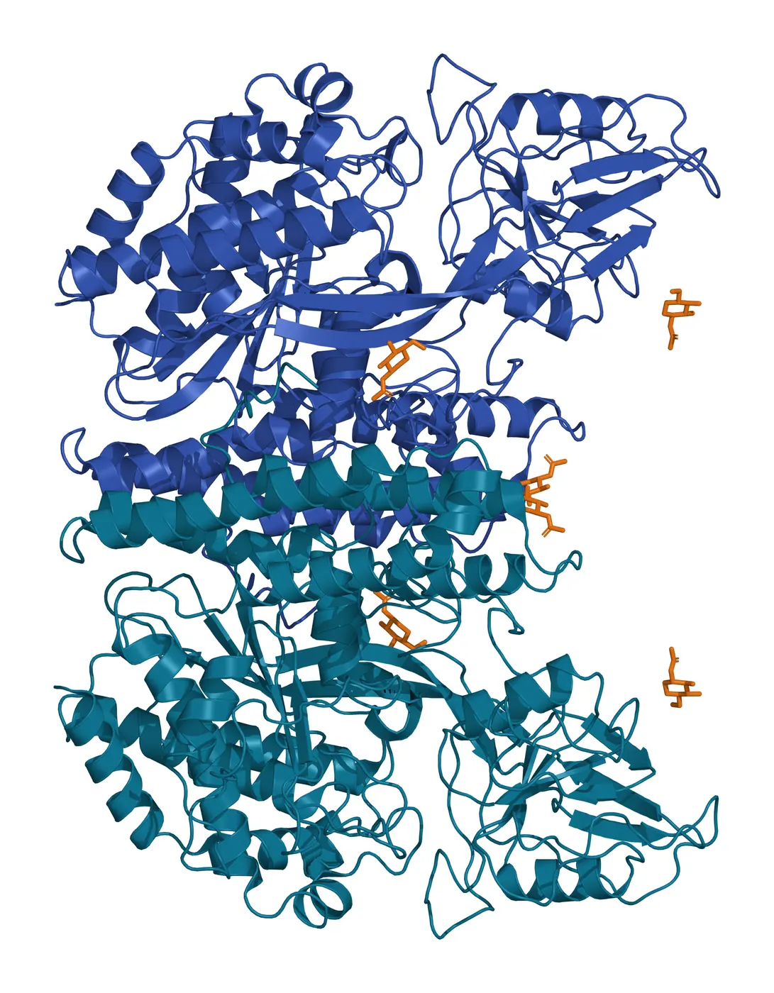
Pan and colleagues then showed by electron microscopy that the receptor sat on round bodies of about 50 nm inside large multivesicular elements, and that these bodies were released when the elements fused with the plasma membrane.[11] Lawrence and colleagues later solved the dimeric receptor ectodomain at 3.2 Å (Fig. 2).[9]
Johnstone and colleagues named the reticulocyte vesicles exosomes in 1987. The vesicles pelleted at 100,000 g and carried acetylcholinesterase, nucleoside and glucose transport activities and the transferrin receptor, all functions the cell loses as it matures.[12] Consensus around the endosomal meaning formed from that paper onward.[4]
Exosomes are the extracellular vesicles that originate in the multivesicular body
MISEV2023 defines exosomes as EVs from internal compartments of the cell that are released through the multivesicular body (MVB).[1] MeSH gives the same definition: a type of EV secreted by exocytosis when multivesicular bodies fuse with the plasma membrane.[5]
Raposo and colleagues extended the concept beyond red cells in 1996. In B lymphoblastoid cells, the limiting membrane of the MHC class II compartment fused with the plasma membrane and released its internal vesicles, which the authors named exosomes and which carried peptide-loaded MHC class II.[13]
Colombo, Raposo and Théry reviewed how the definition developed from these reticulocyte and immune cell studies. Their review also recorded the growing difficulty of separating exosomes from other EVs, which limits the attribution of a function to one vesicle type.[14]
Ectosomes bud directly from the plasma membrane
Ectosomes are EVs assembled at and released from the plasma membrane, and MISEV2023 lists microvesicle and microparticle as synonyms.[1][15] Cocucci and Meldolesi described them as ubiquitous vesicles whose functions may be largely analogous to those of exosomes.[15]
Van Niel, D’Angelo and Raposo framed the EV population as two main classes, exosomes from the endosomal system and microvesicles shed from the plasma membrane.[16]
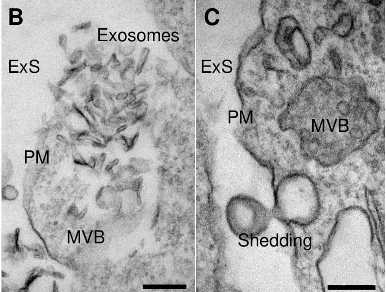
Both routes have been imaged in tissue. In the chick trigeminal ganglion, Bernardi and colleagues recorded multivesicular bodies releasing exosome-like structures and a vesicle shedding from the plasma membrane (Fig. 3).[17]
The conventional model links each route to a size and a membrane signature. Brisson and colleagues summarized it as microvesicles of 100 nm to 1 µm that expose phosphatidylserine and exosomes of 50 to 100 nm that carry CD63, then found CD63 on most EVs from activated platelets, in both a large and a small population.[18]
Molecular markers for the two routes are being defined. Jeppesen and colleagues identified annexin A1 as a specific marker of microvesicles shed from the plasma membrane, and Mathieu and colleagues proposed LAMP1 for exosomes and BSG and SLC3A2 for ectosomes.[19][20]
Biogenesis names require evidence of the compartment of origin
MISEV2018 described the assignment of an EV to a biogenesis pathway as extraordinarily difficult unless the vesicle is caught in the act of release, for example by live imaging.[2] MISEV2023 adds that most separation techniques mix EVs formed by different mechanisms and that universal markers of exosomes or ectosomes have yet to be found.[1]
Kowal and colleagues measured the overlap directly in human dendritic cell EVs. Several classic exosome markers, including MHC, flotillin and HSP70, were similarly present in all EV populations, and small EVs held both exosomal and nonexosomal subpopulations.[21]
Mathieu and colleagues tracked CD9 and CD63 from synthesis to residence in living HeLa cells. CD9, resident at the plasma membrane, was released in EVs more abundantly than CD63, and ectosomes outnumbered exosomes in that cell line.[20]
The same group reviewed how secretion and uptake differ between exosomes and other EVs, a question the field is still mapping.[22] Together these studies show that a small EV fraction usually mixes vesicles of both origins.[20][21]
Operational terms describe size, density, composition and source
MISEV2018 proposed operational terms that refer to what was measured. Small EVs were defined as below 100 nm or below 200 nm and medium or large EVs as above 200 nm, alongside density classes and marker-based names.[2]
- Size: small EVs and medium or large EVs, each with its range stated.[2]
- Density: low, middle or high, with each range defined.[2]
- Composition: a marker profile such as CD63+/CD81+ EVs or Annexin A5-stained EVs.[2]
- Source or condition: the cell of origin or the state of the producing cells, such as podocyte EVs or hypoxic EVs.[2]
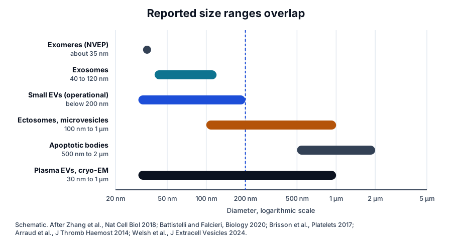
MISEV2023 keeps these terms and adds a caution on size. Small generally means below 200 nm, the upper and lower cut-offs remain a matter of definition, and differential ultracentrifugation yields populations with overlapping size profiles (Fig. 4).[1]
Measurements in human material show how broad these distributions are. Arraud and colleagues found spherical EVs of 30 nm to 1 µm in plasma from healthy donors by cryo-electron microscopy, together with tubular EVs and membrane fragments.[23]
Brisson and colleagues found that EVs from activated platelets formed one broad size peak from 50 nm to 1 µm, with about half between 50 and 400 nm.[18] Zabeo and colleagues sorted EVs from a single mast cell line into nine morphological categories by size and shape.[24]
Electron micrograph shape needs careful interpretation. Gould and Raposo noted that small EVs span a broader size and density range than early reports suggested, and that their cup shape in micrographs is a preparation artifact.[25]
Apoptotic bodies, migrasomes and exomeres extend the vocabulary
Some EV names refer to a cellular process. Apoptotic bodies of about 500 nm to 2 µm are released by cells during programmed cell death, and MISEV2023 lists them with migrasomes as process-linked EV subtypes.[1][26]
Ma and colleagues described migrasomes in 2015. Migrating cells leave retraction fibers behind them, and large vesicles holding numerous smaller vesicles grow at the fiber tips and intersections before release.[27]
Zhang and colleagues separated extracellular particles by asymmetric flow field-flow fractionation into small exosomes of 60 to 80 nm and large exosomes of 90 to 120 nm. They also identified abundant non-membranous nanoparticles of about 35 nm, which they named exomeres.[28]
Exomeres and supermeres are amembranous particles, and several proteins once assigned to exosomes are enriched in them.[29] MISEV2023 treats exomeres, supermeres, lipoproteins and ribonucleoprotein particles as NVEPs and notes that NVEPs may greatly outnumber EVs in biological matrices.[1]
Jeppesen and colleagues resolved this boundary with high-resolution density gradients and immunoaffinity capture. Extracellular RNA, RNA-binding proteins and other cellular proteins differed between exosomes and non-vesicle compartments, and Argonaute 1 to 4, glycolytic enzymes and cytoskeletal proteins fell below detection in exosomes.[19]
Most EP separation methods co-isolate EVs and NVEPs, and many characterization methods detect both. When the two classes cannot be distinguished, MISEV2023 suggests the term EP, or the phrases EV preparation and EV-containing preparation.[1]
MISEV and EV-TRACK pair each term with the evidence behind it
György and colleagues proposed "extracellular vesicles" as a collective term in a widely cited 2011 review of membrane vesicles.[30] Witwer and Théry credited that review, and the founding of ISEV the same year, with the return of the generic term to the literature.[4]
Gould and Raposo, writing after the first ISEV meeting in 2012, asked authors to state how they use each term and how their vesicles were collected. They also encouraged extracellular vesicle as the generic term and as a keyword in every publication.[25]
Lötvall and colleagues published MISEV2014, an ISEV position statement setting minimal biochemical, biophysical and functional standards for attributing cargo or functions to EVs.[31] MISEV2018 expanded it with protocols, tables and a checklist, and 94 percent of its survey respondents endorsed the nomenclature recommendation.[2]
Witwer and colleagues canvassed MISEV authors and ISEV members on how the guidelines should evolve.[32] MISEV2023 compiled feedback from ISEV task forces and more than 1000 researchers, and 79.5 percent of respondents agreed completely and 19.9 percent mostly with its nomenclature section.[1]
EV-TRACK puts the reporting principle into practice. Van Deun and colleagues built it as a crowdsourced knowledgebase of EV biology and methodology,[33] with a checklist for authors and editors that makes the methods behind each EV experiment transparent and searchable.[34]
MISEV2023 summarizes its spirit in a handful of questions that a report should answer.[1]
- Which terms are used, and what do they mean?[1]
- From what source were the EVs obtained, and how was it collected?[1][25]
- How were the EVs separated, concentrated, characterized and stored?[1]
- How confidently can a function or biomarker be attributed to EVs over other components?[1]
- Are the methods and data shared in enough detail for others to replicate the work?[1][33]
A precise report pairs each name with its basis. Witwer and Théry recommended that any term other than EV be defined clearly at each use, and MISEV2023 suggests presenting a study of unproven origin as an EV study.[1][4]
Where to go from here
The exosome biogenesis reference covers the ESCRT and ceramide machinery behind the multivesicular body, and the protein marker reference covers the tetraspanins and other markers used to characterize EV preparations. The exomeres and supermeres reference covers the non-vesicular particles in detail.
The quality reference on MISEV2023 compliance explains how these naming rules appear in supplier documentation. Verified practices can request the certificate of analysis for any lot, and our team can explain which terms a characterization report uses and the measurements behind them.
Referencias comentadas
Numeradas en el orden en que se citan en el texto. Cada fuente lleva una nota sobre lo que respalda en esta página; los artículos revisados por pares se listan con revista, volumen, número y páginas, y las fuentes primarias son regulaciones, documentos de agencias y bases de datos de referencia.
- 1.Peer-reviewedWelsh JA, Goberdhan DCI, O’Driscoll L, et al. Minimal information for studies of extracellular vesicles (MISEV2023): From basic to advanced approaches. Journal of Extracellular Vesicles. 2024;13(2):e12404. https://pubmed.ncbi.nlm.nih.gov/38326288/Annotation: ISEV position statement and current guideline. Cited for the EV definition, the caution on biogenesis terms, operational terms, the EP and NVEP hierarchy, and survey agreement with the nomenclature section.
- 2.Peer-reviewedThéry C, Witwer KW, Aikawa E, et al. Minimal information for studies of extracellular vesicles 2018 (MISEV2018): a position statement of the International Society for Extracellular Vesicles and update of the MISEV2014 guidelines. Journal of Extracellular Vesicles. 2018;7(1):1535750. https://pubmed.ncbi.nlm.nih.gov/30637094/Annotation: ISEV position statement updating MISEV2014. Cited for the operational size, density and marker terms, the difficulty of assigning biogenesis, and 94 percent endorsement of the nomenclature.
- 3.DatabaseNCBI MeSH D000067128, Extracellular Vesicles. Accessed 20 October 2026. https://www.ncbi.nlm.nih.gov/mesh/2009687
- 4.Peer-reviewedWitwer KW, Théry C. Extracellular vesicles or exosomes? On primacy, precision, and popularity influencing a choice of nomenclature. Journal of Extracellular Vesicles. 2019;8(1):1648167. https://pubmed.ncbi.nlm.nih.gov/31489144/Annotation: Editorial history of the terms. Cited for the earlier DNA and RNA-complex meanings of exosome, consensus after Johnstone 1987, the 2011 return of the generic term, and defining terms at each use.
- 5.DatabaseNCBI MeSH D055354, Exosomes. Accessed 20 October 2026. https://www.ncbi.nlm.nih.gov/mesh/68055354
- 6.Peer-reviewedTrams EG, Lauter CJ, Salem N Jr, et al. Exfoliation of membrane ecto-enzymes in the form of micro-vesicles. Biochimica et Biophysica Acta. 1981;645(1):63-70. https://pubmed.ncbi.nlm.nih.gov/6266476/Annotation: First use of exosome for extracellular vesicles. Cited for exfoliated 500 to 1000 nm vesicles carrying ecto-5′-nucleotidase and the proposal to call them exosomes.
- 7.Peer-reviewedPan BT, Johnstone RM. Fate of the transferrin receptor during maturation of sheep reticulocytes in vitro: selective externalization of the receptor. Cell. 1983;33(3):967-978. https://pubmed.ncbi.nlm.nih.gov/6307529/Annotation: Antibody tracking of the transferrin receptor in maturing reticulocytes. Cited for selective release of the receptor in vesicles carrying few other membrane proteins.
- 8.DatabaseNCBI Gene 7037, TFRC transferrin receptor (Homo sapiens). Accessed 20 October 2026. https://www.ncbi.nlm.nih.gov/gene/7037
- 9.Peer-reviewedLawrence CM, Ray S, Babyonyshev M, et al. Crystal structure of the ectodomain of human transferrin receptor. Science. 1999;286(5440):779-782. https://pubmed.ncbi.nlm.nih.gov/10531064/Annotation: Crystal structure of the dimeric transferrin receptor ectodomain at 3.2 Å. Cited for the receptor’s endocytic cycle and for the structure shown in Fig. 2.
- 10.Peer-reviewedHarding C, Heuser J, Stahl P. Receptor-mediated endocytosis of transferrin and recycling of the transferrin receptor in rat reticulocytes. Journal of Cell Biology. 1983;97(2):329-339. https://pubmed.ncbi.nlm.nih.gov/6309857/Annotation: Electron microscopy of gold-labeled transferrin in reticulocytes. Cited for multivesicular endosomes that fuse with the plasma membrane and release their contents.
- 11.Peer-reviewedPan BT, Teng K, Wu C, et al. Electron microscopic evidence for externalization of the transferrin receptor in vesicular form in sheep reticulocytes. Journal of Cell Biology. 1985;101(3):942-948. https://pubmed.ncbi.nlm.nih.gov/2993317/Annotation: Immunoelectron microscopy of reticulocyte maturation. Cited for receptor-bearing bodies of about 50 nm inside multivesicular elements, released on fusion with the plasma membrane.
- 12.Peer-reviewedJohnstone RM, Adam M, Hammond JR, et al. Vesicle formation during reticulocyte maturation. Association of plasma membrane activities with released vesicles (exosomes). Journal of Biological Chemistry. 1987;262(19):9412-9420. https://pubmed.ncbi.nlm.nih.gov/3597417/Annotation: Biochemistry of vesicles released by sheep reticulocytes. Cited for naming them exosomes and for the membrane activities, including the transferrin receptor, that they carry.
- 13.Peer-reviewedRaposo G, Nijman HW, Stoorvogel W, et al. B lymphocytes secrete antigen-presenting vesicles. Journal of Experimental Medicine. 1996;183(3):1161-1172. https://pubmed.ncbi.nlm.nih.gov/8642258/Annotation: Immunoelectron microscopy of B lymphoblastoid cells. Cited for fusion of the MHC class II compartment with the plasma membrane and release of exosomes carrying peptide-loaded MHC class II.
- 14.Peer-reviewedColombo M, Raposo G, Théry C. Biogenesis, secretion, and intercellular interactions of exosomes and other extracellular vesicles. Annual Review of Cell and Developmental Biology. 2014;30:255-289. https://pubmed.ncbi.nlm.nih.gov/25288114/Annotation: Review of exosome definition and biogenesis. Cited for the history of the endosomal definition and the difficulty of separating exosomes from other EVs.
- 15.Peer-reviewedCocucci E, Meldolesi J. Ectosomes and exosomes: shedding the confusion between extracellular vesicles. Trends in Cell Biology. 2015;25(6):364-372. https://pubmed.ncbi.nlm.nih.gov/25683921/Annotation: Review comparing ectosomes and exosomes. Cited for ectosomes as ubiquitous plasma membrane vesicles with functions largely analogous to those of exosomes.
- 16.Peer-reviewedvan Niel G, D’Angelo G, Raposo G. Shedding light on the cell biology of extracellular vesicles. Nature Reviews Molecular Cell Biology. 2018;19(4):213-228. https://pubmed.ncbi.nlm.nih.gov/29339798/Annotation: Review of EV cell biology. Cited for the two main classes, exosomes from the endosomal system and microvesicles shed from the plasma membrane.
- 17.Peer-reviewedBernardi YE, Sanchez-Vasquez E, Márquez RB, et al. miR-203 secreted in extracellular vesicles mediates the communication between neural crest and placode cells required for trigeminal ganglia formation. PLoS Biology. 2024;22(7):e3002074. https://pubmed.ncbi.nlm.nih.gov/39038054/Annotation: Chick embryo study of EV communication during trigeminal ganglion formation. Cited for electron micrographs of multivesicular bodies releasing vesicles and a vesicle shedding at the plasma membrane.
- 18.Peer-reviewedBrisson AR, Tan S, Linares R, et al. Extracellular vesicles from activated platelets: a semiquantitative cryo-electron microscopy and immuno-gold labeling study. Platelets. 2017;28(3):263-271. https://pubmed.ncbi.nlm.nih.gov/28102751/Annotation: Cryo-EM and immunogold analysis of platelet EVs. Cited for a single broad size peak from 50 nm to 1 µm and the conventional 100 nm to 1 µm microvesicle range.
- 19.Peer-reviewedJeppesen DK, Fenix AM, Franklin JL, et al. Reassessment of Exosome Composition. Cell. 2019;177(2):428-445.e18. https://pubmed.ncbi.nlm.nih.gov/30951670/Annotation: High-resolution density gradient and immunoaffinity analysis of exosomes and non-vesicle material. Cited for annexin A1 as a microvesicle marker and for proteins below detection in exosomes.
- 20.Peer-reviewedMathieu M, Névo N, Jouve M, et al. Specificities of exosome versus small ectosome secretion revealed by live intracellular tracking of CD63 and CD9. Nature Communications. 2021;12(1):4389. https://pubmed.ncbi.nlm.nih.gov/34282141/Annotation: Live tracking of CD9 and CD63 in HeLa cells. Cited for ectosomes outnumbering exosomes in that cell line and for LAMP1, BSG and SLC3A2 as candidate route-specific markers.
- 21.Peer-reviewedKowal J, Arras G, Colombo M, et al. Proteomic comparison defines novel markers to characterize heterogeneous populations of extracellular vesicle subtypes. Proceedings of the National Academy of Sciences of the United States of America. 2016;113(8):E968-E977. https://pubmed.ncbi.nlm.nih.gov/26858453/Annotation: Quantitative proteomics of dendritic cell EV populations. Cited for classic exosome markers present in all EVs and for exosomal and nonexosomal subpopulations within small EVs.
- 22.Peer-reviewedMathieu M, Martin-Jaular L, Lavieu G, et al. Specificities of secretion and uptake of exosomes and other extracellular vesicles for cell-to-cell communication. Nature Cell Biology. 2019;21(1):9-17. https://pubmed.ncbi.nlm.nih.gov/30602770/Annotation: Review of exosome and EV secretion and uptake. Cited for the differences between exosomes and other EVs as an open, actively mapped question.
- 23.Peer-reviewedArraud N, Linares R, Tan S, et al. Extracellular vesicles from blood plasma: determination of their morphology, size, phenotype and concentration. Journal of Thrombosis and Haemostasis. 2014;12(5):614-627. https://pubmed.ncbi.nlm.nih.gov/24618123/Annotation: Cryo-electron microscopy of plasma EVs from healthy donors. Cited for spherical EVs of 30 nm to 1 µm, tubular EVs and membrane fragments.
- 24.Peer-reviewedZabeo D, Cvjetkovic A, Lässer C, et al. Exosomes purified from a single cell type have diverse morphology. Journal of Extracellular Vesicles. 2017;6(1):1329476. https://pubmed.ncbi.nlm.nih.gov/28717422/Annotation: Electron microscopy of EVs from the HMC-1 mast cell line. Cited for nine morphological categories within a single-cell-type preparation.
- 25.Peer-reviewedGould SJ, Raposo G. As we wait: coping with an imperfect nomenclature for extracellular vesicles. Journal of Extracellular Vesicles. 2013;2:20389. https://pubmed.ncbi.nlm.nih.gov/24009890/Annotation: Letter following the first ISEV meeting. Cited for stating term use and collection methods, adopting EV as the generic term, and the cup shape as a preparation artifact.
- 26.Peer-reviewedBattistelli M, Falcieri E. Apoptotic Bodies: Particular Extracellular Vesicles Involved in Intercellular Communication. Biology. 2020;9(1):21. https://pubmed.ncbi.nlm.nih.gov/31968627/Annotation: Morpho-functional review of apoptotic bodies. Cited for apoptotic bodies of 500 nm to 2 µm and for the 40 to 120 nm exosome range.
- 27.Peer-reviewedMa L, Li Y, Peng J, et al. Discovery of the migrasome, an organelle mediating release of cytoplasmic contents during cell migration. Cell Research. 2015;25(1):24-38. https://pubmed.ncbi.nlm.nih.gov/25342562/Annotation: Discovery of migrasomes in migrating cells. Cited for large vesicles containing smaller vesicles that form on retraction fibers.
- 28.Peer-reviewedZhang H, Freitas D, Kim HS, et al. Identification of distinct nanoparticles and subsets of extracellular vesicles by asymmetric flow field-flow fractionation. Nature Cell Biology. 2018;20(3):332-343. https://pubmed.ncbi.nlm.nih.gov/29459780/Annotation: Asymmetric flow field-flow fractionation of extracellular particles. Cited for the 60 to 80 nm and 90 to 120 nm exosome subsets and the discovery of exomeres of about 35 nm.
- 29.Peer-reviewedJeppesen DK, Zhang Q, Franklin JL, et al. Extracellular vesicles and nanoparticles: emerging complexities. Trends in Cell Biology. 2023;33(8):667-681. https://pubmed.ncbi.nlm.nih.gov/36737375/Annotation: Review of EV and nanoparticle diversity. Cited for exomeres and supermeres as amembranous particles enriched in proteins once assigned to exosomes.
- 30.Peer-reviewedGyörgy B, Szabó TG, Pásztói M, et al. Membrane vesicles, current state-of-the-art: emerging role of extracellular vesicles. Cellular and Molecular Life Sciences. 2011;68(16):2667-2688. https://pubmed.ncbi.nlm.nih.gov/21560073/Annotation: Comprehensive review of membrane vesicles. Cited as the 2011 review that proposed extracellular vesicles as the collective term.
- 31.Peer-reviewedLötvall J, Hill AF, Hochberg F, et al. Minimal experimental requirements for definition of extracellular vesicles and their functions: a position statement from the International Society for Extracellular Vesicles. Journal of Extracellular Vesicles. 2014;3:26913. https://pubmed.ncbi.nlm.nih.gov/25536934/Annotation: ISEV position statement, MISEV2014. Cited for the minimal biochemical, biophysical and functional standards for attributing cargo or functions to EVs.
- 32.Peer-reviewedWitwer KW, Goberdhan DC, O’Driscoll L, et al. Updating MISEV: Evolving the minimal requirements for studies of extracellular vesicles. Journal of Extracellular Vesicles. 2021;10(14):e12182. https://pubmed.ncbi.nlm.nih.gov/34953156/Annotation: ISEV Board canvass of MISEV authors and society members. Cited for the process that led from MISEV2018 toward MISEV2023.
- 33.Peer-reviewedEV-TRACK Consortium, Van Deun J, Mestdagh P, et al. EV-TRACK: transparent reporting and centralizing knowledge in extracellular vesicle research. Nature Methods. 2017;14(3):228-232. https://pubmed.ncbi.nlm.nih.gov/28245209/Annotation: Description of the EV-TRACK knowledgebase. Cited for the crowdsourced resource that centralizes EV biology and methodology.
- 34.Peer-reviewedVan Deun J, Hendrix A, EV-TRACK consortium. Is your article EV-TRACKed? Journal of Extracellular Vesicles. 2017;6(1):1379835. https://pubmed.ncbi.nlm.nih.gov/29184624/Annotation: Editorial introducing the EV-TRACK checklist. Cited for the checklist that makes the methods of EV experiments transparent and searchable.
Cite this page
ExaVeyra Sciences Editorial Team. Exosome vs Extracellular Vesicle: MISEV2023 Nomenclature. ExaVeyra Sciences; 20 October 2026. Available from: https://exaveyra.com/science/exosome-biochemistry/extracellular-vesicle-nomenclature. Accessed [date accessed].
Fuentes de las figuras
- 35.PDB 1CX8, Crystal structure of the ectodomain of human transferrin receptor. ExaVeyra Sciences, rendered in PyMOL from PDB 1CX8. Structure data: RCSB Protein Data Bank (rcsb.org), CC0. Publicación de la estructura: Lawrence CM, Ray S, Babyonyshev M, et al. Crystal structure of the ectodomain of human transferrin receptor. Science. 1999;286(5440):779-782.