Esta referencia está disponible actualmente en inglés. La versión en español está en preparación.
Exomeres, Supermeres and the Revised Map of Exosome Composition
Revisado por el equipo editorial de ExaVeyra Sciences · Actualizado el 20 de octubre de 2026
ExaVeyra Sciences es un proveedor B2B con sede en Miami que suministra exosomas para uso estético tópico a clínicas con licencia, y remite a los prescriptores con licencia a una farmacia de preparados compuestos 503A que prepara formulaciones según prescripciones individuales.
| Pregunta | Respuesta |
|---|---|
| Qué cubre esta página | How high-resolution separation refined exosome composition: exomeres, supermeres and other non-vesicular particles, and where the classic markers sit. |
| Quién puede hacer pedidos | Clínicas y profesionales con licencia profesional activa y NPI verificados por ExaVeyra. Los pedidos de exosomas requieren además un reconocimiento firmado de uso profesional. |
| Qué se envía y cómo | El material de exosomas para uso estético tópico, caracterizado por lote, se envía en cadena de frío por exprés nocturno. La entrega puede tardar 1 a 3 días hábiles. Se emite un certificado de análisis para cada lote y se entrega a solicitud. |
Exomeres and supermeres are amembranous nanoparticles that cells release alongside exosomes, and their discovery changed the accepted picture of exosome composition.1 High-resolution separation showed that many proteins and RNAs once credited to exosomes travel in these and other non-vesicular extracellular particles.2
This page follows that revision: the separation methods that resolved the particle classes, the markers that moved to non-vesicular fractions, the structure of Argonaute2 as the best-understood non-vesicular RNA carrier, and the MISEV2023 terms that now describe the whole population.
Early exosome preparations captured a mixture of particle classes
Valadi and colleagues reported in 2007 that exosomes from mast cells contain messenger RNA and microRNA, and that the messenger RNA can be translated after transfer to another cell.3 That finding framed exosomes as the main vehicle for extracellular RNA for the following decade.
Kowal and colleagues then compared vesicle populations separated by sedimentation speed, density floatation and immuno-isolation. Several classic exosome markers, including MHC proteins, flotillin and HSP70, were present at similar levels in all vesicle types.4
The MISEV2018 guidelines drew the practical conclusion. Ascribing a function to exosomes in particular requires evidence beyond a crude, heterogeneous preparation, because exosomes are difficult to separate from biophysically similar particles.5
Asymmetric-flow field-flow fractionation resolved exomeres and two exosome subsets
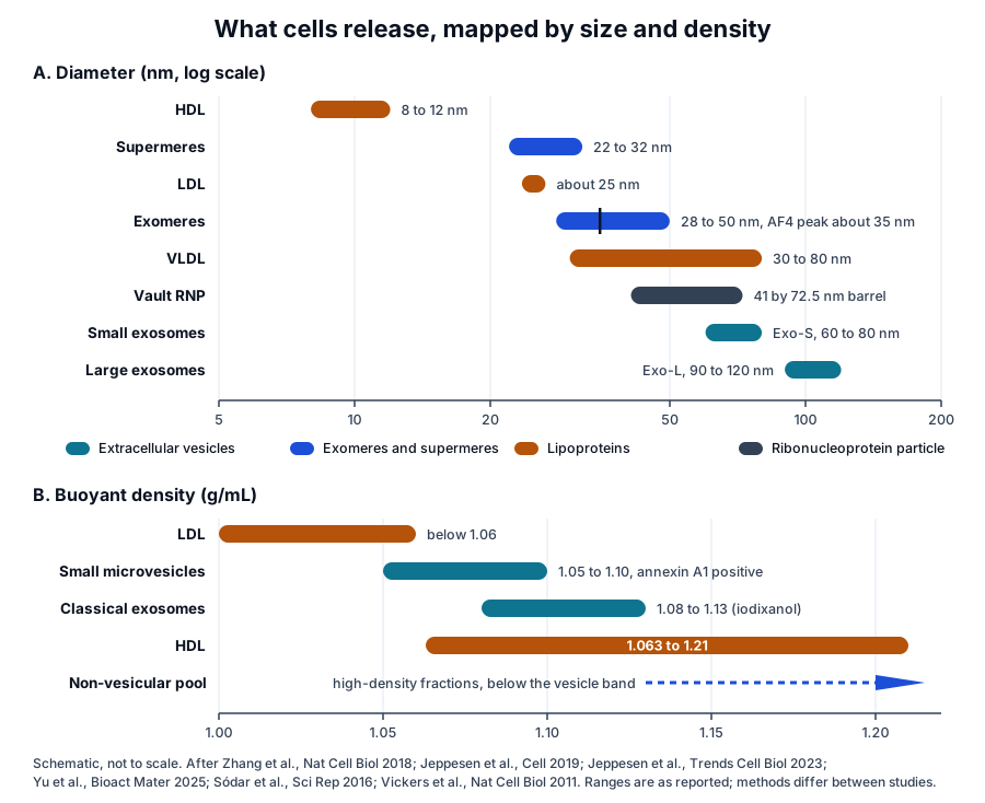
Zhang and colleagues applied asymmetric-flow field-flow fractionation (AF4) to small vesicle preparations in 2018. AF4 sorts particles by hydrodynamic size between a channel flow and a perpendicular cross-flow, and it resolved two exosome subsets and a third, smaller population.6
- Large exosome vesicles (Exo-L), 90 to 120 nm, with proteins of the mitotic spindle and IL-2/STAT5 signalling.6
- Small exosome vesicles (Exo-S), 60 to 80 nm, with proteins of endosomal function and secretion pathways.6
- Exomeres, about 35 nm, which appeared as amembranous particles in electron micrographs and were enriched in metabolic enzymes.6
The companion protocol reports that AF4 separates particles from a few nanometres upward at a resolution of about 1 nm, with the fractionation step itself taking about an hour.7 The size classes it defined anchor the map of extracellular particles used on this page (Fig. 1).
Density gradients and direct capture reassessed exosome composition
Jeppesen and colleagues took a complementary route in 2019. They floated crude small vesicle pellets upward through high-resolution iodixanol gradients, then pulled classical exosomes out of the vesicle fractions with beads carrying antibodies to CD63, CD81 or CD9 (Fig. 2).8
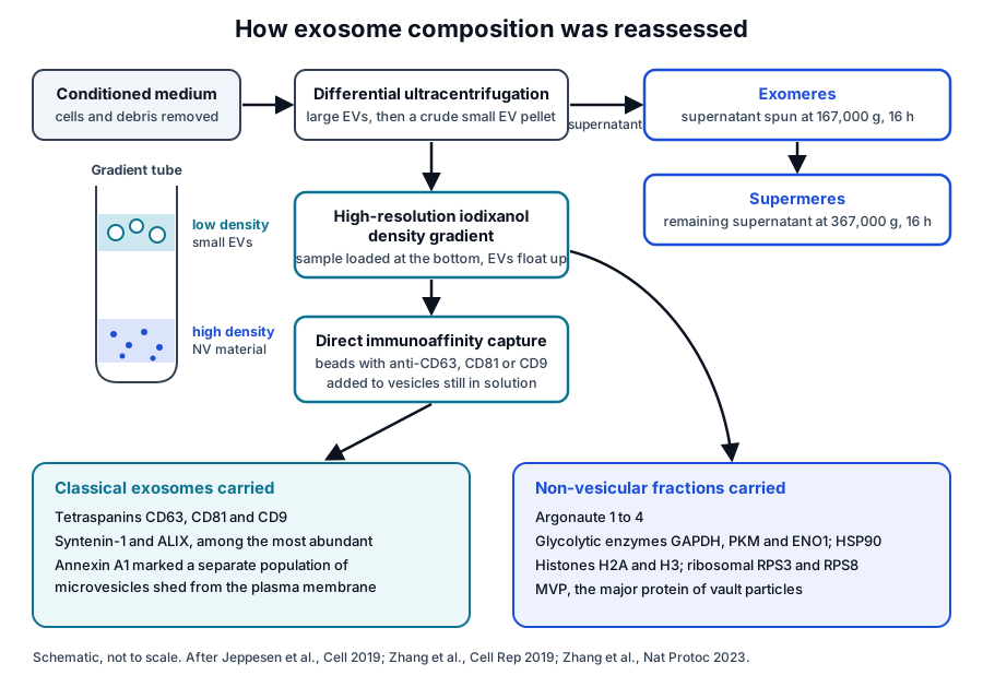
Membrane vesicles collected in low-density fractions, and most classical exosomes floated at 1.08 to 1.13 g/mL in iodixanol. Non-vesicular material stayed in high-density fractions, and a second round of fractionation showed that it was released as such, independent of vesicle damage.8
The two pools differed sharply. Syntenin-1 and ALIX were among the most abundant proteins of the purified vesicles, while the non-vesicular fractions were dominated by metabolic enzymes such as GAPDH, PKM and ENO1, together with HSP90, tubulins and histones H2A and H3.8
Direct capture placed Argonaute 1 to 4, glycolytic enzymes and cytoskeletal proteins outside classical exosomes.8 The same study identified annexin A1 as a specific marker of microvesicles shed directly from the plasma membrane, which float at slightly lower density than exosomes.8
Argonaute complexes carry most extracellular microRNA outside vesicles
Arroyo and colleagues fractionated human plasma and serum by differential centrifugation and size-exclusion chromatography in 2011. Most circulating microRNAs co-fractionated with protein complexes, eluted with Argonaute2, and were recovered by Argonaute2 immunoprecipitation, while a minority associated mainly with vesicles.9
Turchinovich and colleagues reached the same conclusion independently. Most microRNA in plasma and culture medium passed 0.22 micrometre filters, stayed in the supernatant after centrifugation at 110,000 g, and co-immunoprecipitated with the 96 kDa Argonaute2 protein.10
Jeppesen and colleagues later found that microRNAs in non-vesicular fractions showed marked trimming of their 3 prime ends, and that vault RNAs were associated with the same fractions.8 They concluded that cells secrete Argonaute proteins independently of exosomes.8
Lipoproteins, vaults and ribosomes extend the set of RNA carriers
Vickers and colleagues showed that high-density lipoprotein (HDL) carries endogenous microRNAs in plasma and delivers them to cells, where they act on reporter messenger RNAs. Delivery depended on scavenger receptor class B type I, and export to HDL was regulated by neutral sphingomyelinase.11
Vaults are a further ribonucleoprotein class in the non-vesicular pool. MVP makes up more than 70 percent of the protein of these 41 by 72.5 nm particles, and Jeppesen and colleagues found MVP in non-vesicular fractions alongside vault RNAs.8
Tosar and colleagues added full ribosomes and full-length tRNAs to the extracellular RNA map. Their fragmentation outside the cell generates small RNAs that resist degradation, so the extracellular abundance of a fragment reflects processing as well as secretion.12
The structure of Argonaute2 explains how it holds a microRNA
Argonaute2 is the human Argonaute protein encoded by AGO2, a highly basic protein with a PAZ domain and a PIWI domain.13 Liu and colleagues identified it in 2004 as the one mammalian Argonaute that cleaves target messenger RNA, the catalytic engine of RNA interference.14
Schirle and MacRae solved the first crystal structure of human Argonaute2 in 2012. It is a bilobed molecule with a central cleft for guide and target RNA, and guide nucleotides 2 to 6 sit in an A-form conformation ready to base pair with a target.15
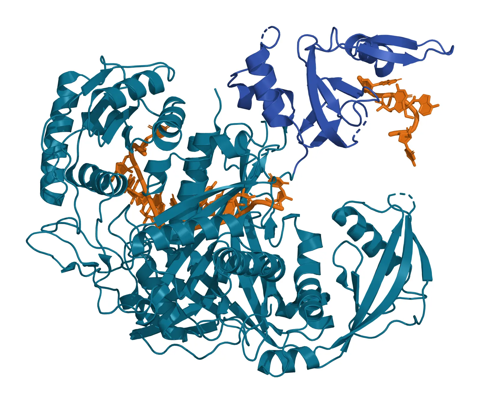
Elkayam and colleagues solved Argonaute2 bound to a single defined microRNA, miR-20a, at 2.2 Å (Fig. 3). The microRNA is anchored at both ends by the MID and PAZ domains and makes several kinks and turns along the binding groove.16
The same study found that microRNA binding locks the otherwise flexible enzyme into a stable conformation.16 Arroyo and colleagues saw plasma microRNAs lost after protease digestion of plasma, indicating that protein complexes protect them from plasma RNases.9
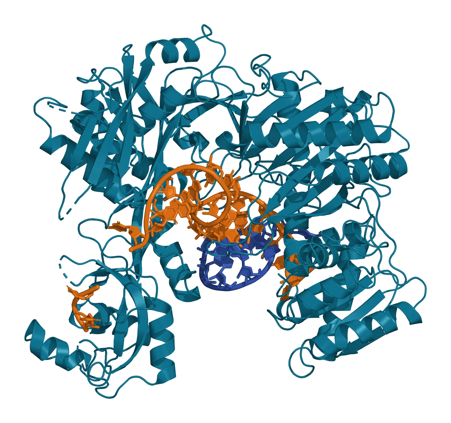
Schirle and colleagues then captured Argonaute2 with guide and target RNA paired across the seed (Fig. 4). Target recognition proceeds stepwise: guide nucleotides 2 to 5 are exposed first, and pairing there opens guide positions 2 to 8 and 13 to 16 for further recognition.17
Exomeres and supermeres carry distinct protein and RNA cargo
Exomeres are small, lipid-poor particles rich in metabolic enzymes
Exomeres in the AF4 study were smaller than 50 nm, weakly negatively charged, stiffer than exosomes, and 5.8 to 7.0 nm in average height by atomic force microscopy.6 They contained fewer lipids than exosomes, with relatively more triglycerides and ceramides, and were enriched in glycolysis and mTOR pathway proteins.6
Zhang and colleagues then pelleted exomeres directly by ultracentrifugation and found them enriched in Argonaute 1 to 3 and amyloid precursor protein.18 Exomeres transferred functional β-galactoside α2,6-sialyltransferase 1 to recipient cells, raising α2,6-sialylation of surface proteins that included β1 integrin.18
Exomeres also carried the EGFR ligand amphiregulin. Amphiregulin-containing exomeres prolonged EGFR signalling in recipient cells and modulated EGFR trafficking in normal intestinal organoids, establishing exomeres as carriers of active cargo.18
Supermeres are the smallest particles in the series
Supermeres take their name from the supernatant of exomeres, the fraction in which they were found. They are smaller than exomeres by atomic force microscopy and are pelleted from that supernatant at higher force.2 Yu and colleagues give their diameter as about 22 to 32 nm, roughly half the volume of an exomere.19
Glycolytic enzymes trace the shift in thinking. Earlier small vesicle preparations contained them, direct capture placed them outside classical exosomes, and they are enriched in exomeres and more highly enriched in supermeres.2
Supermeres from many cell types are rich in transforming growth factor beta-induced protein (TGFBI), while fatty acid synthase is more associated with exomeres. Their membrane-protein content consists of ectodomain cleavage products, including those of APP, glypican-1, MET and amphiregulin.2
Supermeres contain high quantities of extracellular RNA and are enriched in miR-1246.20 Tosar, Cayota and Witwer reanalysed the published data and found that the top-ranking supermere RNAs are footprints of protein complexes protecting fragments of U2 small nuclear RNA and 28S ribosomal RNA.20
The same authors noted that several exomere-enriched proteins self-assemble into defined particles: LGALS3BP forms ring-like decamers of 30 to 40 nm, and VCP forms hexamers 15.6 nm across.20
Jeppesen and colleagues answered with further analyses of their supermere data, isolated from the exomere supernatant after a 16 h ultracentrifugation, that support supermeres as a distinct nanoparticle.21 The same group lists the relationship between exomeres and supermeres among the open questions now driving the field.2
MISEV2023 names non-vesicular extracellular particles
MISEV2023 introduced a nomenclature for this material. Extracellular particles (EPs) is the umbrella term, extracellular vesicles are particles delimited by a lipid bilayer, and non-vesicular extracellular particles (NVEPs) are cell-derived multimolecular assemblies without a delimiting bilayer.22
The guideline leaves the naming of specific classes such as lipoprotein particles, ribonucleoprotein particles, exomeres and supermeres to their own fields. It notes that NVEPs may greatly outnumber EVs and that most separation methods co-isolate the two.22
Its protein reference table builds on the reassessment. MISEV2023 lists Argonautes and 14-3-3 proteins as generally more abundant in NVEPs, and HSP90AA and HSP90AB, TGFBI, HSPA13 and LDHA and LDHB as exomere- or supermere-enriched components for purity control.22
Liu and colleagues summarise the consequence in a 2026 review. Most NVEPs are smaller than 50 nm and carry protein and RNA cargo once thought to belong to exosomes, so some functions attributed to exosomes are being reassigned.23
Separation method defines which particles a preparation contains
Size and density overlap between particle classes, so each method enriches a different mixture (Fig. 1). Jeppesen and colleagues note that EVs and NVEPs of similar size differ in density because their lipid, protein and nucleic acid content differs, which is why density gradients separate them.2
- Size-exclusion chromatography of plasma places vesicle markers in the same fractions as lipoprotein particles; adding a density cushion reduced lipoprotein carry-over about 100-fold.24
- Low-density lipoprotein, about 25 nm across, remained in plasma vesicle preparations after each isolation method tested and associated with vesicles when mixed in vitro.25
- HDL floats at 1.063 to 1.21 g/mL, a range that overlaps the density of exosomes.25
- A sequential protocol now isolates large and small EVs, non-vesicular fractions, exomeres and supermeres from one starting sample by ultracentrifugation, filtration and bottom-loaded density gradients.26
In that protocol, exomeres are pelleted at 167,000 g and supermeres from the remaining supernatant at 367,000 g, each for 16 h.26 Reviews comparing ultracentrifugation, density gradients, size-exclusion chromatography and flow field-flow fractionation describe strategies for combining them.23
Particle counting is equally method-dependent. NVEPs smaller than EVs may fall below the detection range of some characterisation methods, so their share of a preparation can go unmeasured.22 Reporting the separation method, density and marker panel with every composition claim makes results comparable across laboratories.
Where to go from here
The cargo sorting reference covers how proteins and RNAs enter vesicles, and the uptake reference covers what happens when vesicles reach a recipient cell. The quality references on MISEV2023 and on particle counting explain how purity and particle number are measured.
Verified practices can request lot-specific characterization data with the certificate of analysis. Our team can explain how a preparation was separated and what its marker panel represents.
Referencias
Numeradas en el orden en que se citan en el texto. Cada estudio indica su diseño y, cuando incluyó personas, cuántas, con una línea que dice qué respalda en esta página. Las fuentes primarias son normas, documentos de agencias y bases de datos de referencia.
- 1.Jeppesen DK, Zhang Q, Coffey RJ. Extracellular vesicles and nanoparticles at a glance. Journal of Cell Science. 2024. doi:10.1242/jcs.260201. PMID: 39641198.Narrative reviewReference 1 supports: Overview of extracellular vesicle and nanoparticle diversity. Cited for cells releasing non-vesicular nanoparticles, including exomeres and supermeres, alongside vesicles.
- 2.Jeppesen DK, Zhang Q, Franklin JL, et al. Extracellular vesicles and nanoparticles: emerging complexities. Trends in Cell Biology. 2023. doi:10.1016/j.tcb.2023.01.002. PMID: 36737375.Narrative reviewReference 2 supports: Review of vesicle and nanoparticle classes. Cited for supermere naming, isolation and cargo, the gradient of glycolytic enzyme enrichment, and density differences between particle classes.
- 3.Valadi H, Ekström K, Bossios A, et al. Exosome-mediated transfer of mRNAs and microRNAs is a novel mechanism of genetic exchange between cells. Nature Cell Biology. 2007. doi:10.1038/ncb1596. PMID: 17486113.In vitroLandmarkReference 3 supports: Microarray and transfer experiments with mast cell exosomes. Cited for the original report of messenger RNA and microRNA in exosomes.
- 4.Kowal J, Arras G, Colombo M, et al. Proteomic comparison defines novel markers to characterize heterogeneous populations of extracellular vesicle subtypes. Proceedings of the National Academy of Sciences of the United States of America. 2016. doi:10.1073/pnas.1521230113. PMID: 26858453.In vitroReference 4 supports: Quantitative proteomics of separated vesicle populations. Cited for classic markers such as MHC, flotillin and HSP70 being present across all vesicle types.
- 5.Théry C, Witwer KW, Aikawa E, et al. Minimal information for studies of extracellular vesicles 2018 (MISEV2018): a position statement of the International Society for Extracellular Vesicles and update of the MISEV2014 guidelines. Journal of Extracellular Vesicles. 2018. doi:10.1080/20013078.2018.1535750. PMID: 30637094.Clinical guidelineReference 5 supports: ISEV reporting guideline. Cited for the evidence needed before ascribing a function to exosomes in a heterogeneous preparation.
- 6.Zhang H, Freitas D, Kim HS, et al. Identification of distinct nanoparticles and subsets of extracellular vesicles by asymmetric flow field-flow fractionation. Nature Cell Biology. 2018. doi:10.1038/s41556-018-0040-4. PMID: 29459780.In vitroReference 6 supports: AF4 fractionation with proteomic, lipidomic and biophysical profiling. Cited for the identification of exomeres and the Exo-S and Exo-L subsets, their sizes, charge, stiffness and cargo.
- 7.Zhang H, Lyden D. Asymmetric-flow field-flow fractionation technology for exomere and small extracellular vesicle separation and characterization. Nature Protocols. 2019. doi:10.1038/s41596-019-0126-x. PMID: 30833697.In vitroReference 7 supports: Method protocol for AF4. Cited for the separation principle, its size range and resolution, and the duration of the fractionation step.
- 8.Jeppesen DK, Fenix AM, Franklin JL, et al. Reassessment of Exosome Composition. Cell. 2019. doi:10.1016/j.cell.2019.02.029. PMID: 30951670.In vitroReference 8 supports: High-resolution density gradients with direct immunoaffinity capture. Cited for the composition of classical exosomes, the proteins and RNAs assigned to non-vesicular fractions, and annexin A1 as a microvesicle marker.
- 9.Arroyo JD, Chevillet JR, Kroh EM, et al. Argonaute2 complexes carry a population of circulating microRNAs independent of vesicles in human plasma. Proceedings of the National Academy of Sciences of the United States of America. 2011. doi:10.1073/pnas.1019055108. PMID: 21383194.In vitroReference 9 supports: Fractionation of human plasma and serum. Cited for most circulating microRNA co-purifying with Argonaute2 complexes and for protein complexes protecting it from plasma RNases.
- 10.Turchinovich A, Weiz L, Langheinz A, et al. Characterization of extracellular circulating microRNA. Nucleic Acids Research. 2011. doi:10.1093/nar/gkr254. PMID: 21609964.In vitroReference 10 supports: Filtration, ultracentrifugation and immunoprecipitation of plasma and culture medium. Cited for extracellular microRNA remaining in high-speed supernatants and associating with Argonaute2.
- 11.Vickers KC, Palmisano BT, Shoucri BM, et al. MicroRNAs are transported in plasma and delivered to recipient cells by high-density lipoproteins. Nature Cell Biology. 2011. doi:10.1038/ncb2210. PMID: 21423178.PreclinicalReference 11 supports: Isolation of HDL-bound microRNA and delivery assays in cells and mice. Cited for HDL as a microRNA carrier, SR-BI-dependent delivery and neutral sphingomyelinase-regulated export.
- 12.Tosar JP, Segovia M, Castellano M, et al. Fragmentation of extracellular ribosomes and tRNAs shapes the extracellular RNAome. Nucleic Acids Research. 2020. doi:10.1093/nar/gkaa674. PMID: 32785615.In vitroReference 12 supports: Size-exclusion chromatography and sequencing of non-vesicular extracellular RNA. Cited for extracellular ribosomes and full-length tRNAs and their fragmentation into stable small RNAs.
- 13.NCBI Gene 27161, AGO2 argonaute RISC catalytic component 2 (Homo sapiens). Accessed 20 October 2026.Primary source
- 14.Liu J, Carmell MA, Rivas FV, et al. Argonaute2 is the catalytic engine of mammalian RNAi. Science. 2004. doi:10.1126/science.1102513. PMID: 15284456.PreclinicalLandmarkReference 14 supports: Genetic and biochemical analysis of mammalian Argonautes. Cited for Argonaute2 as the single family member with messenger RNA cleavage activity.
- 15.Schirle NT, MacRae IJ. The crystal structure of human Argonaute2. Science. 2012. doi:10.1126/science.1221551. PMID: 22539551.In vitroReference 15 supports: Crystal structure of human Argonaute2 at 2.3 Å. Cited for the bilobed architecture, the central RNA-binding cleft and the A-form guide nucleotides 2 to 6.
- 16.Elkayam E, Kuhn CD, Tocilj A, et al. The structure of human argonaute-2 in complex with miR-20a. Cell. 2012. doi:10.1016/j.cell.2012.05.017. PMID: 22682761.In vitroReference 16 supports: Crystal structure of Argonaute2 bound to miR-20a (PDB 4F3T). Cited for anchoring of the microRNA by the MID and PAZ domains and the stabilising effect of microRNA binding.
- 17.Schirle NT, Sheu-Gruttadauria J, MacRae IJ. Structural basis for microRNA targeting. Science. 2014. doi:10.1126/science.1258040. PMID: 25359968.In vitroReference 17 supports: Crystal structures of Argonaute2 with guide and target RNAs, including PDB 4W5O. Cited for the stepwise mechanism of seed pairing.
- 18.Zhang Q, Higginbotham JN, Jeppesen DK, et al. Transfer of Functional Cargo in Exomeres. Cell Reports. 2019. doi:10.1016/j.celrep.2019.01.009. PMID: 30956133.In vitroReference 18 supports: Ultracentrifugation-based exomere isolation and cargo transfer assays. Cited for Argonaute 1 to 3 and APP enrichment, ST6Gal-I transfer and amphiregulin signalling in normal intestinal organoids.
- 19.Yu L, Shi H, Gao T, et al. Exomeres and supermeres: Current advances and perspectives. Bioactive Materials. 2025. doi:10.1016/j.bioactmat.2025.04.012. PMID: 40276541.Narrative reviewReference 19 supports: Review of exomere and supermere biology and isolation. Cited for the reported supermere diameter and its volume relative to exomeres.
- 20.Tosar JP, Cayota A, Witwer K. Exomeres and Supermeres: monolithic or diverse? Journal of Extracellular Biology. 2022. doi:10.1002/jex2.45. PMID: 36311878.Narrative reviewReference 20 supports: Reanalysis of published supermere proteomic and transcriptomic data. Cited for supermere RNA content, miR-1246 enrichment, protected U2 and 28S RNA fragments and LGALS3BP decamers.
- 21.Jeppesen DK, Zhang Q, Franklin JL, et al. Are Supermeres a Distinct Nanoparticle? Journal of Extracellular Biology. 2022. doi:10.1002/jex2.44. PMID: 36311879.Narrative reviewReference 21 supports: Response with additional analyses of supermere data. Cited for the case that supermeres form a distinct nanoparticle class.
- 22.Welsh JA, Goberdhan DCI, O’Driscoll L, et al. Minimal information for studies of extracellular vesicles (MISEV2023): From basic to advanced approaches. Journal of Extracellular Vesicles. 2024. doi:10.1002/jev2.12404. PMID: 38326288.Clinical guidelineReference 22 supports: ISEV reporting guideline. Cited for the EP, EV and NVEP definitions, co-isolation of NVEPs with EVs, and the NVEP and exomere or supermere components in its protein table.
- 23.Liu H, Chen Z, He H, et al. Beyond Exosomes: Biological Properties, Isolation Challenges, and Functional Redefinition of Non-Vesicular Extracellular Nanoparticles (NVEPs). Biomolecules. 2026. doi:10.3390/biom16091330. PMID: 42793162.Narrative reviewReference 23 supports: Review of NVEP classes and isolation methods. Cited for NVEP size, the reassignment of exosome cargo, and the comparison of separation methods.
- 24.Karimi N, Cvjetkovic A, Jang SC, et al. Detailed analysis of the plasma extracellular vesicle proteome after separation from lipoproteins. Cellular and Molecular Life Sciences. 2018. doi:10.1007/s00018-018-2773-4. PMID: 29441425.In vitroReference 24 supports: Size-exclusion chromatography with a density cushion on plasma from healthy donors. Cited for lipoprotein co-elution with vesicles and its 100-fold reduction.
- 25.Sódar BW, Kittel Á, Pálóczi K, et al. Low-density lipoprotein mimics blood plasma-derived exosomes and microvesicles during isolation and detection. Scientific Reports. 2016. doi:10.1038/srep24316. PMID: 27087061.In vitroReference 25 supports: Flow cytometry, density gradients and electron microscopy of plasma particles. Cited for LDL co-purifying with vesicles, LDL size and the HDL density range.
- 26.Zhang Q, Jeppesen DK, Higginbotham JN, et al. Comprehensive isolation of extracellular vesicles and nanoparticles. Nature Protocols. 2023. doi:10.1038/s41596-023-00811-0. PMID: 36914899.In vitroReference 26 supports: Sequential isolation protocol for vesicles and nanoparticles. Cited for the workflow that yields EVs, non-vesicular fractions, exomeres and supermeres from one sample, and its centrifugation conditions.
Cite this page
ExaVeyra Sciences Editorial Team. Exomeres, Supermeres and the Revised Map of Exosome Composition. ExaVeyra Sciences; 20 October 2026. Available from: https://exaveyra.com/science/exosome-biochemistry/exomeres-supermeres-exosome-composition. Accessed [date accessed].
Fuentes de las figuras
- 27.PDB 4F3T, Human Argonaute2 bound to miR-20a. ExaVeyra Sciences, rendered in PyMOL from PDB 4F3T. Structure data: RCSB Protein Data Bank (rcsb.org), CC0. Publicación de la estructura: Elkayam E, Kuhn CD, Tocilj A, et al. The structure of human argonaute-2 in complex with miR-20a. Cell. 2012;150(1):100-110.
- 28.PDB 4W5O, Human Argonaute2 with guide and target RNA. ExaVeyra Sciences, rendered in PyMOL from PDB 4W5O. Structure data: RCSB Protein Data Bank (rcsb.org), CC0. Publicación de la estructura: Schirle NT, Sheu-Gruttadauria J, MacRae IJ. Structural basis for microRNA targeting. Science. 2014;346(6209):608-613.