Esta referencia está disponible actualmente en inglés. La versión en español está en preparación.
Extracellular Vesicle Glycosylation, Glycans and the Glycocalyx
Revisado por el equipo editorial de ExaVeyra Sciences · Actualizado el 20 de octubre de 2026
ExaVeyra Sciences es un proveedor B2B con sede en Miami que suministra exosomas para uso estético tópico a clínicas con licencia, y remite a los prescriptores con licencia a una farmacia de preparados compuestos 503A que prepara formulaciones según prescripciones individuales.
| Pregunta | Respuesta |
|---|---|
| Qué cubre esta página | The extracellular vesicle glycan coat: N- and O-glycans, sialic acid, glycosaminoglycans and lectins, and how glycosylation shapes vesicle uptake. |
| Quién puede hacer pedidos | Clínicas y profesionales con licencia profesional activa y NPI verificados por ExaVeyra. Los pedidos de exosomas requieren además un reconocimiento firmado de uso profesional. |
| Qué se envía y cómo | El material de exosomas para uso estético tópico, caracterizado por lote, se envía en cadena de frío por exprés nocturno. La entrega puede tardar 1 a 3 días hábiles. Se emite un certificado de análisis para cada lote y se entrega a solicitud. |
Extracellular vesicle glycosylation is the set of sugar chains that coat every vesicle: N-glycans and O-glycans on membrane proteins, glycosphingolipids in the outer leaflet, and heparan sulfate on proteoglycans.1 These exosome glycans form a dense glycocalyx, the outermost layer of the vesicle.2
This page describes how the vesicle glycome is built, how laboratories measure it, how lectins read its terminal sugars, and how changing it alters where vesicles go. Each finding is attributed to the study that reported it.
Extracellular vesicle glycosylation spans proteins, lipids and proteoglycans
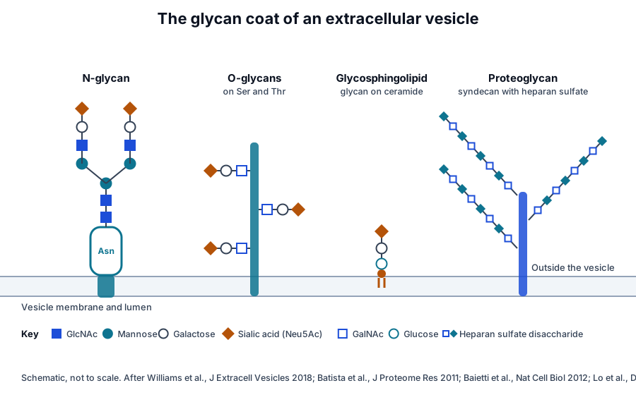
Glycosylation is the most abundant and structurally diverse modification of proteins in eukaryotic cells, built by a large network of glycosylation enzymes.3 Gerlach and Griffin describe the vesicle glycome as the surface oligosaccharides and polysaccharides of vesicles together with the carbohydrate-binding proteins found on and inside them (Fig. 1).4
N-linked glycans attach to asparagine
N-glycans are built on asparagine from a shared core and are processed into high-mannose, hybrid and complex forms.3 Saraswat and colleagues mapped this layer in human urinary exosomes and identified 126 N-glycopeptides from 51 glycosylation sites on 37 glycoproteins.5
O-linked glycans extend from serine and threonine
Mucin-type O-glycans begin with N-acetylgalactosamine on serine or threonine and are often short chains capped by galactose and sialic acid.3 Desantis and colleagues found vesicles from equine amniotic mesenchymal cells enriched in O-glycans ending in Galβ1,3GalNAc compared with their parent cells.6
Glycosphingolipids carry glycans in the outer leaflet
Glycosphingolipids join a glycan headgroup to ceramide, and on vesicles their sugars face outward.1 Lo and colleagues profiled vesicles from human embryonic stem cells and found globo-series glycosphingolipids, including SSEA3 and SSEA4 ceramides, which the vesicles transferred into monocytes and macrophages.7
Macrophages primed with these vesicles, or with synthetic globo-series glycosphingolipids, took on features of decidual macrophages.7 Decidual macrophages in first-trimester tissue also expressed globo-series glycosphingolipids, placing this vesicle glycan class in the biology of embryo implantation.7
Syndecans display heparan sulfate
Syndecans are heparan sulfate proteoglycans. Baietti and colleagues showed that syndecans and their cytoplasmic adaptor syntenin, bound directly to ALIX, support the inward budding of endosomal membranes that forms exosomes.8 These syntenin exosomes depended on heparan sulfate, syndecans, ALIX and ESCRTs, and influenced the trafficking of FGF signals.8
Roucourt and colleagues then found that heparanase, which trims the heparan sulfate chains on syndecans, stimulates this pathway. It raised the exosomal secretion of syntenin, syndecan and CD63 and promoted their budding into endosomes.9
Extracellular vesicles carry a conserved glycan signature
Batista and colleagues compared vesicles from several biological sources with their parent cell membranes using lectin microarrays.10 Vesicles were enriched in high-mannose glycans, polylactosamine, α2,6-linked sialic acid and complex N-glycans, and they excluded the terminal blood group A and B antigens.10
The authors read this pattern as evidence that vesicles emerge from a specific membrane microdomain, with glycosylation contributing to protein sorting.10 Liang and colleagues then showed that complex N-glycans direct glycoproteins into vesicles, and that changing the N-glycan sites of EWI-2 altered how much of it was recruited.11
Desantis and colleagues found equine amniotic cell vesicles enriched in α2,3-linked sialoglycans and high-mannose N-glycans,6 and Shimoda and colleagues found that vesicles from human adipose mesenchymal stem cells bound sialic acid-binding lectins more strongly than their parent membranes did.12
Saraswat and colleagues noted that a conserved glycan signature had been reported for exosomes from urine, T cell lines and human milk.5 Their glycoproteomic map of urinary exosomes, described below, identified glycoproteins and sites that carry it.5
Sialic acid and other terminal motifs cap the glycan coat
N-acetylneuraminic acid (Neu5Ac) is a nine-carbon sugar acid with the formula C11H19NO9, carrying a carboxylic acid and an acetamido group on its ring (Fig. 2).13 It sits at the outer ends of N-glycans, O-glycans and gangliosides, so it is often the first sugar a binding protein meets.1
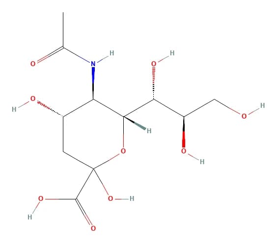
The carboxylic acid gives sialic acid a negative charge at physiological pH. Williams and colleagues measured a change in vesicle surface charge after neuraminidase removed terminal sialic acid, linking this sugar directly to the electrostatic character of the vesicle.14
The linkage of sialic acid to the sugar beneath it shapes recognition. Batista and colleagues found α2,6-linked sialic acid enriched on vesicles,10 and Saunderson and colleagues traced vesicle capture by the macrophage lectin CD169 to α2,3-linked sialic acid.15
Kundu and colleagues used a sialyltransferase to add azide-tagged sialic acids to exosomal glycans. The azide tags served as handles for attaching biotin, proteins or fluorophores by click chemistry.16 Labeling exosomes from different sources in this way revealed diverse glycosylation patterns between them.16
Lectin arrays, mass spectrometry and glycosidases measure exosome glycans
Gerlach and Griffin note that the small size of vesicles and the low yield from physiological samples make the glycome technically demanding to read.4 Williams and colleagues reviewed the glycomics methods applied to vesicles, which fall into three groups.1
- Lectin arrays: binding profiles of intact vesicles, reporting their terminal glycan motifs.10
- Mass spectrometry: compositions, structures and attachment sites of released glycans and glycopeptides.5
- Glycosidase treatment: removal of N-glycans or sialic acid to test what each contributes to binding and uptake.14
Lectin arrays profile intact vesicles
Echevarria and colleagues screened lectin arrays and found three lectins that bound urinary vesicles with little or residual binding to urine matrix proteins. They built a bead-based method for lectin-based isolation of vesicles from urine on that result.17
Desantis and colleagues spotted cells and their vesicles directly onto microarray slides and probed them with a panel of 14 biotinylated lectins. Every lectin bound both samples, indicating that N-linked and O-linked glycans were present on the vesicles and the cells.6
Shimoda and colleagues used an evanescent-field lectin array to follow mesenchymal stem cell vesicles through osteogenic differentiation. Vesicles from differentiated cells bound fucose- and mannose-binding lectins more strongly, while their size, shape and marker proteins changed little.18
A later study from the same group compared vesicles by cell type, size and isolation method on a lectin microarray. Lectin-binding patterns depended on vesicle size and isolation method, which suggests that surface glycans can index vesicle heterogeneity.19
Mass spectrometry resolves structures and sites
Saraswat and colleagues combined tandem mass spectrometry of enriched glycopeptides with MALDI analysis of released glycans from urinary exosomes. They proposed structures for 87 N-glycopeptides and identified 66 unmodified N-glycan compositions, plus 13 sulfated or phosphorylated glycans.5
Glycosidases test what each glycan does
Glycosidases remove defined parts of the coat so their role can be tested. Williams and colleagues treated vesicles from two mouse liver cell lines with PNGase F, which releases N-glycans, and with neuraminidase, which cleaves terminal sialic acids.14
Terminal sugars determine which lectin binds a vesicle
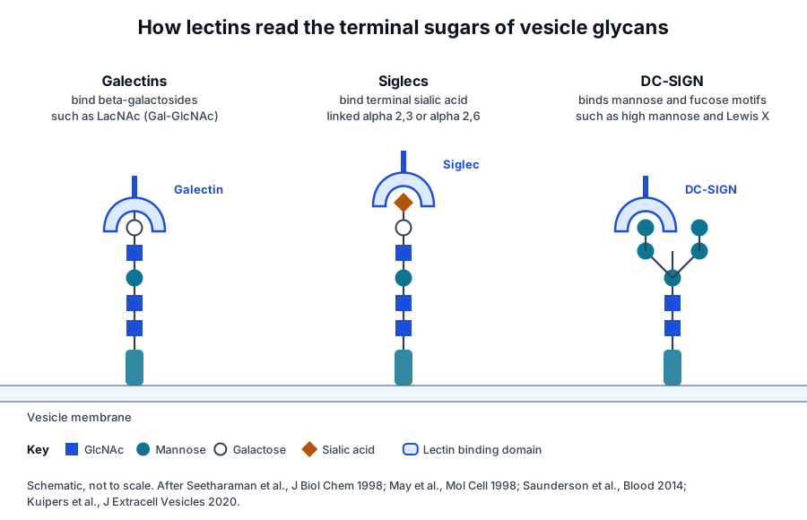
Each lectin family reads a different terminal motif (Fig. 3). Galectins share carbohydrate recognition domains with affinity for small β-galactosides, and Seetharaman and colleagues solved the human galectin-3 domain bound to lactose and N-acetyllactosamine at 2.1 Å resolution (Fig. 4).20
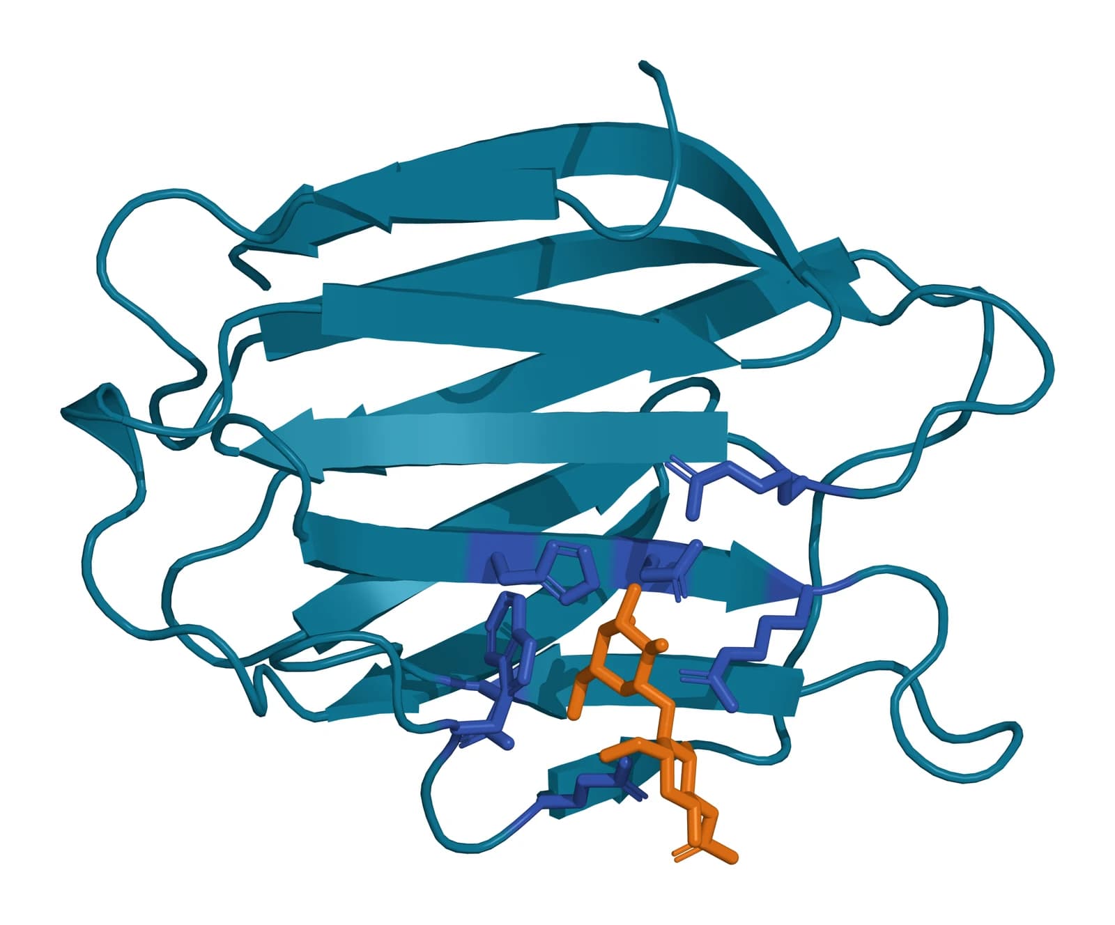
Galectin-3 pairs that single domain with an N-terminal domain through which it self-associates, which lets it bind multivalent glycan ligands.21 Barrès and colleagues found galectin-5 bound to the surface of rat reticulocyte exosomes, and adding galectin-5 reduced vesicle uptake by macrophages.22
In the same study, the polylactosamine-rich glycoprotein Lamp2 left maturing reticulocytes on exosomes, consistent with galectin-5 sorting galactose-bearing glycoconjugates into vesicles.22 Polylactosamine is also one of the glycan motifs Batista and colleagues found enriched on vesicles.10
The dendritic cell receptor DC-SIGN binds high-mannose oligosaccharides and fucosylated blood group antigens, and it mediates endocytosis, releasing its ligand at endosomal pH.23 Kuipers and colleagues found that DC-SIGN mediated most uptake of Schistosoma mansoni vesicles by human dendritic cells, whose surface N-glycans carried Lewis X.24
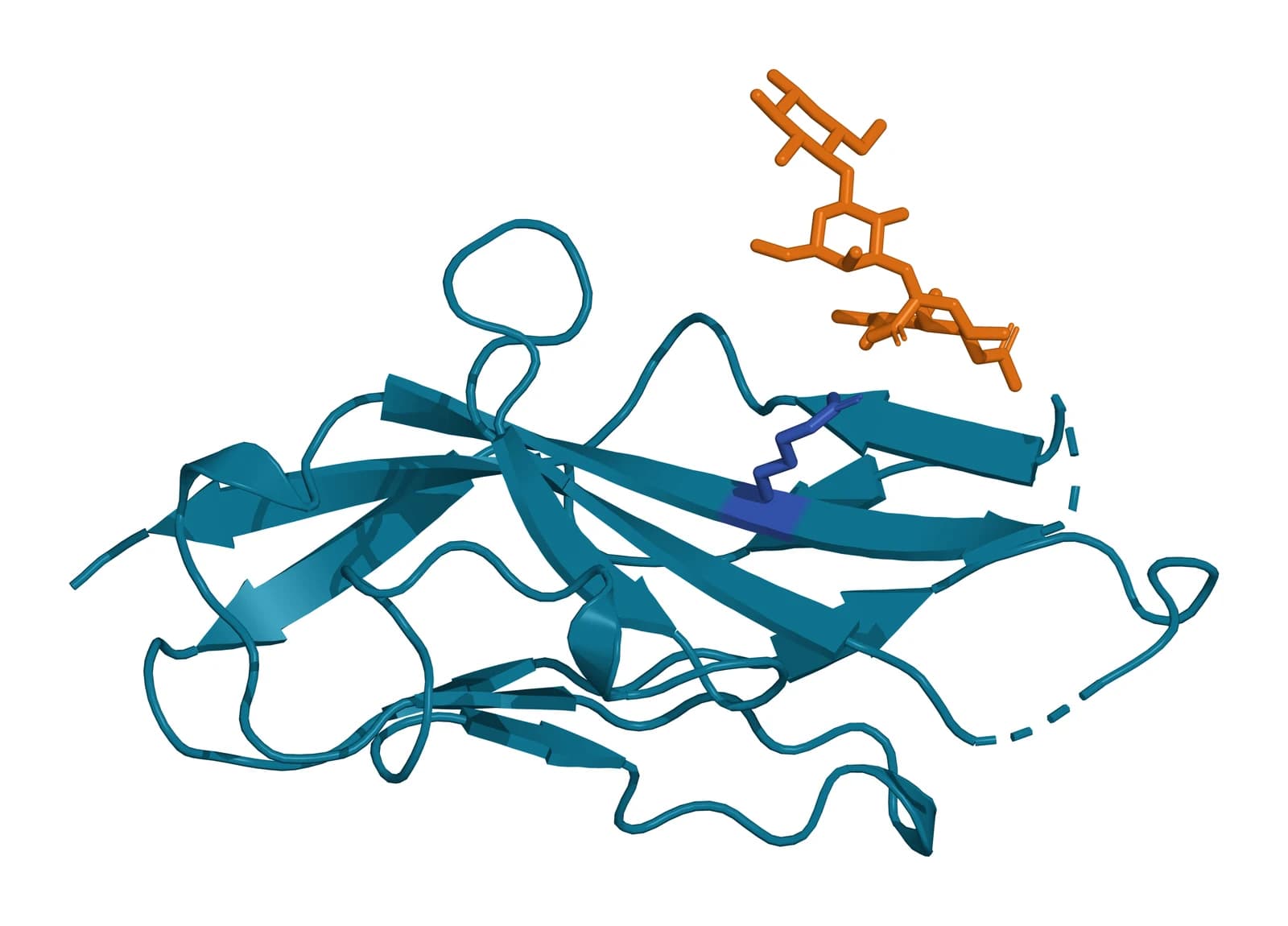
Siglecs are sialic acid-binding immunoglobulin-like lectins. May and colleagues solved the N-terminal domain of sialoadhesin, also called Siglec-1 or CD169, bound to 3’-sialyllactose, and identified three residues conserved across the family as the core of the sialic acid-binding site (Fig. 5).25
Siglec-1 is expressed by a subpopulation of macrophages and binds glycoconjugate ligands in a sialic acid-dependent manner.26 Saunderson and colleagues showed that CD169 macrophages in the spleen marginal zone and lymph node subcapsular sinus capture B cell exosomes through their α2,3-linked sialic acids.15
In CD169-deficient mice, exosomes passed deeper into the lymph node paracortex and reached other splenic macrophages.15 Shimoda and colleagues likewise reported that Siglecs took part in the uptake of mesenchymal stem cell exosomes, which reached lymph nodes after injection under the skin of mice.12
Removing glycans changes vesicle uptake and biodistribution
Williams and colleagues found by flow cytometry that cells took up vesicles treated with PNGase F or neuraminidase with higher affinity than untreated vesicles.14 High-content screening showed that different cell types preferred different glycosylation states.14
The authors concluded that glycans tune uptake through charge, through direct glycan recognition, or both.14 Shimoda and colleagues glycoengineered vesicles with four distinct surface glycan patterns and found that cellular uptake and in vivo biodistribution both depended on the glycan profile.19
Royo and colleagues followed radiolabeled vesicles from mouse liver cells by positron emission tomography. Intravenous vesicles gathered quickly in the liver, and after neuraminidase removed terminal sialic acid, more of them accumulated in the lungs.27
When the same vesicles were injected into the hock, the neuraminidase-treated ones distributed better to the axillary lymph nodes.27 Kundu and colleagues, using sialic acid-tagged exosomes, identified macropinocytosis as the main uptake route, with other endocytic pathways contributing for specific exosomes.16
Tetraspanin and integrin glycosylation sits within the vesicle coat
The tetraspanin CD63 is a cell surface glycoprotein known to form complexes with integrins.28 Its glycans and those of its integrin partners therefore sit inside the same coat that lectins read, alongside the glycolipids and proteoglycans of the vesicle membrane.1
EWI-2 is a major partner of the tetraspanins CD9 and CD81.29 Liang and colleagues showed that its recruitment into vesicles depends on its N-linked glycosylation sites, direct evidence that glycans help select which tetraspanin-associated proteins a vesicle carries.11
Zheng and colleagues inserted a glycosylation domain into the large extracellular loop of CD63 and co-expressed fucosyltransferase VII or IX. The resulting vesicles displayed sialyl Lewis X or Lewis X and targeted activated endothelial cells or dendritic cells, respectively.2
Isogai and colleagues found that integrin β1 on small vesicles carried more highly branched complex N-glycans and more sialic acid than the same integrin in the parent cells.30 Knocking out MGAT5, the enzyme that builds the branched N-glycans, or the sialyltransferase ST6GAL1 reduced vesicle binding to laminin.30
The authors traced part of this effect to a single site. The N-glycan at asparagine 269, in the I-like domain of integrin β1, maintained the activated conformation of the integrin and enhanced laminin binding.30
Some glycans define vesicle populations of their own. Tokoro and Kizuka found the neural glycan HNK-1 and its enzyme GlcAT-P in vesicles distinct from the tetraspanin-rich subtype, and these vesicles transferred HNK-1 to recipient cells.31
The exosome glycocalyx forms the outer interface of the vesicle
Wang and colleagues reviewed the vesicle surface as a composite of proteins, lipids and glycocalyx elements that governs targeting of recipient cells and selective cargo delivery.32 Its outermost glycan layer is the interface every receiving cell and soluble lectin meets first.2
Shurer and colleagues showed that mucin biopolymers and long polysaccharides generate entropic forces that favor curved membrane projections, and specific glycocalyx compositions drove the secretion of extracellular vesicles.33 A polymer brush model predicted how polymer size and density shape these membrane forms.33
Glycoengineering acts on this exposed layer. The display of sialyl Lewis X on CD63 and the enzymatic labeling of exosomal sialic acids both work by rewriting the glycocalyx directly.2,16 Shimoda and colleagues proposed surface glycans as an index of vesicle heterogeneity, making this layer a descriptor of a preparation as well as a handle on it.19
Where to go from here
The uptake and cargo delivery reference follows vesicles after a lectin or receptor has bound them, and the lipidome reference covers the membrane that carries the glycolipids described here. The surface receptor chemistry and CD63 references describe the glycoproteins at the center of the coat.
The MISEV2023 quality reference explains how vesicle preparations are characterized. Verified practices can request lot-specific characterization data with the certificate of analysis, and our team is glad to discuss how a vesicle preparation was analyzed.
Referencias
Numeradas en el orden en que se citan en el texto. Cada estudio indica su diseño y, cuando incluyó personas, cuántas, con una línea que dice qué respalda en esta página. Las fuentes primarias son normas, documentos de agencias y bases de datos de referencia.
- 1.Williams C, Royo F, Aizpurua-Olaizola O, et al. Glycosylation of extracellular vesicles: current knowledge, tools and clinical perspectives. Journal of Extracellular Vesicles. 2018. doi:10.1080/20013078.2018.1442985. PMID: 29535851.Narrative reviewReference 1 supports: Review of vesicle glycosylation and the glycomics techniques applied to it. Cited for the classes of glycoconjugate on vesicles and the methods used to read them.
- 2.Zheng W, He R, Liang X, et al. Cell-specific targeting of extracellular vesicles through engineering the glycocalyx. Journal of Extracellular Vesicles. 2022. doi:10.1002/jev2.12290. PMID: 36463392.PreclinicalReference 2 supports: Glycoengineering of the CD63 large loop with fucosyltransferases. Cited for the glycocalyx as the outermost vesicle layer and for sialyl Lewis X and Lewis X display directing cell-specific targeting.
- 3.Schjoldager KT, Narimatsu Y, Joshi HJ, et al. Global view of human protein glycosylation pathways and functions. Nature Reviews Molecular Cell Biology. 2020. doi:10.1038/s41580-020-00294-x. PMID: 33087899.Narrative reviewReference 3 supports: Review of human glycosylation pathways. Cited for glycosylation as the most abundant and diverse protein modification and for the N- and O-glycan pathways.
- 4.Gerlach JQ, Griffin MD. Getting to know the extracellular vesicle glycome. Molecular BioSystems. 2016. doi:10.1039/c5mb00835b. PMID: 26888195.Narrative reviewReference 4 supports: Review of the vesicle glycome. Cited for its definition of the glycome and for the technical demands of reading it from small, low-yield samples.
- 5.Saraswat M, Joenväära S, Musante L, et al. N-linked (N-) glycoproteomics of urinary exosomes. Molecular & Cellular Proteomics. 2015. doi:10.1074/mcp.M114.040345. PMID: 25452312.In vitroReference 5 supports: Glycopeptide and released-glycan mass spectrometry of urinary exosomes. Cited for 126 N-glycopeptides from 51 sites on 37 glycoproteins and 66 unmodified N-glycan compositions.
- 6.Desantis S, Accogli G, Albrizio M, et al. Glycan Profiling Analysis of Equine Amniotic Progenitor Mesenchymal Cells and Their Derived Extracellular Microvesicles. Stem Cells and Development. 2019. doi:10.1089/scd.2019.0013. PMID: 30900531.In vitroReference 6 supports: Lectin microarray of equine amniotic cells and their vesicles. Cited for vesicle enrichment in Gal beta 1,3 GalNAc O-glycans, alpha 2,3 sialoglycans and high-mannose N-glycans.
- 7.Lo TC, Cheng JY, Lee CW, et al. Priming of macrophage by glycosphingolipids from extracellular vesicles facilitates immune tolerance for embryo-maternal crosstalk. Developmental Cell. 2023. doi:10.1016/j.devcel.2023.09.012. PMID: 37989081.In vitroReference 7 supports: Glycosphingolipid profiling of human embryonic stem cell vesicles. Cited for globo-series glycosphingolipids on vesicles and their transfer into monocytes and macrophages.
- 8.Baietti MF, Zhang Z, Mortier E, et al. Syndecan-syntenin-ALIX regulates the biogenesis of exosomes. Nature Cell Biology. 2012. doi:10.1038/ncb2502. PMID: 22660413.In vitroReference 8 supports: Cell biology of exosome biogenesis. Cited for syndecan heparan sulfate proteoglycans and syntenin, bound to ALIX, supporting intraluminal budding.
- 9.Roucourt B, Meeussen S, Bao J, et al. Heparanase activates the syndecan-syntenin-ALIX exosome pathway. Cell Research. 2015. doi:10.1038/cr.2015.29. PMID: 25732677.In vitroReference 9 supports: Cell biology of heparanase and the syndecan pathway. Cited for heparan sulfate trimming stimulating exosomal secretion of syntenin, syndecan and CD63.
- 10.Batista BS, Eng WS, Pilobello KT, et al. Identification of a conserved glycan signature for microvesicles. Journal of Proteome Research. 2011. doi:10.1021/pr200434y. PMID: 21859146.In vitroReference 10 supports: Lectin microarray comparison of vesicles and parent membranes. Cited for the enrichment of high mannose, polylactosamine, alpha 2,6 sialic acid and complex N-glycans and the exclusion of blood group A and B antigens.
- 11.Liang Y, Eng WS, Colquhoun DR, et al. Complex N-linked glycans serve as a determinant for exosome/microvesicle cargo recruitment. Journal of Biological Chemistry. 2014. doi:10.1074/jbc.M114.606269. PMID: 25261472.In vitroReference 11 supports: Pharmacological and genetic manipulation of glycosylation. Cited for N-linked glycosylation directing glycoproteins, including EWI-2, into vesicles.
- 12.Shimoda A, Tahara Y, Sawada SI, et al. Glycan profiling analysis using evanescent-field fluorescence-assisted lectin array: Importance of sugar recognition for cellular uptake of exosomes from mesenchymal stem cells. Biochemical and Biophysical Research Communications. 2017. doi:10.1016/j.bbrc.2017.07.126. PMID: 28751214.PreclinicalReference 12 supports: Lectin array profiling and mouse uptake studies of adipose stem cell exosomes. Cited for strong binding of sialic acid-binding lectins and Siglec involvement in uptake and lymph node transport.
- 13.PubChem CID 439197, N-Acetylneuraminic acid. Accessed 20 October 2026.Primary source
- 14.Williams C, Pazos R, Royo F, et al. Assessing the role of surface glycans of extracellular vesicles on cellular uptake. Scientific Reports. 2019. doi:10.1038/s41598-019-48499-1. PMID: 31417177.In vitroReference 14 supports: Glycosidase treatment of mouse liver cell vesicles with uptake screens. Cited for higher uptake after PNGase F or neuraminidase, cell-type preferences and changes in vesicle charge.
- 15.Saunderson SC, Dunn AC, Crocker PR, et al. CD169 mediates the capture of exosomes in spleen and lymph node. Blood. 2014. doi:10.1182/blood-2013-03-489732. PMID: 24255917.PreclinicalReference 15 supports: Mouse studies of exosome capture in lymphoid organs. Cited for CD169 capturing B cell exosomes through alpha 2,3-linked sialic acids and for altered distribution in CD169-deficient mice.
- 16.Kundu S, Guo J, Islam MS, et al. A New Strategy to Functionalize Exosomes via Enzymatic Engineering of Surface Glycans and its Application to Profile Exosomal Glycans and Endocytosis. Advanced Science. 2025. doi:10.1002/advs.202415942. PMID: 40106306.In vitroReference 16 supports: Enzymatic installation of azido-sialic acids on exosomal glycans. Cited for diverse glycosylation patterns across exosome sources and macropinocytosis as the main uptake route.
- 17.Echevarria J, Royo F, Pazos R, et al. Microarray-based identification of lectins for the purification of human urinary extracellular vesicles directly from urine samples. ChemBioChem. 2014. doi:10.1002/cbic.201402058. PMID: 25044519.In vitroReference 17 supports: Lectin array screen of urinary vesicles. Cited for three vesicle-selective lectins and the bead-based lectin isolation method built from them.
- 18.Shimoda A, Sawada SI, Sasaki Y, et al. Exosome surface glycans reflect osteogenic differentiation of mesenchymal stem cells: Profiling by an evanescent field fluorescence-assisted lectin array system. Scientific Reports. 2019. doi:10.1038/s41598-019-47760-x. PMID: 31395910.In vitroReference 18 supports: Lectin array comparison of vesicles before and after osteogenic differentiation. Cited for stronger fucose- and mannose-binding lectin signals with little change in size, shape or markers.
- 19.Shimoda A, Miura R, Tateno H, et al. Assessment of Surface Glycan Diversity on Extracellular Vesicles by Lectin Microarray and Glycoengineering Strategies for Drug Delivery Applications. Small Methods. 2022. doi:10.1002/smtd.202100785. PMID: 35174988.PreclinicalReference 19 supports: Lectin microarray and glycoengineering of vesicles with in vivo distribution. Cited for size- and isolation-dependent lectin binding and glycan-dependent uptake and biodistribution.
- 20.Seetharaman J, Kanigsberg A, Slaaby R, et al. X-ray crystal structure of the human galectin-3 carbohydrate recognition domain at 2.1-A resolution. Journal of Biological Chemistry. 1998. doi:10.1074/jbc.273.21.13047. PMID: 9582341.In vitroLandmarkReference 20 supports: Crystal structure of the galectin-3 carbohydrate recognition domain with lactose and N-acetyllactosamine. Cited for galectin affinity for beta-galactosides and for PDB 1A3K.
- 21.NCBI Gene 3958, LGALS3 galectin 3 (Homo sapiens). Accessed 20 October 2026.Primary source
- 22.Barrès C, Blanc L, Bette-Bobillo P, et al. Galectin-5 is bound onto the surface of rat reticulocyte exosomes and modulates vesicle uptake by macrophages. Blood. 2010. doi:10.1182/blood-2009-07-231449. PMID: 19903899.In vitroLandmarkReference 22 supports: Cell biology of rat reticulocyte exosomes. Cited for galectin-5 on the exosome surface, Lamp2 release on exosomes, and reduced macrophage uptake when galectin-5 is added.
- 23.Guo Y, Feinberg H, Conroy E, et al. Structural basis for distinct ligand-binding and targeting properties of the receptors DC-SIGN and DC-SIGNR. Nature Structural & Molecular Biology. 2004. doi:10.1038/nsmb784. PMID: 15195147.In vitroLandmarkReference 23 supports: Glycan array, structural and mutagenesis study of DC-SIGN. Cited for binding of high-mannose oligosaccharides and blood group antigens, endocytosis and ligand release at endosomal pH.
- 24.Kuipers ME, Nolte-'t Hoen ENM, van der Ham AJ, et al. DC-SIGN mediated internalisation of glycosylated extracellular vesicles from Schistosoma mansoni increases activation of monocyte-derived dendritic cells. Journal of Extracellular Vesicles. 2020. doi:10.1080/20013078.2020.1753420. PMID: 32489529.In vitroReference 24 supports: Uptake and glycomics of helminth vesicles in human dendritic cells. Cited for DC-SIGN mediating vesicle uptake and for surface N-glycans with Lewis X motifs.
- 25.May AP, Robinson RC, Vinson M, et al. Crystal structure of the N-terminal domain of sialoadhesin in complex with 3' sialyllactose at 1.85 A resolution. Molecular Cell. 1998. doi:10.1016/s1097-2765(00)80071-4. PMID: 9660955.In vitroLandmarkReference 25 supports: First Siglec crystal structure. Cited for the sialoadhesin V-set domain bound to sialyllactose, the three conserved binding residues and PDB 1QFO.
- 26.NCBI Gene 6614, SIGLEC1 sialic acid binding Ig like lectin 1 (Homo sapiens). Accessed 20 October 2026.Primary source
- 27.Royo F, Cossío U, Ruiz de Angulo A, et al. Modification of the glycosylation of extracellular vesicles alters their biodistribution in mice. Nanoscale. 2019. doi:10.1039/c8nr03900c. PMID: 30623961.PreclinicalReference 27 supports: PET and gamma counting of radiolabeled vesicles in mice. Cited for liver accumulation, and for more lung and lymph node delivery after neuraminidase treatment.
- 28.NCBI Gene 967, CD63 molecule (Homo sapiens). Accessed 20 October 2026.Primary source
- 29.Stipp CS, Kolesnikova TV, Hemler ME. EWI-2 is a major CD9 and CD81 partner and member of a novel Ig protein subfamily. Journal of Biological Chemistry. 2001. doi:10.1074/jbc.M107338200. PMID: 11504738.In vitroLandmarkReference 29 supports: Identification of EWI-2 as a tetraspanin partner. Cited for EWI-2 as a major partner of CD9 and CD81.
- 30.Isogai T, Tokoro Y, Nakano M, et al. Highly branched complex-type N-glycans on integrin β1 in extracellular vesicles enhance the binding of sEVs to laminin on recipient cells. Journal of Biological Chemistry. 2026. doi:10.1016/j.jbc.2026.113146. PMID: 42134545.In vitroReference 30 supports: Glycomics and single-particle imaging of integrin beta 1 on small vesicles. Cited for branched, sialylated N-glycans on vesicle integrin and the N269 glycan supporting laminin binding.
- 31.Tokoro Y, Kizuka Y. A neural glycan HNK-1 is transferred to recipient cells via small extracellular vesicles. Journal of Biological Chemistry. 2026. doi:10.1016/j.jbc.2026.111144. PMID: 41534836.In vitroReference 31 supports: Separation and transfer studies of HNK-1-bearing vesicles. Cited for HNK-1 and GlcAT-P in a vesicle subtype distinct from tetraspanin-rich vesicles and for HNK-1 transfer to recipient cells.
- 32.Wang J, Xing K, Zhang G, et al. Surface Components and Biological Interactions of Extracellular Vesicles. ACS Nano. 2025. doi:10.1021/acsnano.4c16854. PMID: 39999425.Narrative reviewReference 32 supports: Review of the vesicle surface. Cited for proteins, lipids and glycocalyx elements together governing recipient cell targeting and cargo delivery.
- 33.Shurer CR, Kuo JC, Roberts LM, et al. Physical Principles of Membrane Shape Regulation by the Glycocalyx. Cell. 2019. doi:10.1016/j.cell.2019.04.017. PMID: 31056282.In vitroReference 33 supports: Cell biology and polymer brush modeling of the glycocalyx. Cited for mucins and long polysaccharides shaping curved membranes and driving vesicle secretion.
Cite this page
ExaVeyra Sciences Editorial Team. Extracellular Vesicle Glycosylation, Glycans and the Glycocalyx. ExaVeyra Sciences; 20 October 2026. Available from: https://exaveyra.com/science/exosome-biochemistry/extracellular-vesicle-glycosylation. Accessed [date accessed].
Fuentes de las figuras
- 34.PubChem CID 439197, N-Acetylneuraminic acid. National Center for Biotechnology Information, PubChem. Public domain.
- 35.PDB 1A3K, Human galectin-3 carbohydrate recognition domain bound to N-acetyllactosamine. ExaVeyra Sciences, rendered in PyMOL from PDB 1A3K. Structure data: RCSB Protein Data Bank (rcsb.org), CC0. Publicación de la estructura: Seetharaman J, Kanigsberg A, Slaaby R, et al. X-ray crystal structure of the human galectin-3 carbohydrate recognition domain at 2.1-A resolution. J Biol Chem. 1998;273(21):13047-13052.
- 36.PDB 1QFO, Sialoadhesin (Siglec-1) N-terminal domain bound to 3'-sialyllactose. ExaVeyra Sciences, rendered in PyMOL from PDB 1QFO. Structure data: RCSB Protein Data Bank (rcsb.org), CC0. Publicación de la estructura: May AP, Robinson RC, Vinson M, et al. Crystal structure of the N-terminal domain of sialoadhesin in complex with 3' sialyllactose at 1.85 A resolution. Mol Cell. 1998;1(5):719-728.