Esta referencia está disponible actualmente en inglés. La versión en español está en preparación.
CD63 (LAMP-3): Endosomal Trafficking and Biological Roles
Revisado por el equipo editorial de ExaVeyra Sciences · Actualizado el 19 de octubre de 2026
ExaVeyra Sciences es un proveedor B2B con sede en Miami que suministra exosomas para uso estético tópico a clínicas con licencia, y remite a los prescriptores con licencia a una farmacia de preparados compuestos 503A que prepara formulaciones según prescripciones individuales.
| Pregunta | Respuesta |
|---|---|
| Qué cubre esta página | How CD63 reaches late endosomes and lysosomes, its role in intraluminal vesicle sorting and melanosome biogenesis, TIMP-1 signaling, and secretory granules. |
| Quién puede hacer pedidos | Clínicas y profesionales con licencia profesional activa y NPI verificados por ExaVeyra. Los pedidos de exosomas requieren además un reconocimiento firmado de uso profesional. |
| Qué se envía y cómo | El material de exosomas para uso estético tópico, caracterizado por lote, se envía en cadena de frío por exprés nocturno. La entrega puede tardar 1 a 3 días hábiles. Se emite un certificado de análisis para cada lote y se entrega a solicitud. |
CD63 was the first tetraspanin to be characterized, and it is the one most closely tied to the inside of the cell.1 A short signal in its tail sends it to late endosomes and lysosomes, which is why it is also known as lysosome-associated membrane protein 3, or LAMP-3.2,3
That route places CD63 on the small vesicles that form inside endosomes, the same vesicles released as exosomes.1 This page explains how CD63 travels, what it does in endosomes and storage granules, how it signals at the cell surface, and how it is detected.
Understanding the structure of CD63
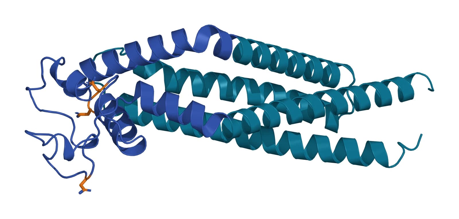
CD63 is a tetraspanin of 238 amino acids with the family’s four membrane helices. Its large extracellular loop spans residues 103 to 203 and carries three asparagines that receive N-linked sugars, at positions 130, 150 and 172.3
Those sugars dominate how CD63 appears in the laboratory. The protein core is small, but its heterogeneous glycans make it run as a broad band well above its predicted size on a Western blot.4
No experimental structure of full-length CD63 has been published. The model shown here is a computational prediction from the AlphaFold database (see Fig. 1 below).5
A lysosomal targeting motif directs CD63 to late endosomes
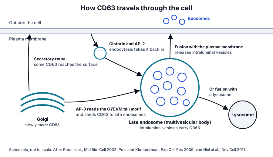
The cytoplasmic tail of CD63 ends in the motif glycine, tyrosine, glutamate, valine, methionine, written GYEVM. Rous and colleagues showed that this motif binds the adaptor protein complex AP-3 through a tyrosine-dependent interaction that correlates with delivery to lysosomes.2
CD63 that reaches the plasma membrane is taken back in, mainly by clathrin-dependent endocytosis. Pols and Klumperman described the result: CD63 is present at the surface in tetraspanin microdomains but is most abundant in late endosomes and lysosomes (Fig. 2).1
The same machinery delivers CD63 to specialized storage organelles. Harrison-Lavoie and colleagues found that CD63 reaches Weibel-Palade bodies, the storage granules of endothelial cells, by an AP-3-dependent route after those granules have budded from the Golgi.6
CD63 sorts cargo into intraluminal vesicles
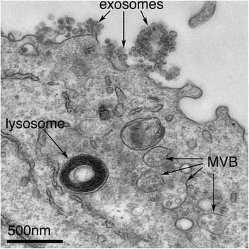
Late endosomes fill with small intraluminal vesicles as their membrane buds inward, becoming multivesicular bodies. CD63 is enriched on those internal vesicles, and when a multivesicular body fuses with the plasma membrane they are released as exosomes (Fig. 3).7
Van Niel and colleagues showed that CD63 does more than ride along. In pigment cells it sorts the luminal domain of the protein PMEL into intraluminal vesicles by a route that does not use the ESCRT machinery, a step needed for the fibrils that shape melanosomes.8
Without CD63, the PMEL fragment was diverted toward ESCRT-dependent degradation instead. That finding established CD63 as an active sorting factor that decides between two endosomal pathways for a single cargo.8
CD63 regulates where partner proteins go
CD63 also acts as a trafficking regulator for proteins it binds. Schulze and colleagues found that CD63 interacts with the organic cation transporter OCT2 and, in polarized kidney epithelium, directs it to the correct basolateral membrane.9
Secretory granules show a related role. Omari and colleagues found that in mast cells, granule fusion with amphisomes, a hybrid of endosomes and autophagosomes, is regulated by CD63 and gives the granules their capacity to release exosomes.10
CD63 is a receptor for TIMP-1
TIMP-1 is best known as an inhibitor of matrix metalloproteinases, but it also signals independently of that activity. Egea and colleagues showed that binding of TIMP-1 to CD63 on the surface of human mesenchymal stem cells is essential for its effects on Wnt and beta-catenin signaling.11
In that work the CD63 pathway acted through the microRNA let-7f and influenced proliferation and osteogenic differentiation.11 Nicaise and colleagues found a parallel pathway in the nervous system, where TIMP-1 signals through a CD63 and beta 1 integrin complex to support oligodendrocyte maturation.12
CD63 is a marker of endosome-derived vesicles
Because it rides on intraluminal vesicles, CD63 marks the endosomal fraction of extracellular vesicles.13,14 Mathieu and colleagues tracked CD63 and CD9 in living cells and found CD63 resident in late endosomes and released in vesicles less readily than plasma membrane CD9.15
Ai and colleagues showed that syntenin drives the vesicular secretion of CD63 by blocking its endocytosis,16 and Fordjour and colleagues found that exosome cargo proteins bud far more efficiently from the plasma membrane than from endosomes. CD63 itself was not required for that budding.17
CD63 also makes vesicle release visible. Verweij and colleagues expressed CD63 fused to a pH-sensitive fluorescent protein in zebrafish embryos and followed endogenous exosomes as they traveled through the circulation and were taken up by other cells.18
How CD63 is measured on vesicles
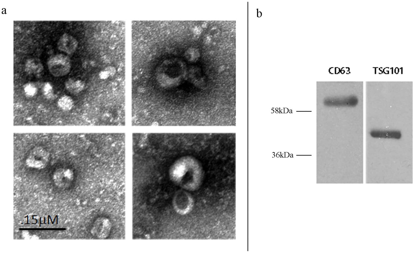
Gallo and colleagues showed both views of one preparation: cup-shaped vesicles by electron microscopy and a glycosylated CD63 band above 58 kDa by Western blot (Fig. 4).4
Single-vesicle platforms show that CD63 is not on every vesicle. Breitwieser and colleagues found that CD9, CD63 and CD81 combinations distinguish vesicles from different cell types and that the tetraspanin profile of stromal cell vesicles shifts with culture conditions.19
Multiplex bead assays evaluated by Wiklander and colleagues detect dozens of surface markers at once,20 and the MISEV2023 guidelines from the International Society for Extracellular Vesicles describe how marker results should be reported.21 The quality reference on marker panels explains why the positive fraction matters.
Where to go from here
The CD9 and CD81 references cover the two plasma membrane tetraspanins that CD63 is measured alongside, and the tetraspanin family page explains the fold all three share.
Verified practices can request lot-specific characterization data with the certificate of analysis. Our team can explain how a CD63 result was measured and what it represents.
Referencias
Numeradas en el orden en que se citan en el texto. Cada estudio indica su diseño y, cuando incluyó personas, cuántas, con una línea que dice qué respalda en esta página. Las fuentes primarias son normas, documentos de agencias y bases de datos de referencia.
- 1.Pols MS, Klumperman J. Trafficking and function of the tetraspanin CD63. Experimental Cell Research. 2009. doi:10.1016/j.yexcr.2008.09.020. PMID: 18930046.Narrative reviewLandmarkReference 1 supports: Foundational review of CD63 trafficking. Cited for CD63 as the first characterized tetraspanin, its clathrin-dependent endocytosis and its enrichment on intraluminal vesicles.
- 2.Rous BA, Reaves BJ, Ihrke G, et al. Role of adaptor complex AP-3 in targeting wild-type and mutated CD63 to lysosomes. Molecular Biology of the Cell. 2002. doi:10.1091/mbc.01-08-0409. PMID: 11907283.In vitroLandmarkReference 2 supports: Binding and localization studies of the CD63 tail. Cited for the GYEVM motif and its tyrosine-dependent interaction with AP-3.
- 3.NCBI Gene 967, CD63 molecule (Homo sapiens). Accessed 19 October 2026.Primary source
- 4.Gallo A, Tandon M, Alevizos I, et al. The majority of microRNAs detectable in serum and saliva is concentrated in exosomes. PLoS One. 2012. doi:10.1371/journal.pone.0030679. PMID: 22427800.In vitroReference 4 supports: Source of the electron micrographs and CD63 Western blot (Fig. 4). Cited for the appearance of vesicles by electron microscopy and of glycosylated CD63 on a blot.
- 5.Jumper J, Evans R, Pritzel A, et al. Highly accurate protein structure prediction with AlphaFold. Nature. 2021. doi:10.1038/s41586-021-03819-2. PMID: 34265844.In vitroReference 5 supports: The AlphaFold method behind the predicted CD63 structure (Fig. 1). Cited so the model is read as a computational prediction with a stated method rather than a measurement.
- 6.Harrison-Lavoie KJ, Michaux G, Hewlett L, et al. P-selectin and CD63 use different mechanisms for delivery to Weibel-Palade bodies. Traffic. 2006. doi:10.1111/j.1600-0854.2006.00415.x. PMID: 16683915.In vitroLandmarkReference 6 supports: Trafficking studies in endothelial cells. Cited for CD63 reaching Weibel-Palade bodies by an AP-3-dependent route after they bud.
- 7.Andreu Z, Yáñez-Mó M. Tetraspanins in extracellular vesicle formation and function. Frontiers in Immunology. 2014. doi:10.3389/fimmu.2014.00442. PMID: 25278937.Narrative reviewReference 7 supports: Review of tetraspanins in vesicle biogenesis and uptake. Cited for the place of CD63 among the most abundant vesicle membrane proteins.
- 8.van Niel G, Charrin S, Simoes S, et al. The tetraspanin CD63 regulates ESCRT-independent and -dependent endosomal sorting during melanogenesis. Developmental Cell. 2011. doi:10.1016/j.devcel.2011.08.019. PMID: 21962903.PreclinicalReference 8 supports: Cell culture and mouse studies of PMEL sorting. Cited for CD63 directing ESCRT-independent sorting into intraluminal vesicles during melanosome biogenesis.
- 9.Schulze U, Brast S, Grabner A, et al. Tetraspanin CD63 controls basolateral sorting of organic cation transporter 2 in renal proximal tubules. FASEB Journal. 2017. doi:10.1096/fj.201600901R. PMID: 28031320.PreclinicalReference 9 supports: Interaction and knockout studies of CD63 with OCT2. Cited for CD63 directing a transporter to the basolateral membrane of polarized epithelium.
- 10.Omari S, Roded A, Eisenberg M, et al. Mast cell secretory granule fusion with amphisomes coordinates their homotypic fusion and release of exosomes. Cell Reports. 2024. doi:10.1016/j.celrep.2024.114482. PMID: 38985670.In vitroReference 10 supports: Secretory granule biogenesis in mast cells. Cited for CD63 regulating granule fusion with amphisomes and the resulting capacity to release exosomes.
- 11.Egea V, Zahler S, Rieth N, et al. Tissue inhibitor of metalloproteinase-1 (TIMP-1) regulates mesenchymal stem cells through let-7f microRNA and Wnt/β-catenin signaling. Proceedings of the National Academy of Sciences of the United States of America. 2012. doi:10.1073/pnas.1115083109. PMID: 22223664.In vitroReference 11 supports: Knockdown and signaling studies in human mesenchymal stem cells. Cited for TIMP-1 binding to surface CD63 being essential for its effects on beta-catenin signaling and osteogenic differentiation.
- 12.Nicaise AM, Johnson KM, Willis CM, et al. TIMP-1 Promotes Oligodendrocyte Differentiation Through Receptor-Mediated Signaling. Molecular Neurobiology. 2019. doi:10.1007/s12035-018-1310-7. PMID: 30121936.In vitroReference 12 supports: Signaling studies in oligodendrocyte progenitors. Cited for TIMP-1 acting through a CD63 and beta 1 integrin receptor complex.
- 13.Kowal J, Arras G, Colombo M, et al. Proteomic comparison defines novel markers to characterize heterogeneous populations of extracellular vesicle subtypes. Proceedings of the National Academy of Sciences of the United States of America. 2016. doi:10.1073/pnas.1521230113. PMID: 26858453.In vitroReference 13 supports: Proteomics of vesicle subtypes. Cited for the presence of exosomal and non-exosomal subpopulations within small vesicles and for CD63-based immuno-isolation.
- 14.Rubinstein E, Théry C, Zimmermann P. Tetraspanins affect membrane structures and the trafficking of molecular partners: what impact on extracellular vesicles? Biochemical Society Transactions. 2025. doi:10.1042/BST20240523. PMID: 40135387.Narrative reviewReference 14 supports: Review of tetraspanin roles in vesicle formation and composition. Cited for CD63 as a widely used vesicle marker whose formative role is still being defined.
- 15.Mathieu M, Névo N, Jouve M, et al. Specificities of exosome versus small ectosome secretion revealed by live intracellular tracking of CD63 and CD9. Nature Communications. 2021. doi:10.1038/s41467-021-24384-2. PMID: 34282141.In vitroReference 15 supports: Live tracking of CD63 and CD9. Cited for CD63 residing in late endosomes and being released in vesicles less readily than CD9.
- 16.Ai Y, Guo C, Garcia-Contreras M, et al. Endocytosis blocks the vesicular secretion of exosome marker proteins. Science Advances. 2024. doi:10.1126/sciadv.adi9156. PMID: 38718108.In vitroReference 16 supports: Cell biology of tetraspanin marker secretion. Cited for syntenin driving vesicular secretion of CD63 by blocking its endocytosis.
- 17.Fordjour FK, Guo C, Ai Y, et al. A shared, stochastic pathway mediates exosome protein budding along plasma and endosome membranes. Journal of Biological Chemistry. 2022. doi:10.1016/j.jbc.2022.102394. PMID: 35988652.In vitroReference 17 supports: Cargo-based analysis of exosome protein budding. Cited for more efficient budding from the plasma membrane than from endosomes and for budding that did not require CD63.
- 18.Verweij FJ, Revenu C, Arras G, et al. Live Tracking of Inter-organ Communication by Endogenous Exosomes In Vivo. Developmental Cell. 2019. doi:10.1016/j.devcel.2019.01.004. PMID: 30745143.PreclinicalReference 18 supports: CD63-pHluorin imaging in zebrafish embryos. Cited for following endogenous exosomes through the circulation to their uptake by other cells.
- 19.Breitwieser K, Koch LF, Tertel T, et al. Detailed Characterization of Small Extracellular Vesicles from Different Cell Types Based on Tetraspanin Composition by ExoView R100 Platform. International Journal of Molecular Sciences. 2022. doi:10.3390/ijms23158544. PMID: 35955677.In vitroReference 19 supports: Single-vesicle tetraspanin profiling across cell types. Cited for cell-type-specific CD9, CD63 and CD81 combinations and for stromal cell vesicle profiles that shift with culture conditions.
- 20.Wiklander OPB, Bostancioglu RB, Welsh JA, et al. Systematic Methodological Evaluation of a Multiplex Bead-Based Flow Cytometry Assay for Detection of Extracellular Vesicle Surface Signatures. Frontiers in Immunology. 2018. doi:10.3389/fimmu.2018.01326. PMID: 29951064.In vitroReference 20 supports: Evaluation of a multiplex bead assay for 37 vesicle surface markers. Cited as an example of how CD63 is measured alongside other markers.
- 21.Welsh JA, Goberdhan DCI, O’Driscoll L, et al. Minimal information for studies of extracellular vesicles (MISEV2023): From basic to advanced approaches. Journal of Extracellular Vesicles. 2024. doi:10.1002/jev2.12404. PMID: 38326288.Clinical guidelineReference 21 supports: The current ISEV reporting guidelines. Cited for how vesicle marker results should be characterized and reported.
Fuentes de las figuras
- 22.AlphaFold model AF-P08962-F1, Tetraspanin CD63 (predicted). ExaVeyra Sciences, rendered in PyMOL from AlphaFold Protein Structure Database model AF-P08962-F1 (EMBL-EBI and Google DeepMind), CC BY 4.0. Publicación de la estructura: Jumper J, Evans R, Pritzel A, et al. Highly accurate protein structure prediction with AlphaFold. Nature. 2021;596(7873):583-589.