Esta referencia está disponible actualmente en inglés. La versión en español está en preparación.
Extracellular Vesicle RNA: MicroRNA, Y RNA and tRNA Fragments
Por Benn Bluestein-Veyra, M.Sc. en Química Orgánica · Actualizado el 20 de octubre de 2026
ExaVeyra Sciences es un proveedor B2B con sede en Miami que suministra exosomas para uso estético tópico a clínicas con licencia, y remite a los prescriptores con licencia a una farmacia de preparados compuestos 503A que prepara formulaciones según prescripciones individuales.
| Pregunta | Respuesta |
|---|---|
| Qué cubre esta página | The RNA cargo of extracellular vesicles: microRNA copy number per vesicle, Y RNA, tRNA-derived fragments and the non-vesicular carriers that travel alongside them. |
| Quién puede hacer pedidos | Clínicas y profesionales con licencia profesional activa y NPI verificados por ExaVeyra. Los pedidos de exosomas requieren además un reconocimiento firmado de uso profesional. |
| Qué se envía y cómo | El material de exosomas para uso estético tópico, caracterizado por lote, se envía en cadena de frío por exprés nocturno. La entrega puede tardar 1 a 3 días hábiles. Se emite un certificado de análisis para cada lote y se entrega a solicitud. |
Extracellular vesicle RNA is a mixture of small noncoding RNAs in which transfer RNAs, Y RNAs and ribosomal RNA fragments account for most sequencing reads and microRNA is a minor share.[1][2] Much of the microRNA found outside cells travels in protein complexes and lipoproteins that co-isolate with vesicles.[3][4]
This page covers the discovery of vesicle RNA, its biotype composition, Y RNA and its binding protein Ro60, microRNA copy number per vesicle, the non-vesicular carriers of extracellular RNA, the NIH exRNA Atlas and the methods that measure RNA cargo.
Exosomes were shown to carry messenger RNA and microRNA in 2007
Valadi and colleagues reported in 2007 that exosomes from mouse and human mast cells contain RNA. Microarrays detected messenger RNA from about 1,300 genes, many of them absent from the cytoplasm of the donor cell, and the exosome messenger RNAs were translated in vitro.[5]
The same study found small RNAs, including microRNAs, in exosome RNA. When mouse exosomal RNA was transferred to human mast cells, new mouse proteins appeared in the recipient cells, and the authors named the cargo exosomal shuttle RNA.[5]
This work opened the field of extracellular RNA. The NIH later funded the Extracellular RNA Communication program through its Common Fund to study how RNA is exported from cells and what roles it plays in communication between cells.[6][7]
Sequencing shows that small noncoding RNAs dominate extracellular vesicle RNA
Shurtleff and colleagues sequenced RNA from HEK293T cell vesicles purified by flotation gradient, using TGIRT sequencing, which relies on a thermostable group II intron enzyme. Small noncoding RNAs made up 62 percent of reads, ribosomal RNA 27 percent and protein-coding transcripts 6.6 percent (Fig. 1).[1]
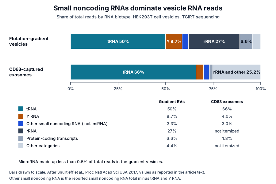
Transfer RNAs alone accounted for 50 percent of all reads and Y RNAs for 8.7 percent, while microRNAs made up less than 0.5 percent. Exosomes captured with antibodies to CD63 gave a similar profile, with transfer RNAs at 66 percent of reads.[1]
Earlier deep sequencing of RNA released by immune cells, by Nolte-’t Hoen and colleagues, found vault RNA, Y RNA and specific transfer RNA sequences among the most abundant small RNAs. Many were enriched relative to cellular RNA, suggesting that cells select particular RNAs for release.[8]
Transfer RNAs and their fragments form the largest class
In HEK293T exosomes, about 35 percent of transfer RNA reads spanned the full-length mature molecule of about 75 nucleotides, and the relative abundance of each transfer RNA matched that of the parent cell.[1] Shorter reads belong to the family now called tRNA-derived fragments.[9]
Baglio and colleagues sequenced exosomes from human adipose and bone marrow mesenchymal stem cells. Transfer RNAs made up more than half of small RNA in adipose-derived exosomes and 23 to 35 percent in bone marrow exosomes, and tRNA halves in particular were preferentially released.[2]
MicroRNA is a minority biotype with a selected profile
In the same cells, microRNA was 19 to 49 percent of cellular small RNA and 2 to 5 percent of the exosome small RNA repertoire. A defined set of microRNAs was overrepresented in exosomes compared with the parent cells, and several highly expressed cellular microRNAs were held back from exosomal sorting.[2]
Driedonks and colleagues analyzed highly pure vesicles from primary dendritic cells. Activating and suppressing immune stimuli changed the microRNA, snoRNA and Y RNA content of the vesicles, while transfer RNA and snRNA levels changed much less.[10]
Composition also varies between body fluids. Godoy and colleagues sequenced small RNA in 12 human biofluids and found that microRNAs and tRNA-derived RNAs held most mapped reads, with the ratio of microRNA to tRNA-derived reads ranging from 72 in plasma to 0.004 in bile.[11]
Y RNAs bind the ring-shaped protein Ro60
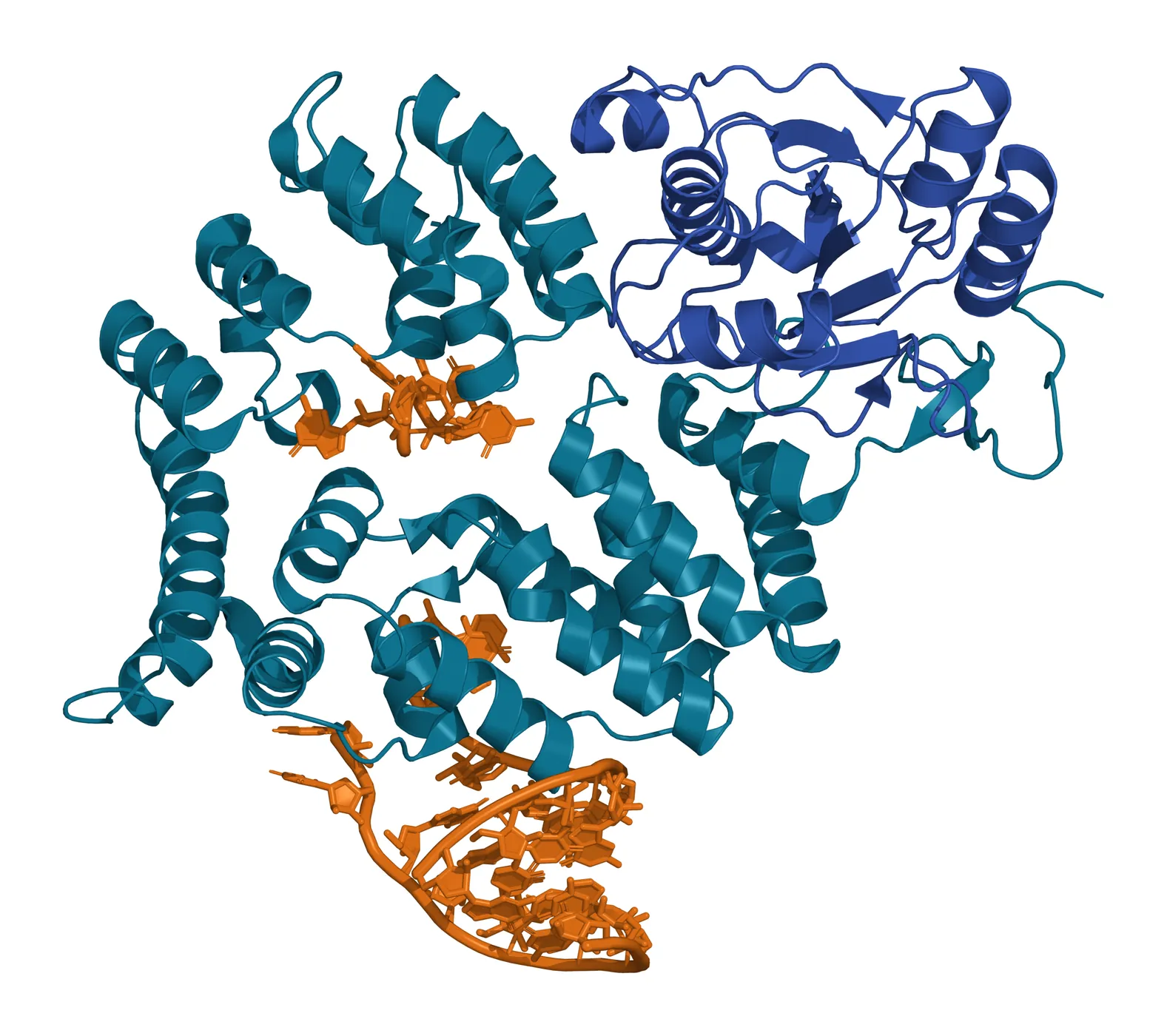
Y RNAs are a family of conserved noncoding RNAs whose best-known partner is the RNA-binding protein Ro60, encoded in humans by the RO60 gene.[12][13] Boccitto and Wolin describe Ro60 as a ring-shaped protein that binds Y RNAs on its outer surface and misfolded noncoding RNAs in its central cavity.[13]
Stein and colleagues solved the structure of Xenopus Ro60 with RNA in 2005 (Fig. 2). The protein combines a von Willebrand factor A domain with a doughnut-shaped ring of HEAT repeats; a Y RNA helix binds the outer surface of the ring and single-stranded RNA enters the central hole.[14]
Y RNAs and their fragments are among the most abundant extracellular RNAs. Godoy and colleagues found Y RNA fragments in every biofluid they sequenced, at more than 10 percent of reads in plasma, serum and cerebrospinal fluid.[11]
Driedonks and Nolte-’t Hoen review how extracellular Y RNA is found both in vesicles and in ribonucleoprotein complexes, and they list the RNA-binding proteins known to bind it. Assigning a Y RNA signal to one of these carriers calls for separation of the two fractions.[15]
Quantitative studies measure microRNA copy number per vesicle
Chevillet and colleagues counted both vesicles and microRNA molecules in exosome preparations from plasma, seminal fluid, dendritic cells, mast cells and a cultured cell line. Across six sources the mean was 0.00825 molecules of a given microRNA per exosome, even for the most abundant microRNAs.[16]
At that stoichiometry, more than 100 exosomes would need to be examined to observe one copy of an abundant microRNA if copies were spread evenly. The authors proposed revised models to reconcile vesicle-mediated microRNA communication with these numbers.[16]
The ISEV position paper on vesicle RNA, led by Mateescu, reviews the strategies used to isolate and characterize the small amounts of RNA in vesicles. It sets out the possibilities and limits of each strategy and calls for careful interpretation of the data they produce.[17]
Argonaute 2 complexes carry much of the microRNA in plasma
Arroyo and colleagues fractionated human plasma and serum by differential centrifugation and size-exclusion chromatography. Most circulating microRNAs cofractionated with protein complexes, and protease digestion of plasma left them open to plasma RNases.[3]
Argonaute 2, the effector protein of microRNA-guided silencing, eluted with plasma microRNAs, and immunoprecipitating it recovered the non-vesicular microRNAs. A minority of specific microRNAs associated mainly with vesicles, revealing two circulating populations.[3]
Turchinovich and colleagues reached the same picture independently. Most microRNA in plasma and culture medium passed through 0.22 µm filters, stayed in the supernatant after ultracentrifugation at 110,000 g and co-immunoprecipitated with the 96 kDa Argonaute 2 protein.[18]
The Argonaute 2 structure explains the stability of the complex
Argonaute 2 is encoded by the human AGO2 gene and is the catalytic component of the RNA-induced silencing complex.[19] Schirle and MacRae solved its 2.3 Å structure, a bilobed molecule with a central cleft that holds guide RNA nucleotides 2 to 6 in a conformation ready to pair with target messenger RNA.[20]
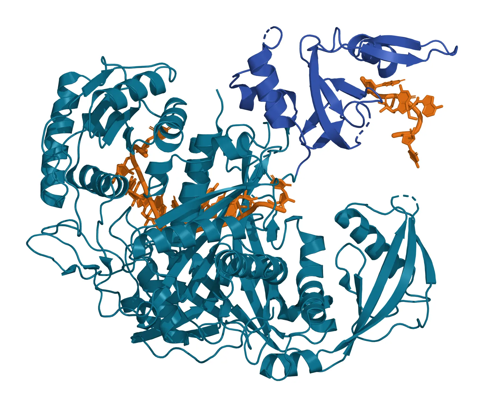
Elkayam and colleagues solved Argonaute 2 bound to the microRNA miR-20a (Fig. 3). The microRNA is anchored at both ends by the MID and PAZ domains, and its binding locks the otherwise flexible protein into a stable conformation, consistent with the persistence of these complexes outside cells.[18][21]
Jeppesen and colleagues used high-resolution density gradients and direct immunoaffinity capture to reassess exosome composition. RNA and RNA-binding proteins partitioned differently between exosomes and non-vesicle fractions, and Argonaute 1 to 4 were absent from the captured exosomes.[22]
Lipoproteins, exomeres and supermeres also carry extracellular RNA
Vickers and colleagues showed in 2011 that high-density lipoprotein (HDL) transports endogenous microRNAs in plasma (Fig. 4). Export of microRNAs from cells to HDL was regulated by neutral sphingomyelinase, and uptake of HDL-bound microRNAs by recipient cells depended on scavenger receptor class B type I.[4]
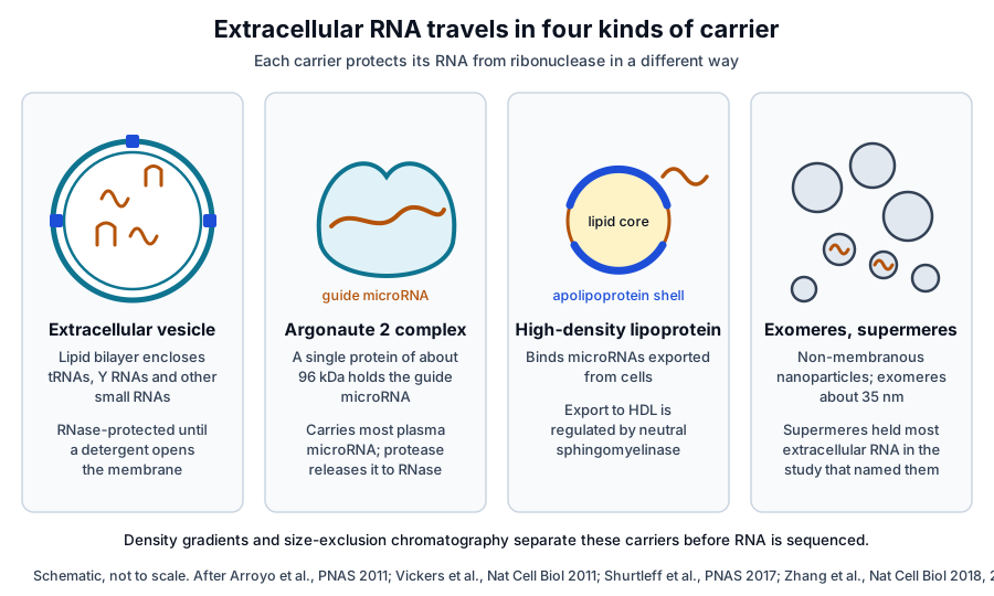
Zhang and colleagues used asymmetric-flow field-flow fractionation to resolve two exosome subsets of 60 to 80 nm and 90 to 120 nm, plus non-membranous nanoparticles of about 35 nm that they named exomeres. Each class carried its own protein, lipid, DNA and RNA profile.[23]
A later study from the Coffey laboratory, led by Zhang, described supermeres, nanoparticles morphologically distinct from exomeres. Their protein and RNA composition differed from that of small vesicles and exomeres, and supermeres held the majority of extracellular RNA.[24]
Extracellular RNA can also change after release. Tosar and colleagues found ribosomes and full-length transfer RNAs in conditioned medium from several mammalian cell lines, and their fragmentation outside the cell generated small RNAs that are highly resistant to degradation.[25]
The non-vesicular fraction is biased toward specific tRNA-derived fragments that form RNase-protecting dimers, and Tosar and Cayota review which fragments dominate each extracellular fraction. Extracellular abundance therefore reflects both secretion and stability.[9][25]
The exRNA Atlas maps RNA cargo types across human biofluids
The Extracellular RNA Communication Consortium (ERCC), funded by the NIH Common Fund, set out to build reference profiles of extracellular RNA from healthy individuals, define how it is made and taken up, and share protocols, data and tools through the exRNA Atlas.[6][7]
Das and colleagues summarized the first phase of the consortium, including collaborative projects on methods for vesicle and extracellular RNA isolation and on tools for sharing and analyzing profiling data.[26]
Murillo and colleagues analyzed 5,309 profiles from 19 studies in the exRNA Atlas by computational deconvolution. The model identified six extracellular RNA cargo types, each detectable in several of serum, plasma, cerebrospinal fluid, saliva and urine, and five of them associated with vesicular, lipoprotein or ribonucleoprotein carriers.[27]
Srinivasan and colleagues compared 10 isolation methods across 5 biofluids. Methods differed markedly in the complexity and reproducibility of small RNA profiles and in how much they captured from vesicle, ribonucleoprotein and HDL carriers, measured by carrier-specific microRNA signatures.[28]
Measuring extracellular vesicle RNA combines separation, protection and sequencing
Each published composition depends on how vesicles were separated from co-isolating carriers. MISEV2023 sets out the approaches for separating vesicles from non-vesicular extracellular particles and for characterizing the resulting preparations.[17][29]
- Density gradient flotation or high-resolution density fractionation, which separates vesicles from protein complexes.[1][22]
- Size-exclusion chromatography, which resolves vesicles, Argonaute 2 complexes and smaller particles by elution volume.[3][25]
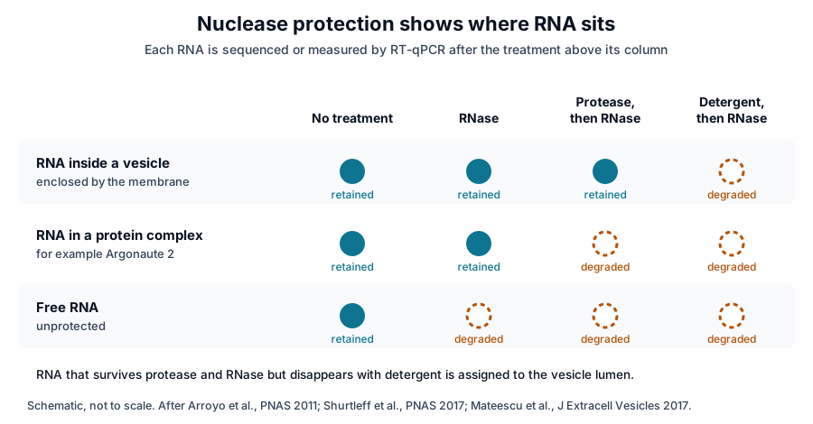
- Nuclease protection, which assigns RNA to the vesicle lumen when it resists RNase until a detergent opens the membrane.[1]
- Small RNA sequencing, where the isolation method and the library chemistry both shape the biotype shares a study reports.[1][28]
The nuclease protection assay reads location from a series of enzyme and detergent steps (Fig. 5). In HEK293T vesicles, transfer RNA and other small noncoding RNAs resisted RNase and protease and were degraded only with detergent, which placed them inside the vesicles.[1]
The same experiments traced many reads mapping to protein-coding genes to DNA on the vesicle surface, and the authors measured messenger RNA in DNase-digested samples.[1] For non-vesicular samples, adding RNase inhibitors revealed ribosomes and full-length transfer RNAs in the extracellular fraction.[25]
Where to go from here
The exomeres and supermeres reference covers the nanoparticles that travel with vesicles, and the cargo sorting reference covers how RNA-binding proteins select RNAs for release. The uptake reference follows vesicles to recipient cells.
The quality reference on MISEV2023 explains what a vesicle preparation can be documented against. Verified practices can request lot-specific characterization data with the certificate of analysis, and our team can explain how a preparation was separated and measured.
Referencias comentadas
Numeradas en el orden en que se citan en el texto. Cada fuente lleva una nota sobre lo que respalda en esta página; los artículos revisados por pares se listan con revista, volumen, número y páginas, y las fuentes primarias son regulaciones, documentos de agencias y bases de datos de referencia.
- 1.Peer-reviewedShurtleff MJ, Yao J, Qin Y, et al. Broad role for YBX1 in defining the small noncoding RNA composition of exosomes. Proceedings of the National Academy of Sciences of the United States of America. 2017;114(43):E8987-E8995. https://pubmed.ncbi.nlm.nih.gov/29073095/Annotation: TGIRT sequencing of gradient-purified and CD63-captured vesicles from HEK293T cells. Cited for the biotype shares in Fig. 1, full-length tRNAs, nuclease protection and surface DNA.
- 2.Peer-reviewedBaglio SR, Rooijers K, Koppers-Lalic D, et al. Human bone marrow- and adipose-mesenchymal stem cells secrete exosomes enriched in distinctive miRNA and tRNA species. Stem Cell Research & Therapy. 2015;6(1):127. https://pubmed.ncbi.nlm.nih.gov/26129847/Annotation: Small RNA sequencing of mesenchymal stem cells and their exosomes. Cited for tRNA dominance, microRNA at 2 to 5 percent of exosome small RNA and selective microRNA release.
- 3.Peer-reviewedArroyo JD, Chevillet JR, Kroh EM, et al. Argonaute2 complexes carry a population of circulating microRNAs independent of vesicles in human plasma. Proceedings of the National Academy of Sciences of the United States of America. 2011;108(12):5003-5008. https://pubmed.ncbi.nlm.nih.gov/21383194/Annotation: Centrifugation, size-exclusion chromatography and immunoprecipitation of human plasma. Cited for most circulating microRNAs cofractionating with Argonaute 2 complexes and for protease sensitivity.
- 4.Peer-reviewedVickers KC, Palmisano BT, Shoucri BM, et al. MicroRNAs are transported in plasma and delivered to recipient cells by high-density lipoproteins. Nature Cell Biology. 2011;13(4):423-433. https://pubmed.ncbi.nlm.nih.gov/21423178/Annotation: Biochemistry of HDL-bound microRNAs in cells, mice and human plasma. Cited for HDL transport of endogenous microRNAs, export regulated by neutral sphingomyelinase and uptake through SR-BI.
- 5.Peer-reviewedValadi H, Ekström K, Bossios A, et al. Exosome-mediated transfer of mRNAs and microRNAs is a novel mechanism of genetic exchange between cells. Nature Cell Biology. 2007;9(6):654-659. https://pubmed.ncbi.nlm.nih.gov/17486113/Annotation: Microarray and transfer studies of mast cell exosomes. Cited for messenger RNA from about 1,300 genes, microRNAs, in vitro translation and the term exosomal shuttle RNA.
- 6.Official sourceNIH Common Fund, Extracellular RNA Communication (ExRNA) program. Accessed 20 October 2026. https://commonfund.nih.gov/exrna
- 7.Peer-reviewedAinsztein AM, Brooks PJ, Dugan VG, et al. The NIH Extracellular RNA Communication Consortium. Journal of Extracellular Vesicles. 2015;4:27493. https://pubmed.ncbi.nlm.nih.gov/26320938/Annotation: NIH program description of the ERCC. Cited for its Common Fund origin and its goals: reference profiles from healthy individuals, biogenesis and uptake, and the exRNA Atlas.
- 8.Peer-reviewedNolte-’t Hoen EN, Buermans HP, Waasdorp M, et al. Deep sequencing of RNA from immune cell-derived vesicles uncovers the selective incorporation of small non-coding RNA biotypes with potential regulatory functions. Nucleic Acids Research. 2012;40(18):9272-9285. https://pubmed.ncbi.nlm.nih.gov/22821563/Annotation: Deep sequencing of small RNA released by immune cells. Cited for vault RNA, Y RNA and tRNA among the most abundant vesicle RNAs and for their enrichment relative to cellular RNA.
- 9.Peer-reviewedTosar JP, Cayota A. Extracellular tRNAs and tRNA-derived fragments. RNA Biology. 2020;17(8):1149-1167. https://pubmed.ncbi.nlm.nih.gov/32070197/Annotation: Review of extracellular tRNAs and tRNA-derived fragments by fraction. Cited for the fragment family and for which fragments dominate vesicle, lipoprotein and ribonucleoprotein fractions.
- 10.Peer-reviewedDriedonks TAP, van der Grein SG, Ariyurek Y, et al. Immune stimuli shape the small non-coding transcriptome of extracellular vesicles released by dendritic cells. Cellular and Molecular Life Sciences. 2018;75(20):3857-3875. https://pubmed.ncbi.nlm.nih.gov/29808415/Annotation: RNA sequencing of highly pure vesicles from primary dendritic cells. Cited for stimulus-specific changes in microRNA, snoRNA and Y RNA content with steadier tRNA and snRNA.
- 11.Peer-reviewedGodoy PM, Bhakta NR, Barczak AJ, et al. Large Differences in Small RNA Composition Between Human Biofluids. Cell Reports. 2018;25(5):1346-1358. https://pubmed.ncbi.nlm.nih.gov/30380423/Annotation: Small RNA sequencing of 12 human biofluid types. Cited for the microRNA to tRNA-derived RNA ratio across fluids and for Y RNA fragments above 10 percent of reads in plasma, serum and CSF.
- 12.DatabaseNCBI Gene 6738, RO60 Ro60, Y RNA binding protein (Homo sapiens). Accessed 20 October 2026. https://www.ncbi.nlm.nih.gov/gene/6738
- 13.Peer-reviewedBoccitto M, Wolin SL. Ro60 and Y RNAs: structure, functions, and roles in autoimmunity. Critical Reviews in Biochemistry and Molecular Biology. 2019;54(2):133-152. https://pubmed.ncbi.nlm.nih.gov/31084369/Annotation: Review of Ro60 and Y RNA biochemistry. Cited for Ro60 as a ring-shaped protein that binds Y RNAs on its outer surface and misfolded RNAs in its cavity.
- 14.Peer-reviewedStein AJ, Fuchs G, Fu C, et al. Structural insights into RNA quality control: the Ro autoantigen binds misfolded RNAs via its central cavity. Cell. 2005;121(4):529-539. https://pubmed.ncbi.nlm.nih.gov/15907467/Annotation: Crystal structures of Ro60 alone and with RNA, rendered in Fig. 2. Cited for the von Willebrand factor A domain, the HEAT-repeat ring and the two RNA binding sites.
- 15.Peer-reviewedDriedonks TAP, Nolte-’t Hoen ENM. Circulating Y-RNAs in Extracellular Vesicles and Ribonucleoprotein Complexes; Implications for the Immune System. Frontiers in Immunology. 2018;9:3164. https://pubmed.ncbi.nlm.nih.gov/30697216/Annotation: Review of extracellular Y RNA. Cited for Y RNA in both vesicles and ribonucleoprotein complexes, its binding proteins and the separation of the two fractions.
- 16.Peer-reviewedChevillet JR, Kang Q, Ruf IK, et al. Quantitative and stoichiometric analysis of the microRNA content of exosomes. Proceedings of the National Academy of Sciences of the United States of America. 2014;111(41):14888-14893. https://pubmed.ncbi.nlm.nih.gov/25267620/Annotation: Counts of exosomes and microRNA molecules from six sources. Cited for a mean of 0.00825 molecules of a given microRNA per exosome and the revised models the authors proposed.
- 17.Peer-reviewedMateescu B, Kowal EJ, van Balkom BW, et al. Obstacles and opportunities in the functional analysis of extracellular vesicle RNA - an ISEV position paper. Journal of Extracellular Vesicles. 2017;6(1):1286095. https://pubmed.ncbi.nlm.nih.gov/28326170/Annotation: ISEV workshop position paper on vesicle RNA. Cited for the strategies used to isolate and characterize vesicle RNA and for careful interpretation of the resulting data.
- 18.Peer-reviewedTurchinovich A, Weiz L, Langheinz A, et al. Characterization of extracellular circulating microRNA. Nucleic Acids Research. 2011;39(16):7223-7233. https://pubmed.ncbi.nlm.nih.gov/21609964/Annotation: Filtration, ultracentrifugation and immunoprecipitation of plasma and culture medium. Cited for non-vesicular microRNA bound to the 96 kDa Argonaute 2 protein and the stability of the complex.
- 19.DatabaseNCBI Gene 27161, AGO2 argonaute RISC catalytic component 2 (Homo sapiens). Accessed 20 October 2026. https://www.ncbi.nlm.nih.gov/gene/27161
- 20.Peer-reviewedSchirle NT, MacRae IJ. The crystal structure of human Argonaute2. Science. 2012;336(6084):1037-1040. https://pubmed.ncbi.nlm.nih.gov/22539551/Annotation: Crystal structure of human Argonaute 2 at 2.3 Å. Cited for the bilobed architecture, the central cleft and guide nucleotides 2 to 6 positioned for target pairing.
- 21.Peer-reviewedElkayam E, Kuhn CD, Tocilj A, et al. The structure of human argonaute-2 in complex with miR-20a. Cell. 2012;150(1):100-110. https://pubmed.ncbi.nlm.nih.gov/22682761/Annotation: Crystal structure of Argonaute 2 bound to miR-20a, rendered in Fig. 3. Cited for anchoring by the MID and PAZ domains and for microRNA binding stabilizing the protein.
- 22.Peer-reviewedJeppesen DK, Fenix AM, Franklin JL, et al. Reassessment of Exosome Composition. Cell. 2019;177(2):428-445.e18. https://pubmed.ncbi.nlm.nih.gov/30951670/Annotation: High-resolution density gradients and direct immunoaffinity capture of exosomes. Cited for the partition of RNA-binding proteins, Argonaute 1 to 4 absent from exosomes, and density fractionation.
- 23.Peer-reviewedZhang H, Freitas D, Kim HS, et al. Identification of distinct nanoparticles and subsets of extracellular vesicles by asymmetric flow field-flow fractionation. Nature Cell Biology. 2018;20(3):332-343. https://pubmed.ncbi.nlm.nih.gov/29459780/Annotation: Field-flow fractionation of secreted particles. Cited for the 60 to 80 nm and 90 to 120 nm exosome subsets, exomeres of about 35 nm and their distinct RNA profiles.
- 24.Peer-reviewedZhang Q, Jeppesen DK, Higginbotham JN, et al. Supermeres are functional extracellular nanoparticles replete with disease biomarkers and therapeutic targets. Nature Cell Biology. 2021;23(12):1240-1254. https://pubmed.ncbi.nlm.nih.gov/34887515/Annotation: Isolation and characterization of supermeres. Cited only for their distinct RNA composition and for supermeres holding the majority of extracellular RNA.
- 25.Peer-reviewedTosar JP, Segovia M, Castellano M, et al. Fragmentation of extracellular ribosomes and tRNAs shapes the extracellular RNAome. Nucleic Acids Research. 2020;48(22):12874-12888. https://pubmed.ncbi.nlm.nih.gov/32785615/Annotation: Sequencing of size-exclusion fractions of non-vesicular samples with RNase inhibitors. Cited for extracellular ribosomes and tRNAs, their fragmentation after release and RNase-protecting fragment dimers.
- 26.Peer-reviewedDas S, Extracellular RNA Communication Consortium, Ansel KM, et al. The Extracellular RNA Communication Consortium: Establishing Foundational Knowledge and Technologies for Extracellular RNA Research. Cell. 2019;177(2):231-242. https://pubmed.ncbi.nlm.nih.gov/30951667/Annotation: Consortium summary of ERCC phase 1. Cited for its collaborative work on isolation methods and on data sharing and analysis tools.
- 27.Peer-reviewedMurillo OD, Thistlethwaite W, Rozowsky J, et al. exRNA Atlas Analysis Reveals Distinct Extracellular RNA Cargo Types and Their Carriers Present across Human Biofluids. Cell. 2019;177(2):463-477.e15. https://pubmed.ncbi.nlm.nih.gov/30951672/Annotation: Computational deconvolution of 5,309 exRNA Atlas profiles. Cited for six cargo types across biofluids, five associated with vesicular, lipoprotein or ribonucleoprotein carriers.
- 28.Peer-reviewedSrinivasan S, Yeri A, Cheah PS, et al. Small RNA Sequencing across Diverse Biofluids Identifies Optimal Methods for exRNA Isolation. Cell. 2019;177(2):446-462.e16. https://pubmed.ncbi.nlm.nih.gov/30951671/Annotation: Comparison of 10 extracellular RNA isolation methods across 5 biofluids. Cited for differences in profile complexity, reproducibility and carrier capture.
- 29.Peer-reviewedWelsh JA, Goberdhan DCI, O’Driscoll L, et al. Minimal information for studies of extracellular vesicles (MISEV2023): From basic to advanced approaches. Journal of Extracellular Vesicles. 2024;13(2):e12404. https://pubmed.ncbi.nlm.nih.gov/38326288/Annotation: ISEV reporting guidance. Cited for approaches to separate vesicles from non-vesicular extracellular particles and to characterize preparations.
Cite this page
ExaVeyra Sciences Editorial Team. Extracellular Vesicle RNA: MicroRNA, Y RNA and tRNA Fragments. ExaVeyra Sciences; 20 October 2026. Available from: https://exaveyra.com/science/exosome-biochemistry/extracellular-rna-biotypes. Accessed [date accessed].
Fuentes de las figuras
- 30.PDB 1YVP, Ro autoantigen complexed with RNAs. ExaVeyra Sciences, rendered in PyMOL from PDB 1YVP. Structure data: RCSB Protein Data Bank (rcsb.org), CC0. Publicación de la estructura: Stein AJ, Fuchs G, Fu C, Wolin SL, Reinisch KM. Structural insights into RNA quality control: the Ro autoantigen binds misfolded RNAs via its central cavity. Cell. 2005;121(4):529-539.
- 31.PDB 4F3T, Human Argonaute2 bound to miR-20a. ExaVeyra Sciences, rendered in PyMOL from PDB 4F3T. Structure data: RCSB Protein Data Bank (rcsb.org), CC0. Publicación de la estructura: Elkayam E, Kuhn CD, Tocilj A, et al. The structure of human argonaute-2 in complex with miR-20a. Cell. 2012;150(1):100-110.