Plastic surgeons meet exosomes from two directions: patients who ask about them after reading about faster recovery from resurfacing or better scars, and suppliers offering vesicle products for use after microneedling and lasers. The literature behind those conversations now spans myofibroblast biology, randomized split-face trials, two plastic surgery flagship journals and a growing set of registered studies, and it addresses problems the specialty works on daily, scar quality, graft retention and skin quality after procedures.
This review is written for aesthetic, reconstructive and facial plastic surgeons and the dermatologists and clinical teams who work with them. It sets out what exosomes are and how they are characterized, what the FDA allows and what its warning letters address, what the literature reports for scar revision, facial aesthetics and reconstruction, the registered trials, and the documentation a surgical practice keeps.
Regulatory position
The FDA states that there are no FDA-approved exosome products, and secreted cell factors are excluded from the HCT/P definition at 21 CFR 1271.3(d)(3), so a vesicle product cannot use the section 361 tissue pathway, and the FDA states that one intended to treat a disease or condition is regulated as a drug and biological product. Exosome material is supplied for topical aesthetic use by licensed practitioners and for IND-authorized clinical research, and ExaVeyra Sciences describes it as biomolecular signaling vesicles for topical aesthetic application and for medical, molecular biology and biochemistry research. Which material, for which patient, after which procedure and by which route are surgeon decisions this article does not make.
Exosomes are lipid-bound signaling vesicles whose cargo reflects the cell and the process that made them
Exosomes are the smallest class of extracellular vesicles, roughly 30 to 150 nm across, formed inside multivesicular bodies and released when those compartments fuse with the plasma membrane. They carry proteins, messenger RNAs, microRNAs and lipids from the parent cell, and the International Society for Extracellular Vesicles now recommends the umbrella term extracellular vesicle unless a preparation's biogenesis has been demonstrated, a convention set out in the MISEV2018 guidelines by Théry and colleagues and updated in MISEV2023 by Welsh and colleagues in the Journal of Extracellular Vesicles.
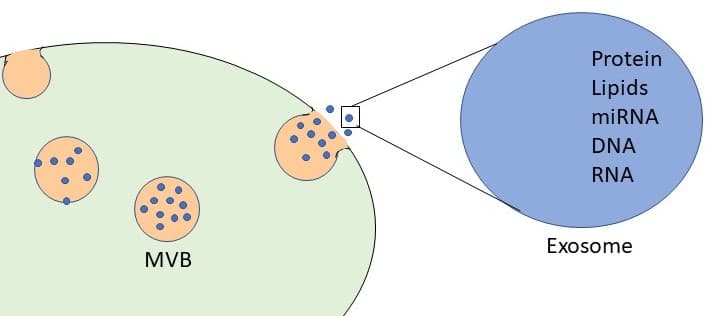
For a surgeon, three properties matter more than the vocabulary. Source cell determines cargo, so adipose, bone marrow, umbilical cord and Wharton's jelly mesenchymal stromal cells, platelets and plant cells each yield a different vesicle population. Manufacturing determines consistency, since culture conditions, isolation by ultracentrifugation, tangential flow filtration or size-exclusion chromatography, and storage change particle count and purity. Characterization determines comparability, which is why MISEV2023 asks for particle concentration and size, at least one transmembrane marker such as CD9, CD63 or CD81, a cytosolic marker, and a measure of non-vesicular contaminants.
| Property | What it determines | What a lot document reports | Standard |
|---|---|---|---|
| Source cell and tissue | Cargo composition and the literature a product can be compared with | Donor tissue, cell type, donor screening | Supplier documentation; MISEV2023 |
| Isolation method | Purity and co-isolated proteins | Ultracentrifugation, TFF, SEC or precipitation | MISEV2023 |
| Particle concentration and size | Lot-to-lot consistency | Nanoparticle tracking or resistive pulse sensing | MISEV2023 |
| Identity markers | That the particles are vesicles | CD9, CD63, CD81 and a cytosolic marker | MISEV2018 and MISEV2023 |
| Safety testing | Fitness for handling in a clinic | Sterility, endotoxin, mycoplasma | Certificate of analysis |
| Storage | Stability through delivery | Temperature and cold-chain log | Supplier documentation |
The plastic surgery literature reviewed below uses all of these sources, and a finding with one vesicle population belongs to that population. The comparisons in each section are therefore described by the source cell and preparation each study used.
What the FDA allows: no approved exosome drug, cleared devices for the procedure, and an IND pathway for therapeutic claims
The FDA's position has been consistent since December 6, 2019, when its public safety notification stated that there are no FDA-approved exosome products and that exosomes used to treat diseases and conditions in humans are regulated as drugs and biological products. The agency's consumer alert on regenerative medicine products, current as of April 2024, repeats that statement, and the International Society for Extracellular Vesicles posted a matching patient safety notice in 2020 noting that no extracellular vesicle therapy had been approved anywhere in the world.
The reason lies in the definitions, since the HCT/P definition at 21 CFR 1271.3(d)(3) excludes secreted or extracted human products such as cell factors, so a cell-free vesicle preparation does not use the section 361 tissue pathway that some minimally manipulated tissues use under the four criteria of 21 CFR 1271.10(a). The drug definition at 21 USC 321(g)(1), which the agency applies, covers a product intended for the diagnosis, cure, mitigation, treatment or prevention of disease, or to affect the structure or function of the body, and a biological product of that kind requires a license under section 351 of the Public Health Service Act or an investigational new drug application for clinical study.
What a plastic surgery practice uses today, and under which rules
| Item | US status | What it permits | Source |
|---|---|---|---|
| Exosome or EV product intended for a disease or condition | No FDA-approved product | Clinical study under an IND with IRB oversight | FDA notification, 2019; consumer alert, 2024 |
| Exosome material supplied for topical aesthetic use | Not an approved drug or licensed biologic; FDA has no category specific to this use | Legal status follows intended use as shown by labeling, promotion and the circumstances of distribution | 21 USC 321(g)(1) and (i); 21 CFR 201.128; PHS Act 351 |
| Microneedling device for aesthetic use | Class II, product code QAI | The needling procedure for its cleared indications, such as facial acne scars | 21 CFR 878.4430 |
| Fractional CO2 laser | Class II | Cutting, destroying or removing tissue by light energy | 21 CFR 878.4810 |
| Autologous fat graft, same surgical procedure | Excepted from Part 1271 when criteria are met | Harvest and reimplantation in one procedure when the fat is only rinsed or cleansed | 21 CFR 1271.15(b); FDA same surgical procedure guidance, 2017 |
| Expanded or enriched adipose stromal cells | Biologic outside 361 criteria | Clinical study under an IND | 21 CFR 1271.10(a) |
The microneedling classification at 21 CFR 878.4430(a) excludes devices intended for transdermal delivery of topical products such as cosmetics, drugs or biologics, and the CO2 laser classification at 21 CFR 878.4810(a)(1) describes a device intended to cut, destroy or remove tissue, so the device clearance covers the needling or the resurfacing, and the material applied afterward stands on its own labeling. A practice therefore keeps two records: the device's 510(k) indication, and the vesicle product's lot documentation.
What the warning letters address
The FDA's Center for Biologics Evaluation and Research has issued warning letters to firms marketing exosome products without an approved application or an IND in effect, including Evolutionary Biologics on December 30, 2024, Supreme Rejuvenation on May 5, 2025, Platinum Biologics on August 15, 2025, New Life Medical Services on September 24, 2025, Blue Horizon International on May 26, 2026 and R3 Medical Companies on August 14, 2026. The letters cite unapproved new drug and unlicensed biologic violations, and the 2026 letter also cited manufacturing practices, including a process the agency said did not appear to isolate exosomes, which is the analytical point that lot-level characterization addresses.
Across these letters, the agency read intended use from the firms' own website and social media claims, including tissue repair, scar and wound healing, hair restoration and skin regeneration claims, and one letter named exosome gel products. A practice can read them as a guide to how labeling and promotional statements determine a product's regulatory status, alongside lot-level characterization of the material it sources, and any therapeutic research question belongs under an IND with institutional review board oversight.
Scar revision research spans myofibroblast biology, randomized acne scar trials and early surgical scar studies
Mechanism: vesicle microRNAs act on the TGF-beta and SMAD pathway that drives fibrosis
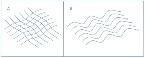
Scar formation is set largely by the myofibroblast, which differentiates under TGF-beta signaling, expresses alpha smooth muscle actin and lays down type I collagen. Fang and colleagues reported in Stem Cells Translational Medicine in 2016 that umbilical cord MSC exosomes reduced scar formation and alpha smooth muscle actin in a mouse full-thickness wound model, and traced the effect to four microRNAs, miR-21, miR-23a, miR-125b and miR-145, that target TGF-beta2, its receptor and SMAD2; blocking those microRNAs with antagomirs removed the effect in cells and in wounded mice, and RNase-treated vesicles lost the effect in vitro.

Adipose-derived vesicles show the same direction. Wang and colleagues reported in Scientific Reports in 2017 that adipose MSC exosomes reduced scar size in mouse full-thickness incisional wounds after intravenous exosome injection, raised the collagen III to I and TGF-beta3 to TGF-beta1 ratios that characterize less fibrotic healing, and increased MMP3 through ERK signaling. Earlier work had shown that adipose MSC exosomes stimulated fibroblast migration, proliferation and collagen synthesis in vitro, as Hu and colleagues reported in Scientific Reports in 2016, and that umbilical cord MSC exosomes acted through Wnt4 signaling in skin repair, as Zhang and colleagues reported in Stem Cells in 2015, a paper later corrected for a duplicated image panel with its conclusions unchanged. In hypertrophic scar models, Li and colleagues reported reduced collagen I and III, alpha smooth muscle actin and phosphorylated Smad2/3 through a miR-192-5p and IL-17RA axis in Stem Cell Research and Therapy in 2021, and Xu and colleagues reported in Burns and Trauma in 2024 that miR-125b-5p carried in adipose stem cell exosomes suppressed Smad2 in scar-derived myofibroblasts.
| Study | Journal | Model | Vesicle source | Reported result |
|---|---|---|---|---|
| Fang 2016 | Stem Cells Transl Med | Mouse full-thickness wound | Umbilical cord MSC | Smaller scars; alpha-SMA down; four microRNAs target TGF-beta/SMAD2 |
| Wang 2017 | Sci Rep | Mouse incisional wound (IV exosomes) | Adipose MSC | Scar size reduced; collagen III:I and TGF-beta3:beta1 up |
| Li 2021 | Stem Cell Res Ther | Hypertrophic scar fibroblasts and mouse wound | Adipose MSC | Collagen I/III, alpha-SMA, p-Smad2/3 down via miR-192-5p |
| Xu 2024 | Burns Trauma | Mouse wound and bleomycin fibrosis | Adipose stem cell | alpha-SMA, COL1, COL3 down via miR-125b-5p and Smad2 |
Atrophic acne scars: a double-blind exosome trial and two trials of related materials
Atrophic acne scar revision with fractional CO2 has the only double-blind randomized exosome trial in this area. Kwon and colleagues ran a double-blind, randomized split-face trial in 25 patients, published in Acta Dermato-Venereologica in 2020, applying adipose stem cell exosomes to one side and a control solution to the other after each of three fractional CO2 sessions. The exosome side showed a 32.5 percent reduction in the échelle d'évaluation clinique des cicatrices d'acné (ECCA) scar score against 19.9 percent on the control side (P < .01), less erythema in the first week (P = .03) and downtime of 4.1 against 4.3 days (P = .03), with no permanent adverse events. The vesicle and control solutions were manufactured and supplied by ExoCoBio, and the authors declared no conflicts of interest.
Helmi and colleagues randomized 100 patients in 2026 to fractional CO2 with polynucleotide and exosome-mimetic nanovesicles or fractional CO2 alone, and reported a 47.3 percent ECCA reduction against 35.9 percent (P < .001), with less post-inflammatory hyperpigmentation, 10 against 24 percent (P = .04), and less prolonged erythema; the randomization method and blinding were not described in the accessible report. Abdel-Maguid and colleagues compared stem cell conditioned medium, saline and PRP after fractional CO2 in 33 patients and found that conditioned medium raised collagen and procollagen I on biopsy (P = .041), while clinical scar scores were similar to laser with saline and PRP gave the larger clinical improvement, a reminder that a conditioned medium and a purified vesicle preparation are different materials.
Surgical, traumatic and hypertrophic scars
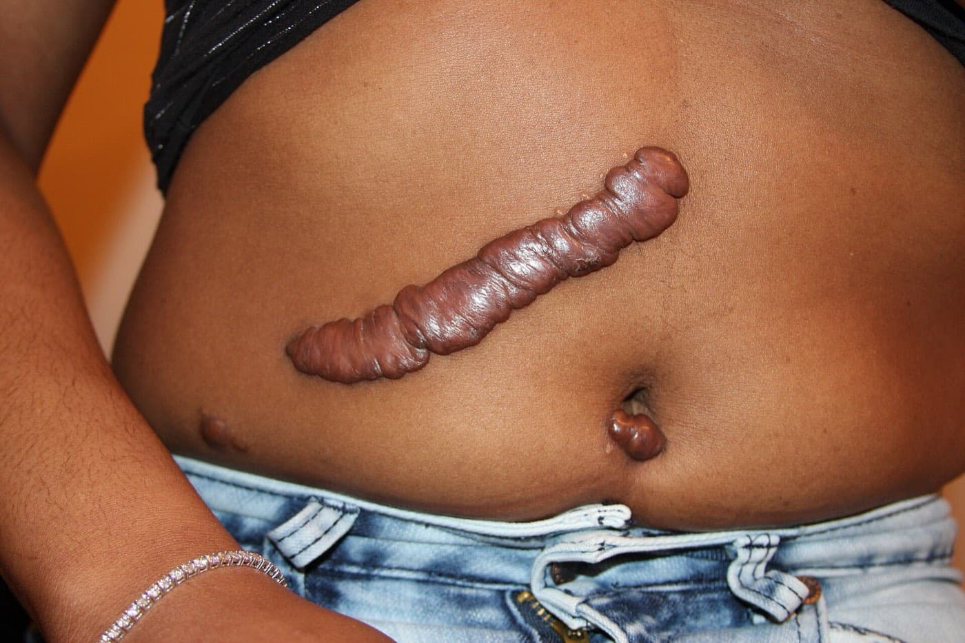
Human data for surgical incision scars are early and growing. Jeong, Park and Jung reported a split-scar study in the Journal of Cosmetic Dermatology in 2026 in which adipose stem cell exosomes and hyaluronic acid were applied to opposite halves of 3 cm chest incision scars from costal cartilage rhinoplasty in 10 patients and scored with the Vancouver Scar Scale and POSAS. Seo and Jung reported in PRS Global Open in 2026 a facial deep dermal injury treated with topical adipose stem cell exosomes after radiofrequency microneedling every two weeks for six months, with the modified Vancouver Scar Scale falling from 9 to 2. Registered trials are now testing the question directly, including a double-blind randomized trial of intralesional exosomes for atrophic scars at Zagazig University (NCT07569666), a quadruple-masked randomized trial comparing umbilical cord MSCs, their conditioned medium and triamcinolone for keloids in Indonesia, completed in June 2022 (NCT05887804), and a phase 1 study of bone marrow MSC extracellular vesicles for burn wounds by Aegle Therapeutics that completed in November 2024 (NCT05078385).
This image shows a clinical procedure.
Facial aesthetics: photoaging, post-procedure recovery and microneedling trials, with Plastic and Reconstructive Surgery and Aesthetic Surgery Journal data
Topical platelet-derived vesicles in Aesthetic Surgery Journal and Plastic and Reconstructive Surgery
The Mayo Clinic group has published topical platelet extract data in both Aesthetic Surgery Journal and Plastic and Reconstructive Surgery. Proffer, Wyles, Behfar and colleagues reported in Aesthetic Surgery Journal in 2022 that 56 adults applying a human platelet extract serum morning and evening for six weeks improved on the Griffiths photoaging score by a mean of 0.214 points (P = .0082), with the top-quartile subgroup showing gains in an imaging skin health score and a fall in erythema, and with dryness the most common side effect in 16.1 percent. Wyles and colleagues then compared the platelet extract with a vitamin C serum on opposite hands in 60 adults in Plastic and Reconstructive Surgery in 2024 and reported the platelet extract non-inferior, with brown-spot area falling about 23 percent against about 26 percent with vitamin C at 26 weeks. Rion Aesthetics, the serum's manufacturer, financed the 2022 study in part and funded the 2024 study, and several authors of both hold founder, employee or consultant roles with the company.
Adipose vesicles after microneedling and radiofrequency microneedling
Park, Kwon and colleagues randomized 28 adults to microneedling with or without an adipose stem cell exosome solution on opposite sides of the face in 2023 and reported a better Global Aesthetic Improvement Scale result on the exosome side at the final visit (P = .005). Estupiñan, Ly and Goldberg compared topical adipose MSC exosomes with PRP after three radiofrequency microneedling sessions in 15 adults with photoaged skin in 2025, and reported equal improvement in investigator scores, a Griffiths score that fell from 4.5 to 3.1 at three months on both sides, and comparable rises in collagen I and glycosaminoglycans on biopsy at six months, in a non-randomized split-face design supported in part by the exosome manufacturer.
| Study | Journal | Design | n | Combined procedure | Main reported result |
|---|---|---|---|---|---|
| Kwon 2020 | Acta Derm Venereol | Double-blind randomized split-face | 25 | Fractional CO2, acne scars | ECCA down 32.5% vs 19.9%; less early erythema |
| Helmi 2026 | Postepy Dermatol Alergol | Randomized comparative | 100 | Fractional CO2, acne scars | ECCA down 47.3% vs 35.9%; less PIH |
| Abdel-Maguid 2021 | Journal of Dermatological Treatment | Randomized split-face | 33 | Fractional CO2, acne scars | Collagen and procollagen I up with conditioned medium |
| Park 2023 | J Cosmet Dermatol | Randomized split-face | 28 | Microneedling, facial aging | Better GAIS on exosome side (P = .005) |
| Estupiñan 2025 | J Cosmet Dermatol | Investigator-blinded split-face | 15 | RF microneedling, photoaging | Equal to PRP; collagen I and GAGs up on both sides |
| Proffer 2022 | Aesthet Surg J | Prospective single arm | 56 | Topical serum alone | Griffiths score improved (P = .0082) |
| Wyles 2024 | Plast Reconstr Surg | Non-randomized within-patient comparison | 60 | Topical serum on the hands | Non-inferior to vitamin C on brown spots |
What the systematic reviews report
Stack and colleagues reviewed 39 human studies of exosome-based aesthetic treatments for an Aesthetic Surgery Journal supplement in 2026, 26 on skin and 13 on hair, and reported, in a largely descriptive synthesis, wrinkle reduction of 20.2 percent, improvement in other skin outcomes of 14.7 to 23.4 percent and a 23.6 percent gain in hair density across the included studies. Dhaliwal and colleagues reviewed eight microneedling studies of 171 patients in 2026 and found no discontinuations for adverse events, with transient erythema and edema the effects reported, while noting that granulomas, hyperpigmentation and hypersensitivity had been described in the wider literature mainly after injection.
What the case reports describe, and the delivery route they share
The published adverse event reports in aesthetic medicine share one feature: intradermal injection. Choi and colleagues described a foreign body granuloma in JAAD Case Reports after injection of an exosome product that had not been officially released, Nahm and colleagues described delayed necrotizing granulomas in seven women after injected exosome treatments, and AlBargawi reported ischemic necrosis of both cheeks three days after intradermal injection of a lyophilized exosome product for acne scars in 2025. The controlled trials above applied characterized material topically, alone or after microneedling or laser procedures, and Soltani-Arabshahi and colleagues described granulomatous reactions to other topical products applied with microneedling in JAMA Dermatology in 2014, so the same adverse event monitoring applies to topical use.
Reconstructive surgery: fat graft retention, flap survival, wound models and lymphatic research
Fat grafting: from cell-assisted lipotransfer in The Lancet to vesicle-assisted grafting in PRS
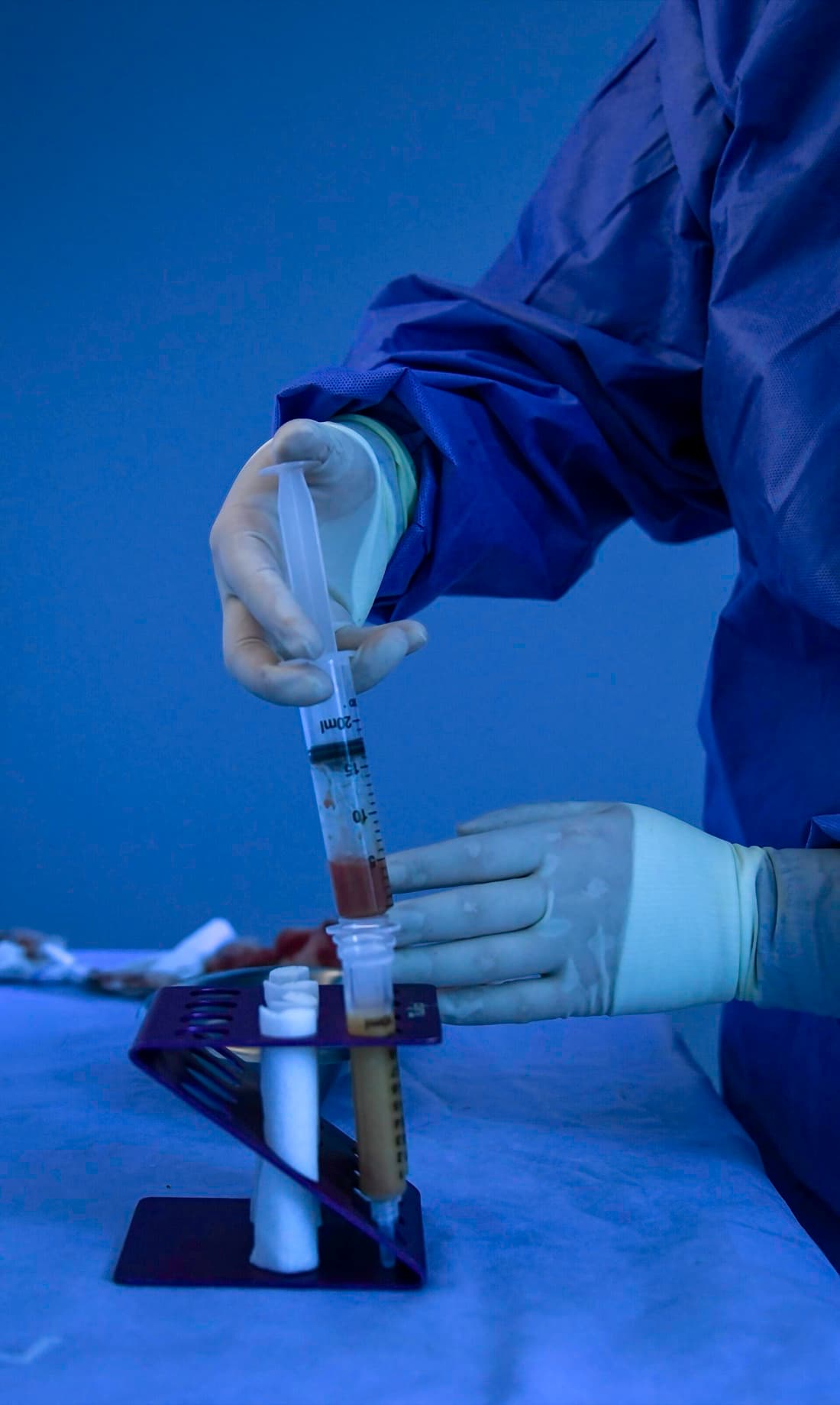
Fat graft volume loss is a recognized limitation in breast and facial reconstruction, and the adipose stromal cell literature set the reference point. Yoshimura and colleagues described cell-assisted lipotransfer for breast augmentation in 40 women in Aesthetic Plastic Surgery in 2008, and Kølle and colleagues reported in The Lancet in 2013 a triple-blind, placebo-controlled trial in 10 healthy volunteers who received upper-arm grafts, in which grafts enriched with ex vivo expanded adipose stem cells retained 80.9 percent of their volume at 121 days on MRI against 16.3 percent for control grafts (P < .0001), with no serious adverse events. In the United States, culture-expanded cells are more than minimally manipulated under 21 CFR 1271.10(a), so clinical use requires an IND.
This image shows a clinical procedure.
Vesicles offer a cell-free version of the same idea. Chen and colleagues reported in Plastic and Reconstructive Surgery in 2019 that adipose stem cell exosomes were comparable to their source cells in a mouse fat graft model, with 10-week retention of 56.4 percent with exosomes, 50.9 percent with cell-assisted grafting and 40.7 percent with saline, together with fewer oil cysts, less fibrosis and higher early angiogenesis and inflammatory signaling. Adding an exosome product to a fat graft in patients would be an investigational use of a drug and biological product.
Flap survival and ischemia-reperfusion
Pu and colleagues reported in the Journal of Investigative Dermatology in 2017 that adipose stem cells, their conditioned medium and their exosomes each improved skin flap survival and capillary density after three hours of ischemia in mice, and blocking IL-6 reduced the benefit of the cells. Bai and colleagues reported in Biochemical and Biophysical Research Communications in 2018 that exosomes from adipose stem cells preconditioned with hydrogen peroxide gave a further gain in flap survival over standard exosomes, an example of how culture conditions change vesicle activity.
Wound models and engineered delivery
Chronic and diabetic wounds are a common preclinical setting, and delivery is now a design question in its own right. Wang and colleagues reported in Theranostics in 2019 that an antibacterial self-healing hydrogel loaded with exosomes closed 88.7 percent of diabetic full-thickness wounds by day 14, against 76.3 percent with exosomes alone, 64.3 percent with hydrogel alone and 36.3 percent in controls. Human wound studies have reached registration, including an open-label pilot of adipose tissue derived exosomes at Shanghai Ninth People's Hospital (NCT05475418) and a randomized open-label study of adipose extracellular vesicles with hyaluronic acid (NCT06253975), and Park and Yi reviewed the plastic surgery wound literature with five clinical cases in PRS Global Open in 2024.
Lymphatic and other reconstructive targets
Wang and colleagues reported in 2018 that exosomes from VEGF-C-treated adipose stem cells promoted lymphangiogenesis in lymphatic endothelial cells in vitro through a miR-132 and TGF-beta pathway, an in vitro rationale for lymphedema research after breast and oncologic reconstruction. We found no published controlled human trial of exosomes in breast reconstruction or irradiated tissue as of October 2026, which makes these settings candidates for registered trials.
| Study | Journal | Model | Material | Reported result |
|---|---|---|---|---|
| Kølle 2013 | Lancet | Human triple-blind RCT, 10 volunteers, upper arm | Expanded adipose stem cells in fat graft | Volume retention 80.9 vs 16.3% at 121 days |
| Yoshimura 2008 | Aesthetic Plast Surg | Human case series, 40 women | Cell-assisted lipotransfer | Breast circumference +4 to 8 cm at 6 months |
| Chen 2019 | Plast Reconstr Surg | Mouse fat graft | Adipose stem cell exosomes | Retention 56.4 vs 50.9 (cells) vs 40.7% (saline) |
| Pu 2017 | J Invest Dermatol | Mouse flap, ischemia-reperfusion | Adipose stem cell exosomes | Flap survival and capillary density improved |
| Bai 2018 | Biochem Biophys Res Commun | Flap, 6 h ischemia-reperfusion | Preconditioned adipose exosomes | Further survival gain over standard exosomes |
| Wang 2019 | Theranostics | Diabetic mouse wound | Exosome-loaded hydrogel | Closure 88.7% at day 14 vs 36.3% control |
| Wang 2018 | Cell Physiol Biochem | Lymphatic endothelial cells | Adipose stem cell exosomes | VEGF-C-dependent lymphangiogenesis |
Six registered trials frame the next round of plastic surgery results
| Registry ID | Title (short) | Sponsor | Design | Enrollment | Status, verified October 2026 |
|---|---|---|---|---|---|
| NCT07569666 | Exosomes in atrophic scars management | Zagazig University | Randomized, double-blind, intralesional exosomes vs saline | 12 | Recruiting from June 2025 |
| NCT06253975 | Extracellular vesicles from human adipose tissue for wound healing | Shanghai Ninth People's Hospital | Randomized, open label, adipose EVs with hyaluronic acid vs hyaluronic acid | 20 planned | Status unknown, last verified February 2024; no results posted |
| NCT05887804 | Umbilical cord MSCs, conditioned medium or triamcinolone for keloids | RSPAD Gatot Soebroto, with Universitas Indonesia | Phase 4, randomized, quadruple-masked | 24 | Completed June 2022; no results posted |
| NCT05457491 | Human platelet extract skin care regimen | Mayo Clinic | Phase 1, single arm, facial regimen | 56 | Terminated; results posted October 2024; corresponds to the 2022 ASJ study |
| NCT05475418 | Adipose tissue derived exosomes for wound healing | Shanghai Ninth People's Hospital | Open label, single group | 5 | Completed October 2023 |
| NCT05078385 | Extracellular vesicles for burn wounds | Aegle Therapeutics | Phase 1, open label | 1 | Completed November 2024; no results posted |
The evidence base is early, specialty-specific and expanding quickly
- Human trials in facial aesthetics and acne scars now include randomized and double-blind split-face designs of 15 to 100 patients, with follow-up of six weeks to six months.
- Animal and cell studies from several groups report anti-fibrotic effects through TGF-beta and SMAD signaling, with different microRNAs named in each study.
- Fat graft, flap and lymphatic data are preclinical, and the strongest human fat retention result comes from expanded cells in The Lancet, which frames the vesicle question for future trials.
- Several positive human studies were supported by the manufacturers of the products they tested, as the annotated references record, and independent replication has yet to be reported.
- Materials differ by source cell, isolation method and dose, so each result belongs to the preparation that produced it, and conditioned medium, platelet extract, plant vesicles and purified MSC exosomes are separate materials.
- The exosome adverse event reports cited here followed intradermal injection, the controlled trials used topical application, and reactions to other topical products applied with microneedling have been reported, so adverse event monitoring applies to every route.
Documentation each surgical practice records
- Product name, source cell and tissue, manufacturer, lot number and expiry, with the certificate of analysis on file
- Particle concentration and size method, identity markers and sterility, endotoxin and mycoplasma results from that lot document
- Storage temperature log from receipt to use, and the time from thaw or reconstitution to application
- The procedure performed, the device and its 510(k) number, settings and anatomical area
- Route of application, kept within the supplier's topical labeling, and the volume applied
- Standardized photographs and a validated scale where one applies: ECCA or Goodman for acne scars, POSAS or Vancouver for surgical scars, Griffiths or GAIS for photoaging
- Adverse events, including delayed papules or nodules, reported to the supplier and to FDA MedWatch
ExaVeyra Sciences supplies exosome material with the lot number and storage requirements on every order and the certificate of analysis for each lot available on request, so that a surgical practice can file them against the record above. Randomized split-face trials, studies in Plastic and Reconstructive Surgery and Aesthetic Surgery Journal and registered scar, wound and keloid trials are adding to this literature each year, and practices that document their materials and procedures carefully will be best placed to read each new result against their own outcomes.
Educational Disclaimer: This article is for educational purposes only and does not constitute medical or legal advice. It reviews published research and registered trials; it does not identify any material, compound, device setting or protocol as safe, effective or appropriate for any patient, and it is not intended to diagnose, treat, cure, or prevent any disease. Conditions named in this article are named as the subjects of the studies cited. Naming a condition studied in a model or a trial does not establish an indication for any product. Exosome material is supplied for topical aesthetic use by licensed practitioners and for laboratory and IND-authorized clinical research. No exosome product is FDA-approved for any use, and ExaVeyra Sciences makes no statement about administration, route, or outcome. Any use of research-grade material in human subjects requires the applicable FDA pathway, such as an investigational new drug application, and institutional review board oversight. Compounded medications are not FDA-approved. They are prepared by a 503A compounding pharmacy licensed by the Texas State Board of Pharmacy for an individual patient against that patient’s prescription, and are available to NPI-verified licensed practitioners only. Discussion of a compound’s literature, including compounds the FDA placed in category 2 of its 503A bulk drug substances policy, is not an offer to supply it. ExaVeyra Sciences is a supplier and takes no part in any diagnosis, treatment decision, study design decision, or plan of care. Findings described are attributed to the peer-reviewed literature, trial registries and agencies cited, and a citation establishes that a question was studied, leaving the suitability of any product for a given patient or protocol to the practitioner’s judgment. The content of these guides is provided for general informational purposes only. It does not constitute legal, medical, or regulatory advice, and does not establish that any particular product or practice is compliant. Regulatory requirements vary by state and depend on the circumstances of each practice. Each practitioner should consult their own legal counsel and the applicable state licensing boards before acting on anything stated here.
References
- 1.Théry C, Witwer KW, Aikawa E, Alcaraz MJ, Anderson JD, Andriantsitohaina R, et al. Minimal information for studies of extracellular vesicles 2018 (MISEV2018): a position statement of the International Society for Extracellular Vesicles and update of the MISEV2014 guidelines. J Extracell Vesicles. 2018;7(1):1535750. PMID 30637094. PMC6322352. doi:10.1080/20013078.2018.1535750.Society guideline, official ISEV journal. Carries in this article: the extracellular vesicle nomenclature and minimal characterization requirements.
- 2.Welsh JA, Goberdhan DCI, O'Driscoll L, Buzas EI, Blenkiron C, Bussolati B, et al. Minimal information for studies of extracellular vesicles (MISEV2023): from basic to advanced approaches. J Extracell Vesicles. 2024;13(2):e12404. doi:10.1002/jev2.12404. PMID 38326288. PMC10850029. Correction published (doi:10.1002/jev2.12451, PMID 38726651).Society guideline compiled with input from more than 1,000 researchers. Carries in this article: particle, marker and contaminant reporting that a lot document can be read against.
- 3.Lener T, Gimona M, Aigner L, Börger V, Buzas E, Camussi G, et al. Applying extracellular vesicles based therapeutics in clinical trials: an ISEV position paper. J Extracell Vesicles. 2015;4:30087. PMID 26725829. doi:10.3402/jev.v4.30087.Society position paper. Carries in this article: manufacturing and quality considerations for vesicle products entering clinical study.
- 4.Witwer KW, Van Balkom BWM, Bruno S, Choo A, Dominici M, Gimona M, et al. Defining mesenchymal stromal cell (MSC)-derived small extracellular vesicles for therapeutic applications. J Extracell Vesicles. 2019;8(1):1609206. PMID 31069028. PMC6493293. doi:10.1080/20013078.2019.1609206.Joint ISEV, ISCT, ISBT and SOCRATES statement. Carries in this article: criteria for cell origin, membrane presence and vesicle integrity in MSC vesicle products.
- 5.International Society for Extracellular Vesicles, Regulatory Affairs Task Force. Patient information and safety notice: extracellular vesicles/exosomes and unproven therapies. isev.org, August 8, 2020.Society safety notice. Carries in this article: no extracellular vesicle or exosome product approved as a therapy worldwide as of June 2020.
- 6.Kwon HH, Yang SH, Lee J, Park BC, Park KY, Jung JY, et al. Combination treatment with human adipose tissue stem cell-derived exosomes and fractional CO2 laser for acne scars: a 12-week prospective, double-blind, randomized, split-face study. Acta Derm Venereol. 2020;100(18):adv00310. PMID 33073298. PMC9309822. doi:10.2340/00015555-3666.Double-blind randomized split-face trial; n = 25; three fractional CO2 sessions; 12 weeks. Carries in this article: ECCA reduction 32.5 vs 19.9 percent (P < .01); less first-week erythema (P = .03); no permanent adverse events. Disclosure: declared: none; exosome and control solutions supplied by the manufacturer.
- 7.Helmi RR, Mohammed RS, Nori W. Fractional CO2 laser augmented by polynucleotide and exosome-mimetic nanovesicles in atrophic acne scars: randomised comparative trial. Postepy Dermatol Alergol. 2026;43(3):314-321. PMID 42621380. PMC13487574. doi:10.5114/ada.2026.161825.Randomized comparative trial; n = 100; 3 months. Carries in this article: ECCA reduction 47.3 vs 35.9 percent (P < .001); PIH 10 vs 24 percent; prolonged erythema 14 vs 30 percent (P = .03); comparator was laser alone. Note: combined polynucleotide and exosome-mimetic product.
- 8.Abdel-Maguid EM, Awad SM, Hassan YS, El-Mokhtar MA, El-Deek HE, Mekkawy MM. Efficacy of stem cell-conditioned medium vs. platelet-rich plasma as an adjuvant to ablative fractional CO2 laser resurfacing for atrophic post-acne scars: a split-face clinical trial. J Dermatolog Treat. 2021;32(2):242-249. PMID 31180258. doi:10.1080/09546634.2019.1630701.Randomized split-face trial; n = 33; three monthly sessions. Carries in this article: collagen and procollagen I up with conditioned medium (P = .041); clinical scores similar to laser with saline (P = .63); PRP ahead of conditioned medium (P = .006).
- 9.Park GH, Kwon HH, Seok J, Yang SH, Lee J, Park BC, Shin E, Park KY. Efficacy of combined treatment with human adipose tissue stem cell-derived exosome-containing solution and microneedling for facial skin aging: a 12-week prospective, randomized, split-face study. J Cosmet Dermatol. 2023;22(12):3418-3426. PMID 37377400. doi:10.1111/jocd.15872.Randomized split-face trial, saline with microneedling on the control side; n = 28; three sessions; 12 weeks. Carries in this article: better GAIS on the exosome side at the final visit (P = .005).
- 10.Estupiñan B, Ly K, Goldberg DJ. Adipose mesenchymal stem cell-derived exosomes versus platelet-rich plasma treatment for photoaged facial skin: an investigator-blinded, split-face, non-inferiority trial. J Cosmet Dermatol. 2025;24(5):e70208. PMID 40414798. PMC12104007. doi:10.1111/jocd.70208.Investigator-blinded, non-randomized split-face trial; n = 15; RF microneedling ×3; 6 months. Carries in this article: equal improvement with exosomes and PRP; Griffiths 4.5 to 3.1 at 3 months on both sides; collagen I and GAGs up in both arms. Disclosure: supported in part by a grant from the exosome manufacturer.
- 11.Proffer SL, Paradise CR, DeGrazia E, Halaas Y, Durairaj KK, Somenek M, et al. Efficacy and tolerability of topical platelet exosomes for skin rejuvenation: six-week results. Aesthet Surg J. 2022;42(10):1185-1193. PMID 35689936. doi:10.1093/asj/sjac149.Prospective single-arm study, flagship Aesthetic Society journal; n = 56; 6 weeks; registered as NCT05457491. Carries in this article: Griffiths score improved (P = .0082); top-quartile erythema and skin health gains; dryness in 16.1 percent. Disclosure: partly funded by the product manufacturer; several authors hold founder, employee or consultant roles.
- 12.Wyles SP, Eaton Jankov L, Copeland K, Bucky LP, Paradise C, Behfar A. A comparative study of two topical treatments for photoaging of the hands. Plast Reconstr Surg. 2024;154(5):978-984. PMID 38086019. PMC11512612. doi:10.1097/PRS.0000000000011240.Non-randomized within-patient comparison, flagship ASPS journal; n = 60; to 26 weeks. Carries in this article: platelet extract non-inferior to vitamin C at 12 weeks (primary endpoint); brown-spot area down 23.4 vs 26.1 percent at 26 weeks. Disclosure: funded by Rion Aesthetics; several authors disclosed ties to the company.
- 13.Stack ER, Spongberg C, Braud SC, Stanton WN, Elway M. Clinical advances in exosome-based therapies for aesthetic medicine: a systematic review and meta-analysis of human clinical trials. Aesthet Surg J. 2026;46(Suppl 1):S13-S25. PMID 41800726. doi:10.1093/asj/sjaf178.PRISMA systematic review; 39 studies (26 skin, 13 hair); PROSPERO CRD420250655353; the methods state that a formal pooled meta-analysis was not performed. Carries in this article: wrinkle reduction 20.2 percent; other skin outcomes 14.7 to 23.4 percent; hair density +23.6 percent.
- 14.Dhaliwal NK, Moothathamby T, Sokhal BS, Gkini MA. The use of microneedling with exosomes in dermatology: a systematic review. J Cosmet Dermatol. 2026;25(4):e70881. PMID 42027180. PMC13107235. doi:10.1111/jocd.70881.Systematic review; 8 studies, 171 patients. Carries in this article: no discontinuations for adverse events; transient erythema and edema; injection-related events in the wider literature.
- 15.Park SY, Yi KH. Exosome-mediated advancements in plastic surgery: navigating therapeutic potential in skin rejuvenation and wound healing. Plast Reconstr Surg Glob Open. 2024;12(8):e6021. PMID 39534068. PMC11556992. doi:10.1097/GOX.0000000000006021.Narrative review with five clinical cases, ASPS open-access journal. Carries in this article: the plastic surgery overview and call for clinical trials.
- 16.Seo MG, Jung DW. Scar remodeling with adipose-derived stem cell exosomes after facial deep dermal injury. Plast Reconstr Surg Glob Open. 2026;14(6):e7802. PMID 42266813. PMC13246081. doi:10.1097/GOX.0000000000007802.Case report; n = 1; topical adipose stem cell exosomes after radiofrequency microneedling every 2 weeks for 6 months; product supplied by ExoCoBio. Carries in this article: modified Vancouver Scar Scale 9 at week 4, 6 at 3 months and 2 at 6 months. Disclosure: declared: none.
- 17.Jeong KH, Park KY, Jung DW. A split-scar study: study of surgical scar using exosomes. J Cosmet Dermatol. 2026;25(3):e70756. PMID 41748500. PMC12945872. doi:10.1111/jocd.70756.Split-scar comparison; n = 10 chest incision scars after costal cartilage rhinoplasty; adipose stem cell exosomes vs hyaluronic acid on opposite halves; VSS and POSAS. Carries in this article: a controlled split-scar human study of exosomes on surgical incisions.
- 18.Choi H, Kwak JH, Shin BS, Na CH, Kim MS. Foreign body granuloma caused by an injection of exosomes. JAAD Case Rep. 2025;64:19-21. PMID 40918543. PMC12410482. doi:10.1016/j.jdcr.2024.03.026.Case report, AAD journal family. Carries in this article: granuloma in a 50-year-old woman after intradermal cheek injection of an exosome product that had not yet been officially released. Disclosure: declared: none.
- 19.Nahm WJ, Thunga S, Yoo J. Complications after exosome treatment for aesthetic skin rejuvenation. Dermatol Rev. 2024;5(4):e242. doi:10.1002/der2.242.Case series; n = 7 women aged 26 to 37 (corrected February 2025). Carries in this article: delayed necrotizing granulomas after injections of exosome products.
- 20.AlBargawi S. Necrosis following dermal injection of lyophilized exosomes: a case report. J Cosmet Dermatol. 2025;24(8):e70387. PMID 40820962. PMC12359287. doi:10.1111/jocd.70387.Case report; a 38-year-old man. Carries in this article: ischemic necrosis of both cheeks three days after multiple intradermal injections for acne scars. Disclosure: declared: none.
- 21.Soltani-Arabshahi R, Wong JW, Duffy KL, Powell DL. Facial allergic granulomatous reaction and systemic hypersensitivity associated with microneedle therapy for skin rejuvenation. JAMA Dermatol. 2014;150(1):68-72. doi:10.1001/jamadermatol.2013.6955.Case series, AMA dermatology journal; n = 3. Carries in this article: foreign body granulomas after topical products applied with microneedling, the basis for monitoring every route.
- 22.Kølle SF, Fischer-Nielsen A, Mathiasen AB, Elberg JJ, Oliveri RS, Glovinski PV, et al. Enrichment of autologous fat grafts with ex-vivo expanded adipose tissue-derived stem cells for graft survival: a randomised placebo-controlled trial. Lancet. 2013;382(9898):1113-1120. PMID 24075051. doi:10.1016/S0140-6736(13)61410-5.Triple-blind RCT; 13 randomized and 10 analyzed healthy volunteers; upper-arm grafts; MRI at day 121. Carries in this article: residual volume 80.9 vs 16.3 percent (P < .0001); no serious adverse events.
- 23.Yoshimura K, Sato K, Aoi N, Kurita M, Hirohi T, Harii K. Cell-assisted lipotransfer for cosmetic breast augmentation: supportive use of adipose-derived stem/stromal cells. Aesthetic Plast Surg. 2008;32(1):48-55. PMID 17763894. PMC2175019. doi:10.1007/s00266-007-9019-4.Case series; n = 40. Carries in this article: the founding description of cell-assisted lipotransfer; circumference +4 to 8 cm at 6 months (pre-2011 landmark).
- 24.Fang S, Xu C, Zhang Y, Xue C, Yang C, Bi H, et al. Umbilical cord-derived mesenchymal stem cell-derived exosomal microRNAs suppress myofibroblast differentiation by inhibiting the TGF-β/SMAD2 pathway during wound healing. Stem Cells Transl Med. 2016;5(10):1425-1439. PMID 27388239. PMC5031180. doi:10.5966/sctm.2015-0367.Mouse wound model plus in vitro. Carries in this article: miR-21, miR-23a, miR-125b and miR-145 target TGF-beta2, TGFbetaR2 and SMAD2; smaller scars; antagomirs removed the effect in vitro and in vivo, RNase in vitro. Disclosure: declared: none.
- 25.Wang L, Hu L, Zhou X, Xiong Z, Zhang C, Shehada HMA, et al. Exosomes secreted by human adipose mesenchymal stem cells promote scarless cutaneous repair by regulating extracellular matrix remodelling. Sci Rep. 2017;7:13321. PMID 29042658. PMC5645460. doi:10.1038/s41598-017-12919-x.Mouse full-thickness incisional wound model, intravenous exosomes; MMP3 and ERK shown in fibroblasts in vitro. Carries in this article: reduced scar size; higher collagen III:I and TGF-beta3:beta1; MMP3 via ERK. Note: corrections published 2018 and 2021.
- 26.Hu L, Wang J, Zhou X, Xiong Z, Zhao J, Yu R, et al. Exosomes derived from human adipose mesenchymal stem cells accelerates cutaneous wound healing via optimizing the characteristics of fibroblasts. Sci Rep. 2016;6:32993. PMID 27615560. PMC5018733. doi:10.1038/srep32993.In vitro and mouse study. Carries in this article: dose-dependent stimulation of fibroblast migration, proliferation and collagen synthesis. Note: author corrections published 2020 and 2026.
- 27.Zhang B, Wang M, Gong A, Zhang X, Wu X, Zhu Y, et al. HucMSC-exosome mediated-Wnt4 signaling is required for cutaneous wound healing. Stem Cells. 2015;33(7):2158-2168. PMID 24964196. doi:10.1002/stem.1771.Rat skin burn model and in vitro. Carries in this article: Wnt4 signaling in umbilical cord MSC exosome activity. Note: corrigendum 2021 (doi:10.1002/stem.3466), conclusions unchanged.
- 28.Li Y, Zhang J, Shi J, Liu K, Wang X, Jia Y, et al. Exosomes derived from human adipose mesenchymal stem cells attenuate hypertrophic scar fibrosis by miR-192-5p/IL-17RA/Smad axis. Stem Cell Res Ther. 2021;12:221. PMID 33789737. doi:10.1186/s13287-021-02290-0. Correction published 2021.Hypertrophic scar fibroblasts and mouse wound model. Carries in this article: collagen I/III, alpha-SMA and p-Smad2/3 down. Disclosure: declared: none.
- 29.Xu C, Zhang H, Yang C, Wang Y, Wang K, Wang R, et al. miR-125b-5p delivered by adipose-derived stem cell exosomes alleviates hypertrophic scarring by suppressing Smad2. Burns Trauma. 2024;12:tkad064. PMID 38765787. PMC11102599. doi:10.1093/burnst/tkad064.Mouse wound and bleomycin fibrosis models. Carries in this article: alpha-SMA, COL1 and COL3 down via Smad2.
- 30.Chen B, Cai J, Wei Y, Jiang Z, Desjardins HE, Adams AE, et al. Exosomes are comparable to source adipose stem cells in fat graft retention with up-regulating early inflammation and angiogenesis. Plast Reconstr Surg. 2019;144(5):816e-827e. PMID 31385891. doi:10.1097/PRS.0000000000006175.Mouse fat graft model, flagship ASPS journal; 16 per group; 10 weeks. Carries in this article: retention 56.4 vs 50.9 vs 40.7 percent; fewer oil cysts.
- 31.Pu CM, Liu CW, Liang CJ, Yen YH, Chen SH, Jiang-Shieh YF, et al. Adipose-derived stem cells protect skin flaps against ischemia/reperfusion injury via IL-6 expression. J Invest Dermatol. 2017;137(6):1353-1362. PMID 28163069. doi:10.1016/j.jid.2016.12.030.Mouse flap model. Carries in this article: cells, conditioned medium and exosomes each improved flap survival; IL-6 blockade reduced the cells' benefit.
- 32.Bai Y, Han YD, Yan XL, Ren J, Zeng Q, Li XD, et al. Adipose mesenchymal stem cell-derived exosomes stimulated by hydrogen peroxide enhanced skin flap recovery in ischemia-reperfusion injury. Biochem Biophys Res Commun. 2018;500(2):310-317. PMID 29654765. doi:10.1016/j.bbrc.2018.04.065.Flap model, 6 h ischemia. Carries in this article: preconditioned exosomes improved survival over standard exosomes.
- 33.Wang C, Wang M, Xu T, Zhang X, Lin C, Gao W, et al. Engineering bioactive self-healing antibacterial exosomes hydrogel for promoting chronic diabetic wound healing and complete skin regeneration. Theranostics. 2019;9(1):65-76. PMID 30662554. doi:10.7150/thno.29766. Erratum published.Diabetic mouse wound model. Carries in this article: closure 88.67 vs 76.3 vs 64.3 vs 36.3 percent at day 14. Disclosure: declared: none.
- 34.Wang X, Wang H, Cao J, Ye C. Exosomes from adipose-derived stem cells promotes VEGF-C-dependent lymphangiogenesis by regulating miRNA-132/TGF-β pathway. Cell Physiol Biochem. 2018;49(1):160-171. PMID 30134228. doi:10.1159/000492851.In vitro lymphatic endothelial study; exosomes from adipose stem cells exposed to VEGF-C. Carries in this article: miR-132 targets Smad7; lymphangiogenesis in vitro.
- 35.Mayo Clinic. Novel antiaging regenerative skin care regimen containing human platelet extract (HPE). ClinicalTrials.gov NCT05457491. clinicaltrials.gov/study/NCT05457491.PI Saranya P. Wyles; phase 1, single group, facial regimen, primary outcomes at 6 weeks; 56 enrolled. Status verified October 2026: terminated at principal investigator discretion; results posted October 2024; corresponds to entry 11.
- 36.Aegle Therapeutics. Safety of extracellular vesicles for burn wounds. ClinicalTrials.gov NCT05078385. clinicaltrials.gov/study/NCT05078385.Phase 1, single group, open label, AGLE-102 bone marrow MSC extracellular vesicles; 1 enrolled. Status: completed November 2024; no results posted.
- 37.Shanghai Ninth People's Hospital. Pilot study of human adipose tissue derived exosomes promoting wound healing. ClinicalTrials.gov NCT05475418. clinicaltrials.gov/study/NCT05475418.Single group, open label, vesicles in hydrogel applied topically; 5 enrolled. Status: completed October 2023; no results posted.
- 38.Zagazig University. Exosomes in atrophic scars management (registry spelling: Exososmes). ClinicalTrials.gov NCT07569666. clinicaltrials.gov/study/NCT07569666.Randomized, double-blind (participant and outcome assessor), intralesional exosomes vs intralesional saline; 12 planned. Status: recruiting from June 2025.
- 39.Gatot Soebroto Army Hospital. Comparison of keloid volume and symptoms reduction between intralesional umbilical cord MSCs, its conditioned medium, and triamcinolone acetonide: a randomised controlled trial. ClinicalTrials.gov NCT05887804. clinicaltrials.gov/study/NCT05887804.Sponsor RSPAD Gatot Soebroto with Universitas Indonesia; phase 4, randomized, quadruple-masked; 24 enrolled; outcomes keloid volume and POSAS. Status: completed June 2022; no results posted.
- 40.Shanghai Ninth People's Hospital. Randomized, controlled, multicenter study of extracellular vesicles from human adipose tissue promoting wound healing. ClinicalTrials.gov NCT06253975. clinicaltrials.gov/study/NCT06253975.Randomized, open label, adipose EVs with hyaluronic acid vs hyaluronic acid; one site listed; 20 planned. Status: unknown, last verified February 2024; no results posted.
- 41.U.S. Food and Drug Administration. Public Safety Notification on Exosome Products. December 6, 2019. fda.gov/vaccines-blood-biologics/safety-availability-biologics/public-safety-notification-exosome-products.Agency safety notification. Carries in this article: no FDA-approved exosome products; exosomes for disease are regulated as drugs and biological products.
- 42.Nebraska Department of Health and Human Services. Health advisory: risk associated with unapproved products derived from stem cells, placenta, and umbilical cord blood. December 6, 2019. dhhs.ne.gov.State health advisory. Carries in this article: the state advisory that accompanied the FDA notification.
- 43.U.S. Food and Drug Administration. Consumer Alert on Regenerative Medicine Products Including Stem Cells and Exosomes. Content current as of April 9, 2024. fda.gov/vaccines-blood-biologics/consumers-biologics/consumer-alert-regenerative-medicine-products-including-stem-cells-and-exosomes.Agency consumer alert. Carries in this article: no FDA-approved exosome products.
- 44.U.S. Food and Drug Administration. Regulatory Considerations for Human Cells, Tissues, and Cellular and Tissue-Based Products: Minimal Manipulation and Homologous Use. Final guidance, July 2020 (Docket FDA-2017-D-6146). fda.gov.Final FDA guidance. Carries in this article: how minimal manipulation and homologous use are judged for tissue products.
- 45.U.S. Food and Drug Administration. 21 CFR 1271.3(d), definition of HCT/Ps, including paragraph (d)(3), which excludes secreted or extracted human products such as milk, collagen and cell factors. ecfr.gov/current/title-21/section-1271.3.Federal regulation. Carries in this article: cell-free secreted products fall outside the HCT/P definition.
- 46.U.S. Food and Drug Administration. 21 CFR 1271.10(a), criteria for regulation solely under section 361 of the PHS Act. ecfr.gov/current/title-21/section-1271.10.Federal regulation. Carries in this article: the four criteria that place expanded cells outside the 361 pathway.
- 47.U.S. Food and Drug Administration. 21 CFR 1271.15(b), the same surgical procedure exception. ecfr.gov/current/title-21/section-1271.15.Federal regulation. Carries in this article: autologous tissue removed and implanted in the same individual in the same surgical procedure.
- 48.U.S. Food and Drug Administration. Same Surgical Procedure Exception under 21 CFR 1271.15(b): Questions and Answers Regarding the Scope of the Exception. Final guidance, November 2017 (Docket FDA-2014-D-1584). fda.gov/regulatory-information/search-fda-guidance-documents/same-surgical-procedure-exception-under-21-cfr-127115b-questions-and-answers-regarding-scope.Final FDA guidance. Carries in this article: autologous adipose that is only rinsed or cleansed may fall within the exception, while further processing such as isolating cells does not.
- 49.Federal Food, Drug, and Cosmetic Act. 21 USC 321(g)(1) drug and 321(i) cosmetic definitions. govinfo.gov/app/details/USCODE-2023-title21/USCODE-2023-title21-chap9-subchapII-sec321.Federal statute. Carries in this article: the intended-use definitions that separate disease claims from appearance claims.
- 50.Public Health Service Act section 351, 42 USC 262, regulation of biological products. govinfo.gov/app/details/USCODE-2023-title42/USCODE-2023-title42-chap6A-subchapII-partF-subpart1-sec262.Federal statute. Carries in this article: the licensure requirement cited in exosome warning letters.
- 51.U.S. Food and Drug Administration. 21 CFR 878.4430, microneedling device for aesthetic use, product code QAI, Class II. ecfr.gov/current/title-21/section-878.4430.Federal regulation. Carries in this article: the classification does not include devices intended for transdermal delivery of topical products such as cosmetics, drugs or biologics; cleared indications cover the needling itself, such as facial acne scars.
- 52.U.S. Food and Drug Administration. 21 CFR 878.4810, laser surgical instrument for use in general and plastic surgery and in dermatology, Class II. ecfr.gov/current/title-21/section-878.4810.Federal regulation. Carries in this article: the classification of fractional CO2 lasers.
- 53.U.S. Food and Drug Administration. 21 CFR 201.128, meaning of intended uses (drugs). ecfr.gov/current/title-21/section-201.128.Federal regulation. Carries in this article: a drug's intended use is shown by labeling claims, advertising and the circumstances surrounding distribution, the rule that governs how exosome material is described.
- 54.U.S. Food and Drug Administration, CBER. Warning letter to Evolutionary Biologics Inc., December 30, 2024 (CBER-24-681586). fda.gov/inspections-compliance-enforcement-and-criminal-investigations/warning-letters.FDA warning letter. Carries in this article: an exosome RNA product marketed without approval.
- 55.U.S. Food and Drug Administration, CBER. Warning letter to Supreme Rejuvenation, LLC, May 5, 2025 (CBER 25-700749). fda.gov.FDA warning letter. Carries in this article: umbilical cord MSC exosome products and gels; unapproved drug and unlicensed biologic violations.
- 56.U.S. Food and Drug Administration, CBER. Warning letter to Platinum Biologics LLC, August 15, 2025 (CBER 25-705090). fda.gov.FDA warning letter. Carries in this article: exosome products marketed without approval.
- 57.U.S. Food and Drug Administration, CBER. Warning letter to New Life Medical Services, LLC, September 24, 2025 (CBER 25-711102). fda.gov.FDA warning letter. Carries in this article: an extracellular vesicle product with cord and amniotic products.
- 58.U.S. Food and Drug Administration, CBER. Warning letter to Blue Horizon International, LLC, May 26, 2026 (CBER 26-728085). fda.gov/inspections-compliance-enforcement-and-criminal-investigations/warning-letters/blue-horizon-international-llc-728085-05262026.FDA warning letter. Carries in this article: a Wharton's jelly MSC exosome product marketed without approval.
- 59.U.S. Food and Drug Administration, CBER. Warning letter to R3 Medical Companies, August 14, 2026 (CBER 26-726330). fda.gov.FDA warning letter. Carries in this article: products marketed as exosomes whose process the agency said did not appear to isolate exosomes; CGMP findings.
Image credits
- Fig. 1Kalamedits, Wikimedia Commons, CC BY-SA 4.0. ↩
- Fig. 2Mikael Häggström, Wikimedia Commons, rendered from SVG, Public domain. ↩
- Fig. 3FaridehR, Wikimedia Commons, CC BY-SA 4.0. ↩
- Fig. 4Htirgan, Wikimedia Commons, resized, CC BY-SA 3.0. ↩
- Fig. 5Dr. Richard Balikian (BalikianFacialPlasticSurgery), Wikimedia Commons, resized, CC BY 4.0. ↩