Fractional CO2 lasers, RF microneedling, non-ablative fractional lasers and picosecond lasers each wound the skin in a different pattern, and those patterns decide what a topically applied material meets afterward. The published studies of exosomes after laser and RF microneedling sessions are small, recent and mostly split-face, and they report outcomes on scars, photoaging and the recovery days that follow a session.
This review sets out what each device class does to tissue, how long the channels stay open, what the trials of exosomes, secretome and conditioned medium after each device reported, what the systematic reviews conclude about certainty, and where the US regulatory lines sit. It is written for dermatology and aesthetic practices and for investigators designing combination studies.
Regulatory position, stated once
The FDA stated in its 2019 public safety notification that there are no FDA-approved exosome products. ExaVeyra supplies exosome material for topical aesthetic use by licensed practitioners and for laboratory and IND-authorized clinical research, described by its available formats. The federal identification for aesthetic microneedling devices at 21 CFR 878.4430 excludes devices intended to deliver cosmetics, drugs or biologics through the skin, so a cleared device is cleared to create channels. Device settings, materials and patient selection are practitioner decisions this article does not make.
Each device class leaves a different kind of channel
Manstein and colleagues introduced fractional photothermolysis in 2004 and reported microscopic zones about 100 micrometers wide and 300 micrometers deep, with re-epithelialization complete within a day. Shin and colleagues compared devices on porcine skin in 2014 and reported that fractional CO2 produced a narrow, deep, cone-shaped zone, Er:YAG a superficial broad zone, fractional Er:glass a dermoepidermal separation without ablation, and fractional RF a superficial crater.
Wang and colleagues reported in 2024 that RF microneedling coagulation volume depended on power and pulse duration, with a stronger tissue reaction in bipolar mode and rising TGF-beta, MMP3 and elastin over 90 days. Zhou, Hamblin and Wen reviewed fractional picosecond lasers in 2023 and described laser-induced optical breakdown in the epidermis and cavitation in the dermis, largely without an open channel.
| Device class | Tissue pattern reported | Open channel | Key source |
|---|---|---|---|
| Ablative fractional CO2 | Narrow, deep, cone-shaped ablation with a coagulation rim | Yes, closing over 1 to 2 days | Shin 2014; Banzhaf 2016 |
| Ablative fractional Er:YAG | Superficial, broad ablation with thin coagulation | Yes, closure within about 36 h in a model | Shin 2014; Lee WR 2026 |
| RF microneedling | Needle channels with a coagulation zone set by power and pulse | Yes, closure within about 12 h in a model | Wang 2024; Lee WR 2026 |
| Mechanical microneedling | Puncture channels without a coagulation rim | Yes, barrier recovery within about 2 h unoccluded | Gupta 2011; Banzhaf 2019 |
| Non-ablative fractional (1540 to 1570 nm) | Stratum-corneum-sparing to semi-ablative, by fluence | Largely no | Erlich 2026 |
| Fractional picosecond | Optical breakdown and cavitation beneath intact surface | Largely no | Zhou 2023 |
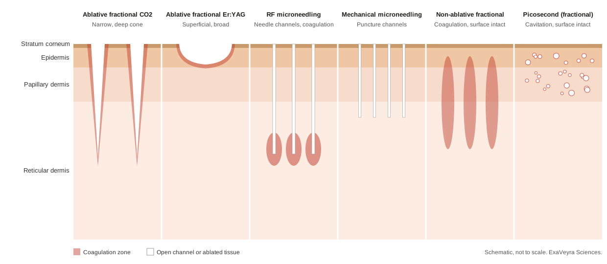
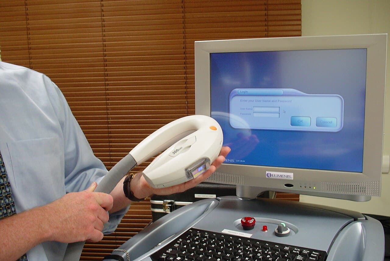
Hybrid fractional platforms pair a non-ablative wavelength with an ablative one, so they carry features of two rows at once, and 1927 nm thulium platforms sit in the non-ablative row, as in the dual 1550 and 1927 nm device Gao and colleagues studied in 2025. Intense pulsed light is a non-fractional light source that leaves the surface intact. Reading a device by its wavelength, mode and energy places it in the table, and the table predicts whether a surface-applied material meets an open channel or an intact surface.
Channels close within hours to a day, which sets the window
Banzhaf and colleagues imaged fractional CO2 channels in six volunteers in 2016 with optical coherence tomography and confocal microscopy. At a low energy, 87 percent of channels were open at one hour, 27 percent at 12 hours and 20 percent at 24 hours; at a higher energy the figures were 100, 53 and 40 percent, with re-epithelialization visible from day two.
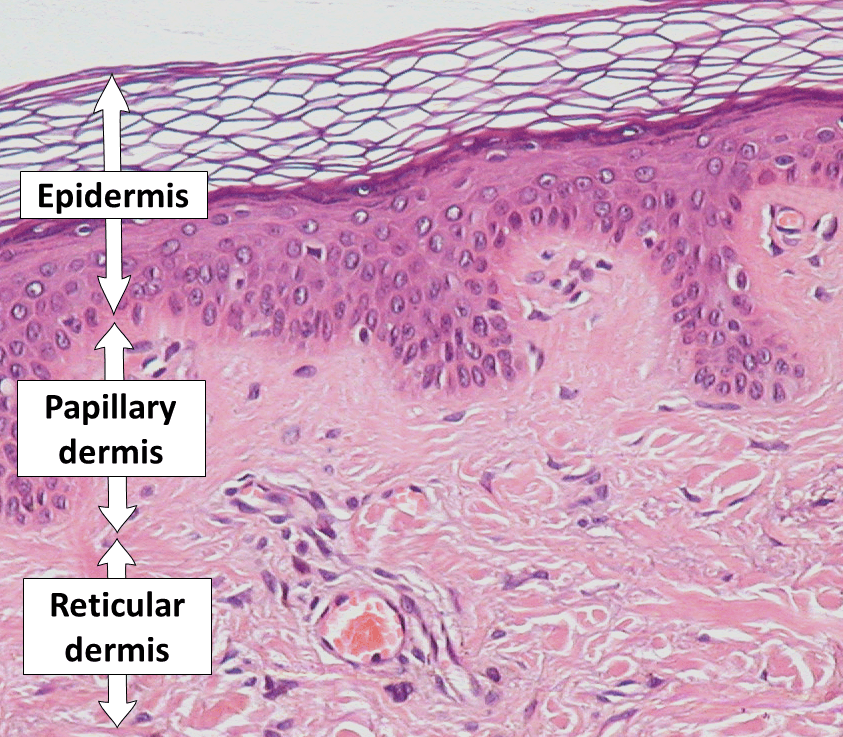
Lee WR and colleagues reported in 2026 that water-loss measurements indicated channel closure within about 36 hours after fractional Er:YAG laser and about 12 hours after RF microneedling in their models. They also reported that lipid nanocarriers of 76 to 81 nanometers, close to the size of small vesicles, were delivered less efficiently than free drug and sat mainly in the channels before spreading into surrounding dermis.
Mechanical microneedle channels close faster still. Gupta and colleagues measured skin resealing in human volunteers in 2011 and reported that unoccluded microneedle sites recovered barrier properties within two hours, while occluded sites stayed permeable for 3 to 40 hours depending on needle length, number and cross-section.
What the channel data mean for vesicle studies
No human study has yet measured how deep vesicles travel through laser or RF channels; the timing comes from small-molecule and nanocarrier models. Hsieh and colleagues reported in 2026 that labeled small vesicles passed intact skin only to a limited extent, and Wang T and colleagues reported in 2023 that dye-labeled vesicles reached the deep dermis after microneedling, 1565 nm non-ablative fractional laser or a plasma device in an animal model. Those findings are why the published combination studies apply material in the window after a device session.
Banzhaf and colleagues reported in 2019 that a thin coagulation rim of about 20 micrometers gave the highest uptake of a hydrophilic test molecule, and Haedersdal and colleagues reviewed 34 laser-assisted delivery studies in 2016, all 16 preclinical studies reporting enhanced uptake. Labadie and colleagues published evidence-based guidelines for laser-assisted drug delivery in 2022 from 54 studies and recommended settings that give a channel wider than the delivered molecule, a principle that matters for particles the size of small vesicles.
Vesicle trials after fractional CO2 report scar and downtime endpoints
Kwon and colleagues ran a double-blind, randomized split-face trial in 25 adults with atrophic acne scars in 2020, applying an adipose stem cell exosome gel to one side after each of three fractional CO2 sessions. They reported a greater reduction in scar score on the exosome side, 32.5 against 19.9 percent, with milder erythema and shorter downtime; the gels were supplied by their manufacturer.
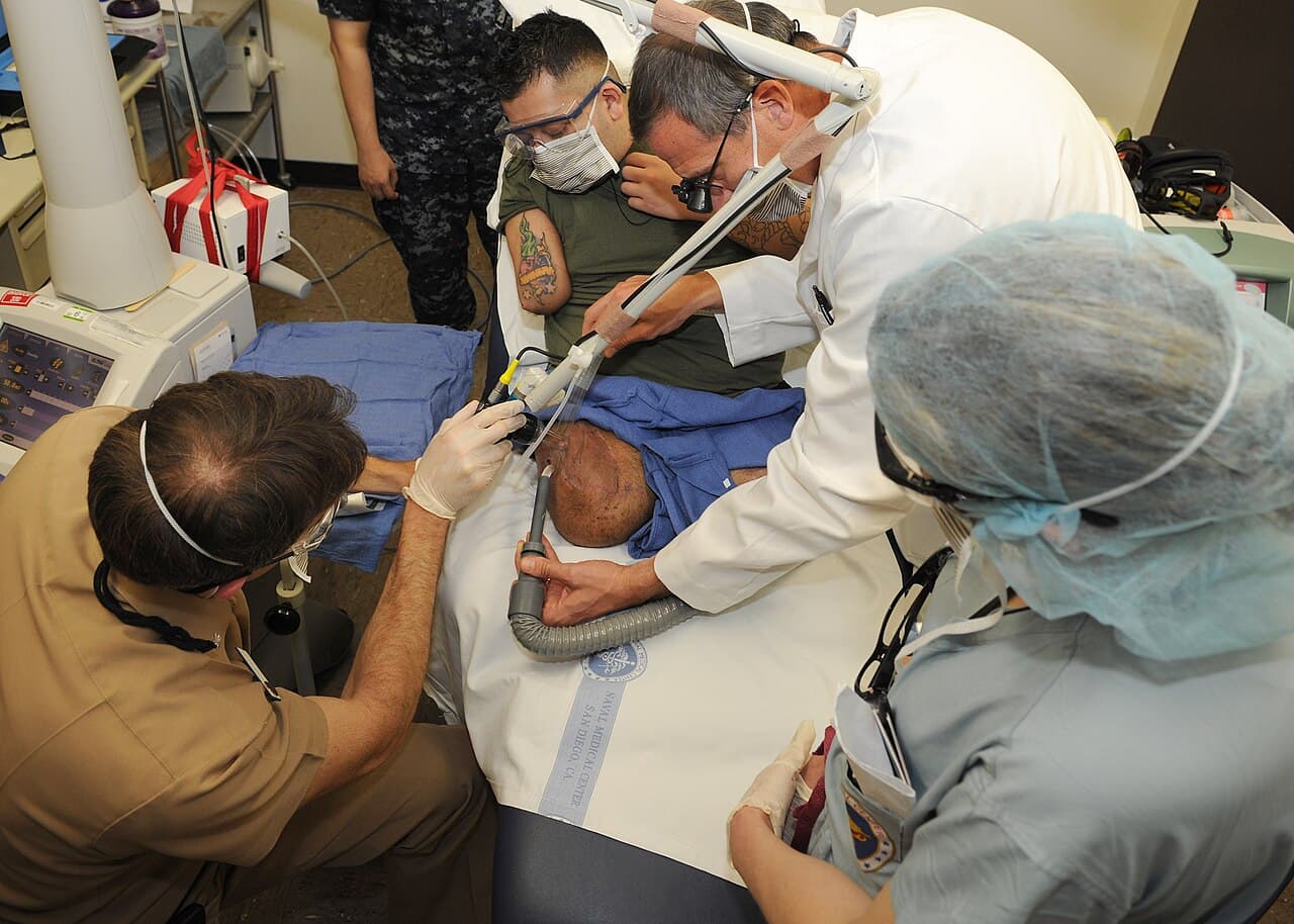
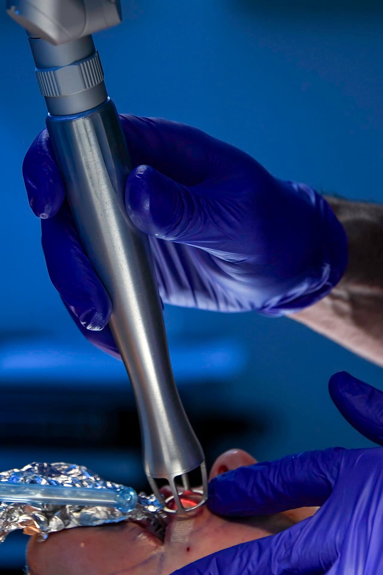
Helmi and colleagues randomized 100 adults with atrophic acne scars in 2026 to fractional CO2 alone or with topical exosome-mimetic nanovesicles and polynucleotide, reporting 47.3 against 35.9 percent scar reduction and fewer cases of prolonged erythema, 14 against 30 percent. Zhou and colleagues reported lower water loss and higher hydration with adipose stem cell conditioned medium after fractional CO2 in 22 adults in 2016.
The literature also holds a clear null result. Abdel-Maguid and colleagues ran a split-face trial with biopsies in 33 adults in 2021 and reported no significant clinical difference between fractional CO2 with stem cell conditioned medium and fractional CO2 alone, while both adjuncts increased dermal collagen expression.
RF microneedling and microneedling studies compare vesicles with PRP, saline and control conditions
Estupiñan, Ly and Goldberg published an investigator-blinded split-face non-inferiority trial in 2025 comparing topical adipose MSC exosomes with PRP after three RF microneedling sessions in 15 adults. Both halves improved equally in wrinkling, dyschromia, erythema and texture, with histological increases in collagen I and glycosaminoglycans at six months and no significant difference between arms.
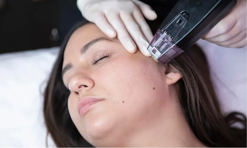
Yen, Huang and Lin reported a 60-participant unblinded split-face study in 2026, finding a 3.32-point wrinkle-severity difference favoring RF microneedling with a topical vesicle preparation over RF microneedling alone at twelve weeks, and cautioning about the unblinded design and short follow-up. Huang and colleagues followed six adults for six months after RF microneedling with a plant-derived vesicle formulation on one side and reported wrinkle differences at one and three months, with texture responses that varied by skin phenotype.
Mechanical microneedling has a saline-controlled comparison. Park GH and colleagues randomized 28 adults in 2023 to an adipose stem cell exosome solution or saline on opposite sides with three microneedling sessions, and reported higher aesthetic improvement scores and instrument-measured gains in wrinkles, elasticity, hydration and pigmentation on the exosome side.
Route comparisons are appearing as well. Yusharyahya and colleagues randomized 30 women in 2023 to microneedling or fractional CO2 as the delivery step for concentrated adipose MSC secretome and reported comparable improvement, with the laser rated higher for satisfaction and lower for comfort. Park JY and Park reported in 2026 that human- and plant-derived exosome products after non-ablative fractional Nd:YAG laser gave numerically greater short-term scar improvement than laser alone in 75 adults, and Vitale and colleagues observed smaller differences after picosecond laser than after microneedling or CO2 in nine women in 2025.
Summarizing the human combination studies
The table sets the key human studies side by side by design, size, what was measured and how long participants were followed. Results are summarized in the sections above in the authors’ own terms, and the funding notes sit in the references.
| Study | Device and material | Design | n | What was measured | Follow-up |
|---|---|---|---|---|---|
| Kwon 2020 | Fractional CO2; adipose stem cell exosome gel | Double-blind randomized split-face | 25 | Acne scar score, erythema, downtime | 12 weeks |
| Helmi 2026 | Fractional CO2; exosome-mimetic nanovesicles plus polynucleotide | Randomized, parallel arms | 100 | Scar score and subtype, hyperpigmentation, prolonged erythema, satisfaction | 3 months after last session |
| Abdel-Maguid 2021 | Fractional CO2; stem cell conditioned medium or PRP | Randomized split-face with biopsies | 33 | Clinical scar grade, dermal collagen, procollagen I | 3 months |
| Zhou BR 2016 | Fractional CO2; adipose stem cell conditioned medium | Randomized split-face | 22 | Satisfaction, roughness, elasticity, hydration, water loss, erythema and melanin index | 1 month after each of 3 sessions |
| Estupiñan 2025 | RF microneedling; adipose MSC exosomes vs PRP | Investigator-blinded split-face non-inferiority | 15 | Wrinkling, dyschromia, erythema, texture, collagen I and III, glycosaminoglycans | 6 months, with biopsies |
| Yen 2026 | RF microneedling; topical vesicle preparation | Randomized split-face, unblinded | 60 | Wrinkle severity, texture, pores, erythema, melanin, hydration, GAIS | 12 weeks |
| Huang 2026 | RF microneedling; plant-derived vesicle formulation | Split-face pilot, random side | 6 | Wrinkles, texture, pores by 3D imaging | 6 months |
| Park GH 2023 | Microneedling; adipose stem cell exosome solution vs saline | Randomized split-face | 28 | GAIS, wrinkles, elasticity, hydration, pigmentation, histology | 12 weeks |
| Yusharyahya 2023 | Microneedling vs fractional CO2; adipose MSC secretome | Single-blind randomized split-face | 30 | Photoaging scale, wrinkles, satisfaction, comfort | 6 weeks |
| Park JY 2026 | Non-ablative fractional Nd:YAG; human- or plant-derived exosome products | Randomized, three arms | 75 | Modified Vancouver and POSAS scar scales, imaging | Short term, after five sessions |
| Vitale 2025 | Microneedling, CO2 or picosecond; commercial exosome product | Observational split-face | 9 | 3D imaging, GAIS, satisfaction | 180 days |
| Lacerna Kimbrell 2026 | Helium RF plasma resurfacing; fibroblast-derived exosomes | Retrospective case series | 101 | Days to re-epithelialization, complications | Until re-epithelialization |
Systematic reviews map a promising and growing evidence base
Gaebe and colleagues applied risk-of-bias tools and GRADE to 16 studies of vesicle-based facial rejuvenation with 503 participants in 2026. They reported that most studies found improvement in at least one skin-quality outcome with no serious adverse events. The review graded certainty as low or very low, the rating GRADE gives to small and early study designs, and the larger randomized trials now registered are designed to build on that foundation.
Stack and colleagues pooled 39 human studies in a 2026 meta-analysis and reported an average facial wrinkle reduction of 20.2 percent, while noting heterogeneity and nonstandardized protocols. Lin and colleagues restricted a 2023 meta-analysis to five randomized trials of stem cell conditioned medium added to laser, peel or microneedling resurfacing and reported differences in wrinkles, pigmentation and pores in favor of the combination.
Wang, Bajwa and Rivers mapped seventeen human dermatology studies from 2020 to 2025 in a 2026 scoping review and reported improvement in 76 percent, with delivery by topical application, microneedling, fractional CO2 and injection, and evidence limited by small samples, single-arm designs and potential conflicts of interest. Knauer and colleagues reviewed 18 topical exosome studies alongside 20 commercial products in 2026 and reported that the products carried minimal information on isolation, processing or shelf life.
Recovery is measured by erythema, water loss and time to re-epithelialization
Erlich and colleagues reviewed 27 non-ablative laser studies in 2026 and described an inflammatory phase, re-epithelialization within 24 hours and remodeling over weeks two to twelve. Recovery studies measure that first stretch: Kim and Gallo reported less erythema and edema after fractional CO2 with PRP than with saline in 15 adults in 2015, and Gao and colleagues reported lower erythema and water loss at days 3, 7 and 14 with a panthenol mask after dual-wavelength non-ablative laser in 60 adults in 2025.
Wu and colleagues compared a chitosan hydrogel with a steroid, growth factor and antibiotic regimen after fractional CO2 in 2026 and reported comparable outcomes by days 7 and 28, measuring erythema, edema, pain, time to decrustation, barrier function and melanin. Lacerna Kimbrell reported in a 2026 manufacturer-funded retrospective series of 101 patients that topical fibroblast-derived exosomes after deep helium RF plasma resurfacing were associated with a median of 10 days to full re-epithelialization against 15.5 days without them.
Skin of color changes the calculation. Dreifus and colleagues reviewed device use in Fitzpatrick types IV to VI in 2026 and reported that epidermal melanin competes as a chromophore, raising the risk of post-inflammatory hyperpigmentation, and that non-ablative fractional lasers can be used with conservative settings while ablative modalities carry higher complication rates. Helmi and colleagues counted post-inflammatory hyperpigmentation as a secondary endpoint, which gives future combination studies a model to follow.
Characterization and careful handling keep vesicle material consistent
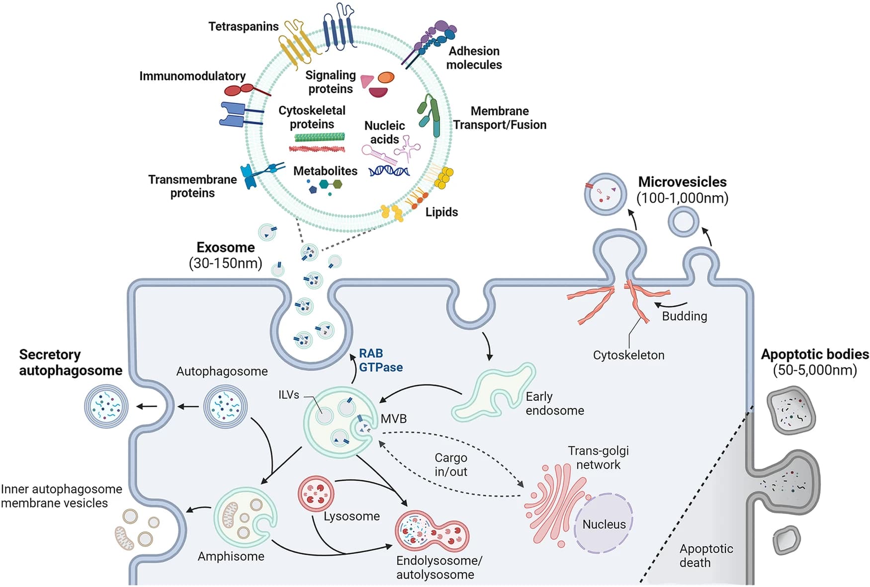
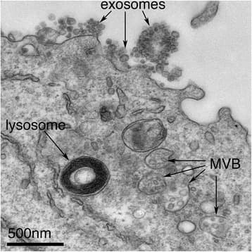
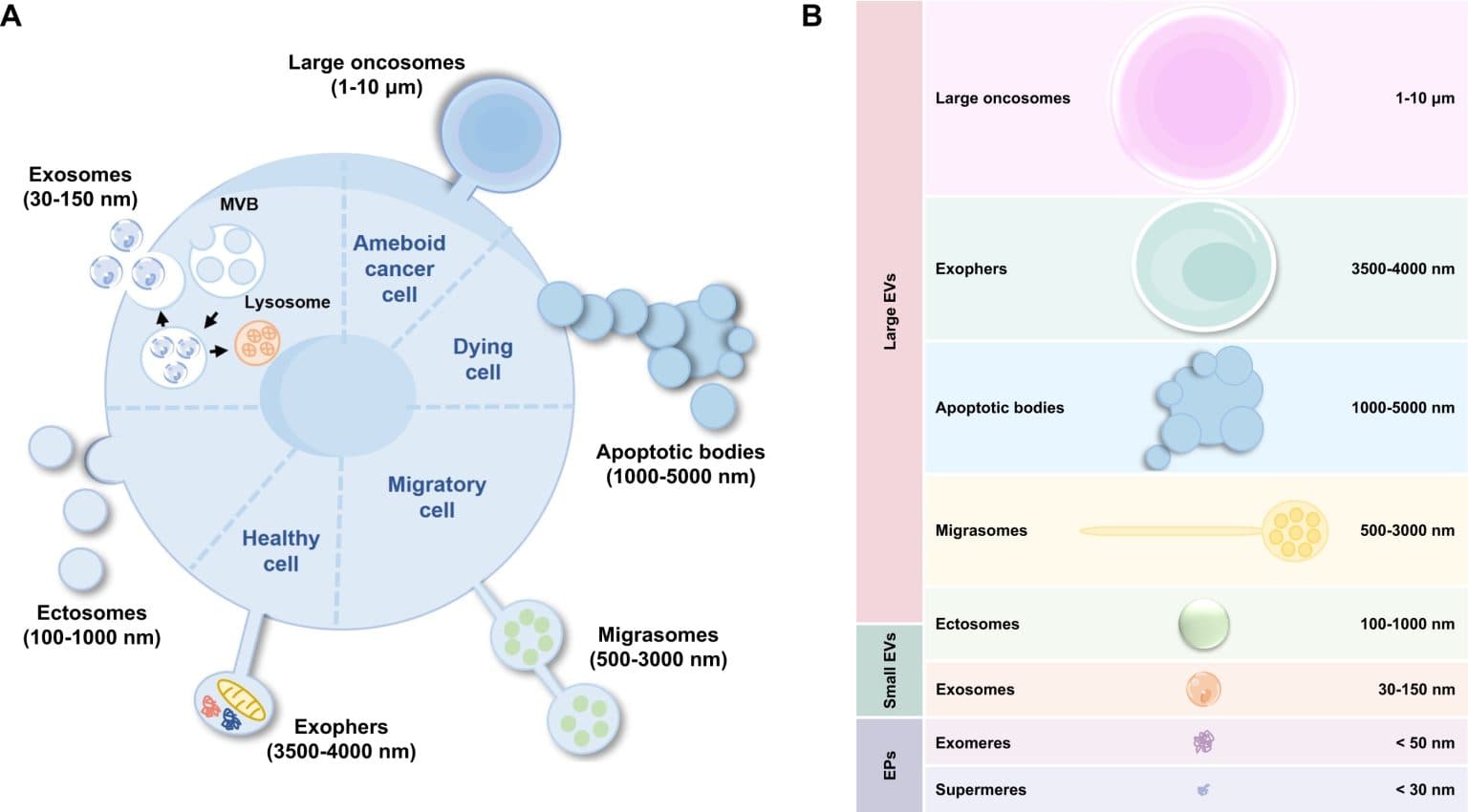
The International Society for Extracellular Vesicles updated its minimal reporting guideline as MISEV2023, setting out how vesicles should be separated from non-vesicular particles and characterized by size, concentration, marker proteins and morphology. A certificate of analysis that maps to those headings lets a practice compare one lot with the material used in a published study.
Görgens and colleagues studied storage for up to two years in 2022 and reported that plain phosphate-buffered saline drastically reduced vesicle recovery within days, and within minutes when used as a diluent, while buffer with human albumin and trehalose preserved vesicles at minus 80 degrees and across freeze-thaw cycles. The authors were affiliated with an EV therapeutics company, as the references note.
For a practice, those findings turn reconstitution and handling into part of the protocol. The supplier’s instructions, the diluent, the time between preparation and application, and the cold-chain record belong in the same file as the device settings.
Published safety reports center on injection, outside the topical workflow
AlBargawi reported ischemic necrosis of both cheeks three days after multiple intradermal injections of an exosome product in 2025, and Park KY reported persistent erythema, nodules and granulomatous inflammation in four women after intradermal injection of exosome-containing cosmetic products in nonclinical settings. Both describe injection rather than surface application after a device session.
Ng and Smith reviewed safety across 70 laser-assisted delivery studies in 2022 and reported erythema, edema, pain and crusting as common, with less frequent urticaria, infection, scarring and dyspigmentation and no life-threatening events. Haedersdal and colleagues reviewed laser-assisted delivery in 2016 and flagged the potential for systemic absorption when large areas are covered.
How the next generation of trials is building on this work
The combination literature is promising and developing quickly, and each design choice below is one the next generation of trials is already refining.
- Small samples: the controlled studies range from 6 to 100 participants, and only four of the twelve studies in the summary table enrolled more than 50.
- Split-face designs share one circulation, so an effect on one side can reach the other; parallel-arm trials such as Helmi 2026 and Park JY 2026 avoid that.
- Blinding is uneven: Kwon 2020 was double-blind, Estupiñan 2025 blinded the investigator, and Yen 2026 was unblinded.
- Comparators vary from saline and vehicle gel to PRP to untreated control conditions, which makes pooling difficult; Gaebe 2026 synthesized results narratively for that reason.
- Industry ties are common: the material in Kwon 2020 came from its manufacturer, Estupiñan 2025 was supported in part by an exosome manufacturer, and Lacerna Kimbrell 2026 was manufacturer-funded.
- Materials differ by source, isolation and concentration, and few papers report characterization to MISEV2023 headings, so results for one product may not transfer to another.
- No human study has measured vesicle depth after a device session; Wang T 2023 tracked dye-labeled vesicles in animals, and MISEV2023 cautions that lipophilic dyes can label non-vesicular particles.
- Follow-up is short, mostly 6 weeks to 3 months, and darker skin types are underrepresented, as Dreifus 2026 noted for device research generally.
- An early randomized split-face study of RF microneedling with conditioned medium was retracted by its journal, so reviews written before 2021 may still count it.
- Several registered studies, including NCT07510295 and NCT07849556, were completed without a located publication, so the published record may lean toward positive results.
Understanding the US regulatory status of devices and vesicle material
Each part of a combination session sits under a different regulation. Fractional CO2 lasers are classified at 21 CFR 878.4810 as laser surgical instruments, and laser products also carry the radiation performance standard at 21 CFR 1040.10. RF microneedling systems have been cleared as electrosurgical cutting and coagulation devices under 21 CFR 878.4400, and mechanical microneedling devices for aesthetic use are Class II under 21 CFR 878.4430, classified by a 2018 final order.
None of those classifications covers a topical payload. The microneedling identification excludes devices intended to deliver cosmetics, drugs or biologics through the skin, and the FDA’s microneedling page states that the devices are not approved for delivery of cosmetics, topical medications, drugs or blood products into the skin and that the risks of combining the devices with other products are not known. The FDA’s 2020 guidance on microneedling products explains when such a product is a device.
For the material itself, the FDA stated in 2019 that there are no FDA-approved exosome products and that exosomes used to treat diseases and conditions in humans are regulated as drugs and biological products, subject to premarket review. The agency’s consumer alert on regenerative medicine products, current as of 2024, describes its concern about statements on the conditions these products can address, and its cosmetics guidance explains that a product’s category follows its intended use, established in part by the claims made for it.
| A supplier can | A supplier stays away from |
|---|---|
| Describe the material by class, source, available format and characterization | Describing a device as cleared to deliver the material |
| Label every vial with its lot number and provide the certificate of analysis and cold-chain record on request | Presenting the material as intended for any named condition |
| Cite published studies with the authors named and the design stated | Restating a study result as a product property |
| State the FDA position on exosome products and device classifications | Suggesting injection or any route outside the material’s labeling |
Documenting a combination protocol in practice
A practice that records the same fields the published studies report can compare its own outcomes with the literature and answer a regulator’s or an investigator’s questions from one file. The fields below follow the studies and guidance cited above.
- The device by model and its FDA clearance or De Novo number, with wavelength or mode, energy, density, needle depth and number of passes
- The channel type the session produces, read against the device table above
- The time from device session to surface application, recorded against the closure data above
- The material by name and class: exosomes, secretome, conditioned medium, plant-derived vesicles or another preparation
- The lot number, expiry, certificate of analysis and characterization panel, mapped where possible to MISEV2023 headings
- The cold-chain record from receipt to use, the diluent, and the time between reconstitution and application
- Fitzpatrick type and pigmentary history, with conservative settings where the literature advises them
- Recovery endpoints: erythema, edema, water loss, time to crusting or re-epithelialization, and patient-reported downtime
- Any adverse event, with the lot number, and its report to the FDA through MedWatch
A combination study documented to that standard can be compared with the trials above, and the registered studies now completing will add to the record. ExaVeyra supplies exosome material to NPI-verified practices and investigators with the certificate of analysis, characterization panel and cold-chain records available on request for each lot, alongside procedural devices and a needle-free sterile applicator for surface application whose classification covers the applicator itself. The field is moving quickly toward larger, better characterized and properly registered studies, and practices that keep careful records are well placed to contribute to it.
Educational Disclaimer: This article is for educational purposes only and does not constitute medical or legal advice. It reviews published research and registered trials; it does not identify any material, compound, device setting or protocol as safe, effective or appropriate for any patient, and it is not intended to diagnose, treat, cure, or prevent any disease. Conditions named in this article are named as the subjects of the studies cited. Naming a condition studied in a model or a trial does not establish an indication for any product. Exosome material is supplied for topical aesthetic use by licensed practitioners and for laboratory and IND-authorized clinical research. No exosome product is FDA-approved for any use, and ExaVeyra Sciences makes no statement about administration, route, or outcome. Any use of research-grade material in human subjects requires the applicable FDA pathway, such as an investigational new drug application, and institutional review board oversight. Compounded medications are not FDA-approved. They are prepared by a 503A compounding pharmacy licensed by the Texas State Board of Pharmacy for an individual patient against that patient’s prescription, and are available to NPI-verified licensed practitioners only. Discussion of a compound’s literature, including compounds the FDA placed in category 2 of its 503A bulk drug substances policy, is not an offer to supply it. ExaVeyra Sciences is a supplier and takes no part in any diagnosis, treatment decision, study design decision, or plan of care. Findings described are attributed to the peer-reviewed literature, trial registries and agencies cited, and a citation establishes that a question was studied rather than that a product suits a given patient or protocol. The content of these guides is provided for general informational purposes only. It does not constitute legal, medical, or regulatory advice, and does not establish that any particular product or practice is compliant. Regulatory requirements vary by state and depend on the circumstances of each practice. Each practitioner should consult their own legal counsel and the applicable state licensing boards before acting on anything stated here.
Peer-Reviewed References
- 1.Kwon HH, Yang SH, Lee J, Park BC, Park KY, Jung JY, et al. Combination Treatment with Human Adipose Tissue Stem Cell-derived Exosomes and Fractional CO2 Laser for Acne Scars: A 12-week Prospective, Double-blind, Randomized, Split-face Study. Acta Derm Venereol. 2020;100(18):adv00310. PMID 33073298. PMC9309822. doi:10.2340/00015555-3666Prospective, double-blind, randomized, split-face trial over 12 weeks with three fractional CO2 sessions; adults with atrophic acne scars (Korea); n = 25. Carries in this article: the primary controlled human evidence for topical vesicle material applied after ablative fractional CO2, including the erythema and downtime endpoints. Disclosure: the exosome and control gels were manufactured and supplied by ExoCoBio Inc., the exosome manufacturer; the authors declared no conflicts of interest.
- 2.Estupiñan B, Ly K, Goldberg DJ. Adipose Mesenchymal Stem Cell-Derived Exosomes Versus Platelet-Rich Plasma Treatment for Photoaged Facial Skin: An Investigator-Blinded, Split-Face, Non-Inferiority Trial. J Cosmet Dermatol. 2025;24(5):e70208. PMID 40414798. PMC12104007. doi:10.1111/jocd.70208Investigator-blinded, split-face, non-inferiority trial; three RF microneedling sessions with topical adipose MSC exosomes on one half of the face and PRP on the other; adults with mild to moderate facial photoaging (USA); n = 15 enrolled, 14 completed; biopsies in the first 10 participants at baseline, 3 and 6 months. Carries in this article: the only located head-to-head comparison of exosomes and PRP after RF microneedling; equivalence between arms. Disclosure: supported in part by a research grant from Benev Company Inc., an exosome product manufacturer, to the investigators' practice (full text).
- 3.Yen C, Huang H, Lin C. Radiofrequency Microneedling Combined With Topical Extracellular Vesicle Preparation for Facial Rejuvenation: A Randomized Split-Face Controlled Study. J Craniofac Surg. 2026. Published online ahead of print. PMID 42635327. doi:10.1097/SCS.0000000000013266Randomized split-face controlled study, unblinded, assessed at baseline, week 4 and week 12; adults seeking facial rejuvenation (Taiwan), mean age 43.6, 83 percent women; n = 60. Carries in this article: the largest split-face RF microneedling plus vesicle study; paired wrinkle-score difference of 3.32 points (95% CI 2.55 to 4.08) and the authors' own caveats on blinding, follow-up and missing biomarkers. Disclosure: the authors reported no conflicts of interest.
- 4.Helmi RR, Mohammed RSH, Nori W. Fractional CO2 laser augmented by polynucleotide and exosome-mimetic nanovesicles in atrophic acne scars: randomised comparative trial. Postepy Dermatol Alergol. 2026;43(3):314-321. PMID 42621380. PMC13487574. doi:10.5114/ada.2026.161825Randomized comparative trial, three sessions four weeks apart, evaluated 3 months after the last session; adults with atrophic acne scars (Iraq); n = 100 (50 per arm). Carries in this article: scar-score and prolonged-erythema endpoints after fractional CO2; the intervention combines exosome-mimetic nanovesicles with polynucleotide, so effects cannot be attributed to vesicles alone. Disclosure: no external funding; the authors declared no conflict of interest.
- 5.Park JY, Park JH. Randomized Clinical Study of Laser-Assisted Delivery of Exosome Boosters for Postoperative Facial Scars and Facial Rejuvenation. Life (Basel). 2026;16(2):217. PMID 41752855. PMC12942482. doi:10.3390/life16020217Randomized controlled clinical study, three arms, five sessions at two-week intervals; adults with postoperative facial scars (Korea); n = 75. Carries in this article: fractional non-ablative Nd:YAG laser alone or with human-derived or plant-derived topical exosome products; numerically greater short-term gains with either product. Disclosure: hospital research fund grant; the two commercial products came from one Korean manufacturer; the authors declared no conflicts of interest.
- 6.Park GH, Kwon HH, Seok J, Yang SH, Lee J, Park BC, et al. Efficacy of combined treatment with human adipose tissue stem cell-derived exosome-containing solution and microneedling for facial skin aging: A 12-week prospective, randomized, split-face study. J Cosmet Dermatol. 2023;22(12):3418-3426. PMID 37377400. doi:10.1111/jocd.15872Prospective, randomized, split-face comparative study over 12 weeks, three sessions three weeks apart, saline control side; adults with facial skin aging (Korea); n = 28. Carries in this article: controlled microneedling comparison with instrument endpoints (wrinkles, elasticity, hydration, pigmentation) and histology. Disclosure: National Research Foundation of Korea grants listed in the PubMed record; no COI statement in the record.
- 7.Abdel-Maguid EM, Awad SM, Hassan YS, El-Mokhtar MA, El-Deek HE, Mekkawy MM. Efficacy of stem cell-conditioned medium vs. platelet-rich plasma as an adjuvant to ablative fractional CO2 laser resurfacing for atrophic post-acne scars: a split-face clinical trial. J Dermatolog Treat. 2021;32(2):242-249. PMID 31180258. doi:10.1080/09546634.2019.1630701Randomized split-face clinical trial in two groups, three monthly sessions, final assessment 3 months later, with biopsies; adults with atrophic post-acne scars (Egypt); n = 33. Carries in this article: the null clinical comparison of conditioned medium against laser alone (p = .63), with increased dermal collagen in both adjunct arms; balances the positive reports. Disclosure: no COI statement in the PubMed record; academic authors.
- 8.Yusharyahya SN, Japranata VV, Sitohang IBS, Legiawati L, Novianto E, Suseno LS, et al. A Comparative Study on Adipose-Derived Mesenchymal Stem Cells Secretome Delivery Using Microneedling and Fractional CO2 Laser for Facial Skin Rejuvenation. Clin Cosmet Investig Dermatol. 2023;16:387-395. PMID 36798539. PMC9926994. doi:10.2147/CCID.S401839Single-blind, randomized split-face trial comparing delivery routes, sessions at weeks 2 and 4, assessed at weeks 4 and 6; Indonesian women aged 35 to 59 with facial photoaging; n = 30. Carries in this article: head-to-head comparison of microneedle and laser channels for secretome delivery (registered as NCT05508191). Disclosure: funded by a government matching fund and PT Kimia Farma; two authors declared pending patents.
- 9.Zhou BR, Zhang T, Bin Jameel AA, Xu Y, Xu Y, Guo SL, et al. The efficacy of conditioned media of adipose-derived stem cells combined with ablative carbon dioxide fractional resurfacing for atrophic acne scars and skin rejuvenation. J Cosmet Laser Ther. 2016;18(3):138-48. PMID 26735291. doi:10.3109/14764172.2015.1114638Randomized split-face comparative study, three fractional CO2 sessions, assessed 1 week after the first session and 1 month after each session; adults with atrophic acne scars (n=13) or rejuvenation goals (n=9) (China); n = 22. Carries in this article: biophysical recovery endpoints (water loss, hydration, erythema and melanin index) with a conditioned-medium adjunct after CO2. Disclosure: no COI statement in the PubMed record; one affiliation is a regenerative medicine engineering research center.
- 10.Huang PP, Chih PL, OuYang SY. Preliminary Insights From a Split-Face Study on Skin Quality Changes After Needling Radiofrequency With or Without a Plant-Derived Exosome-Based Formulation Over 6 Months. Aesthet Surg J. 2026;46:S62-S72. PMID 41800727. doi:10.1093/asj/sjaf253Prospective split-face pilot with random side assignment, follow-up at 1, 3 and 6 months; Asian adults, Fitzpatrick III to IV; n = 6. Carries in this article: RF microneedling with a rose stem cell-derived vesicle formulation; wrinkle differences at 1 and 3 months and texture responses that varied by skin phenotype. Disclosure: published in an Aesthetic Surgery Journal supplement on exosomes; the PubMed record carries no funding or COI statement, so full-text disclosures should be checked.
- 11.Vitale M, Ruri P, Marini S, Zappia E, Yi KH. Clinical Outcomes of Exosome-Augmented Microneedling and Laser Therapies in Full-Face Skin Rejuvenation: A Split-Face Observational Study. J Craniofac Surg. 2025. Published online ahead of print. PMID 40892048. doi:10.1097/SCS.0000000000011807Split-face observational study with 30, 90 and 180-day follow-up; women aged 30 to 50 (Italy); n = 9 across microneedling, fractional CO2 and picosecond laser groups. Carries in this article: the only located human comparison that includes picosecond laser plus a commercial exosome product; the authors suggested pinpoint bleeding may limit uptake after picosecond laser. Disclosure: the authors reported no conflicts of interest.
- 12.Lacerna Kimbrell M. Enhanced Healing and Outcomes With Human Dermal Fibroblast Exosomes: Case Series Combining Deep Helium Radiofrequency Plasma Skin Resurfacing, Facelifts, and Exosomes. Aesthet Surg J. 2026;46(12 Suppl 2):S73-S81. PMID 41800725. doi:10.1093/asj/sjaf214Retrospective single-practice case series, July 2017 to September 2025, four groups with and without topical fibroblast-derived exosomes after deep helium RF plasma resurfacing; n = 101. Carries in this article: days to full re-epithelialization (median 10 with exosomes against 15.5 without, resurfacing only) as a recovery endpoint; nonrandomized and retrospective. Disclosure: funded by ExoCoBio Inc., an exosome manufacturer, per the PubMed grant record.
- 13.Gaebe K, Lowe K, Bosnalie D, Khoury S, Villemure Poliquin N, Nichols A. Effectiveness and Safety of Extracellular Vesicle-Based Therapies for Nonsurgical Facial Rejuvenation: A Systematic Review. Facial Plast Surg Aesthet Med. 2026. Published online ahead of print. PMID 42487416. doi:10.1177/26893614261457879PRISMA systematic review with risk-of-bias and GRADE assessment, searches to June 2025; 16 studies, 503 participants. Carries in this article: the most rigorous located certainty rating for facial EV studies; most studies reported improvement, no serious adverse events were reported, risk of bias was moderate to critical and GRADE certainty was low or very low. Disclosure: academic authors (Canada); no COI statement in the PubMed record.
- 14.Stack ER, Spongberg C, Braud SC, Stanton WN, Elway M. Clinical Advances in Exosome-Based Therapies for Aesthetic Medicine: A Systematic Review and Meta-analysis of Human Clinical Trials. Aesthet Surg J. 2026;46(12 Suppl 2):S13-S25. PMID 41800726. doi:10.1093/asj/sjaf178Systematic review and random-effects meta-analysis, searches to February 2025; 39 human studies (26 skin, 13 hair), nine ongoing registered trials identified. Carries in this article: pooled facial wrinkle reduction of 20.2 percent (95% CI 15.3 to 25.2) with heterogeneity and nonstandardized protocols noted by the authors. Disclosure: published in an Aesthetic Surgery Journal supplement on exosomes; the PubMed record carries no funding or COI statement.
- 15.Lin TJ, Huang YL, Kang YN, Chen C. Effectiveness of Topical Conditioned Medium of Stem Cells in Facial Skin Nonsurgical Resurfacing Modalities for Antiaging: Systematic Review and Meta-Analysis of Randomized Controlled Trials. Aesthetic Plast Surg. 2023;47(2):799-807. PMID 36396862. doi:10.1007/s00266-022-03168-zSystematic review and meta-analysis restricted to randomized controlled trials (Cochrane RoB 2); five RCTs included, four pooled. Carries in this article: the only located meta-analysis limited to RCTs of a secretome-class adjunct after resurfacing (laser, peel or microneedling), reporting wrinkle, pigmentation and pore differences. Disclosure: academic authors (Taiwan); no COI statement in the PubMed record.
- 16.Wang OJE, Bajwa BB, Rivers JK. Exosome-Based Therapies in Dermatology: A Scoping Review. J Drugs Dermatol. 2026;25(4):368-376. PMID 41931695. doi:10.36849/jdd.9610Scoping review (PRISMA-ScR) including US and EU trial registries; 17 human studies from 2020 to 2025. Carries in this article: neutral evidence map; delivery routes reported were topical application, microneedling, fractional CO2 and injection; limitations statement on small samples, single-arm designs and potential conflicts of interest. Disclosure: no COI statement in the PubMed record.
- 17.Knauer G, Chang A, Gupta A, Brownstone N, Mejia N, Bauman S, et al. Clinical Evidence and Commercial Landscape of Topical Exosomes for Skin Rejuvenation: A Scoping Review. Aesthet Surg J. 2026. Published online ahead of print. PMID 42411735. doi:10.1093/asj/sjag141Scoping review with librarian-reviewed search (2,394 records screened) plus a market survey of 20 commercial products; 18 human studies, 10 of them pairing exosomes with a delivery modality. Carries in this article: commercial products carried minimal information on isolation, processing or shelf life, the reason this article asks practices to document characterization and lot data. Disclosure: academic authors (USA); no COI statement in the PubMed record.
- 18.Labadie JG, Ibrahim SA, Worley B, Kang BY, Rakita U, Rigali S, et al. Evidence-Based Clinical Practice Guidelines for Laser-Assisted Drug Delivery. JAMA Dermatol. 2022;158(10):1193-1201. PMID 35976634. doi:10.1001/jamadermatol.2022.3234Systematic review plus two-round Delphi consensus guideline; 54 studies of ablative and non-ablative fractional laser-assisted delivery. Carries in this article: the consensus framework for laser-assisted delivery, including the recommendation that laser settings give a channel diameter greater than the delivered molecule; no recommendation addresses vesicle payloads. Disclosure: multi-author expert panel; individual disclosures are in the full text.
- 19.Haedersdal M, Erlendsson AM, Paasch U, Anderson RR. Translational medicine in the field of ablative fractional laser (AFXL)-assisted drug delivery: A critical review from basics to current clinical status. J Am Acad Dermatol. 2016;74(5):981-1004. PMID 26936299. doi:10.1016/j.jaad.2015.12.008Systematic review of 34 articles (16 preclinical, 18 clinical including 9 RCTs). Carries in this article: the foundational map of laser-assisted delivery; all 16 preclinical studies reported enhanced uptake, and the authors flagged systemic absorption risk over large areas. Disclosure: no COI statement in the PubMed record; academic authors.
- 20.Ng WHS, Smith SD. Laser-Assisted Drug Delivery: A Systematic Review of Safety and Adverse Events. Pharmaceutics. 2022;14(12):2738. PMID 36559233. PMC9787022. doi:10.3390/pharmaceutics14122738Systematic review of 70 human studies reporting safety outcomes. Carries in this article: the adverse-event profile of applying topicals through laser channels (erythema, edema, pain and crusting common; urticaria, infection, scarring and dyspigmentation less frequent; no life-threatening events). Disclosure: the authors declared no competing interest.
- 21.Manstein D, Herron GS, Sink RK, Tanner H, Anderson RR. Fractional photothermolysis: a new concept for cutaneous remodeling using microscopic patterns of thermal injury. Lasers Surg Med. 2004;34(5):426-38. PMID 15216537. doi:10.1002/lsm.20048Prospective clinical and histological study with two prototype 1.5 um devices; healthy forearm skin (n=15) and periorbital photoaged skin (n=30); n = 45. Carries in this article: origin of the microscopic thermal zone concept (about 100 um wide, 300 um deep) and the one-day re-epithelialization benchmark (pre-2011 landmark). Disclosure: authors from Wellman Laboratories; no COI statement in the PubMed record.
- 22.Shin MK, Choi JH, Ahn SB, Lee MH. Histologic comparison of microscopic treatment zones induced by fractional lasers and radiofrequency. J Cosmet Laser Ther. 2014;16(6):317-23. PMID 25148410. doi:10.3109/14764172.2014.957216Preclinical histology in domestic swine, one pass per device (fractional Er:glass, Er:YAG, CO2 and ablative microplasma RF); n = 3 animals. Carries in this article: device-specific zone geometry (cone, broad shallow, cleft, crater), which decides where a surface-applied material can pool. Disclosure: no COI statement in the PubMed record; academic authors.
- 23.Banzhaf CA, Wind BS, Mogensen M, Meesters AA, Paasch U, Wolkerstorfer A, et al. Spatiotemporal closure of fractional laser-ablated channels imaged by optical coherence tomography and reflectance confocal microscopy. Lasers Surg Med. 2016;48(2):157-65. PMID 26266688. doi:10.1002/lsm.22386In vivo imaging study (OCT and reflectance confocal microscopy) after fractional CO2 at 5 to 25 mJ per microbeam; healthy volunteers, inner forearm; n = 6. Carries in this article: channel-closure kinetics by energy (87, 27 and 20 percent open at 1, 12 and 24 hours at 5 mJ) and re-epithelialization from day 2. Disclosure: no COI statement in the PubMed record; academic authors (Copenhagen, Amsterdam, Leipzig).
- 24.Banzhaf CA, Ortner VK, Philipsen PA, Haedersdal M. The ablative fractional coagulation zone influences skin fluorescence intensities of topically applied test molecules-An in vitro study with fluorescence microscopy and fluorescence confocal microscopy. Lasers Surg Med. 2019;51(1):68-78. PMID 30584842. doi:10.1002/lsm.23034Ex vivo skin study (fluorescence and confocal microscopy) with 700 um channels made by microneedles or ablative fractional laser, coagulation zones of 0, 20 and 80 um. Carries in this article: a thin 20 um coagulation rim gave the highest uptake of a hydrophilic test molecule, so laser channels and mechanical needle channels differ. Disclosure: no COI statement in the PubMed record; academic authors.
- 25.Lee WR, Alalaiwe A, Tsai MT, Hu S, Chang FH, Chien MY, et al. Fractional microporation-guided delivery of nanoencapsulated drugs for enhanced cutaneous and follicular absorption: a comparison of ablative laser and radiofrequency microneedling. Drug Deliv Transl Res. 2026;16(1):248-267. PMID 40439860. doi:10.1007/s13346-025-01885-xPreclinical study (porcine skin permeation testing plus a mouse model) comparing fractional Er:YAG laser and RF microneedling. Carries in this article: the closest vesicle-sized model (76 to 81 nm lipid carriers), which sat mainly in the channels and was delivered less efficiently than free drug; water-loss closure within about 36 h after laser and 12 h after RF microneedling. Disclosure: the authors declared no competing interests.
- 26.Gupta J, Gill HS, Andrews SN, Prausnitz MR. Kinetics of skin resealing after insertion of microneedles in human subjects. J Control Release. 2011;154(2):148-55. PMID 21640148. PMC3164267. doi:10.1016/j.jconrel.2011.05.021Human in vivo study using electrical impedance spectroscopy across microneedle geometries, with and without occlusion. Carries in this article: unoccluded microneedle sites recovered barrier properties within 2 hours, while occluded sites resealed over 3 to 40 hours depending on needle length, number and cross-section. Disclosure: academic authors (Georgia Institute of Technology); no COI statement in the PubMed record.
- 27.Wang H, Hamblin MR, Zhang Y, Xu Y, Wen X. Histological evaluation of monopolar and bipolar radiofrequency microneedling treatment in a porcine model. Lasers Surg Med. 2024;56(3):288-297. PMID 38334177. doi:10.1002/lsm.23768Preclinical histology in porcine dorsal skin, monopolar against bipolar RF microneedling, follow-up at 0, 14, 30 and 90 days. Carries in this article: coagulation volume set by power and pulse duration, a stronger tissue reaction in bipolar mode, and rising TGF-beta, MMP3 and elastin over 90 days. Disclosure: no COI statement in the PubMed record; co-author Hamblin declared device-industry ties in the Zhou 2023 review listed below.
- 28.Zhou Y, Hamblin MR, Wen X. An update on fractional picosecond laser treatment: histology and clinical applications. Lasers Med Sci. 2023;38(1):45. PMID 36658259. PMC9852188. doi:10.1007/s10103-022-03704-yNarrative review of histology and clinical literature on fractional picosecond lasers. Carries in this article: laser-induced optical breakdown in the epidermis and cavitation in the dermis, largely without an open channel, which bears on surface application. Disclosure: one author (Hamblin) declared advisory, consulting and equity ties to light-device companies.
- 29.Erlich G, Paasch U, Jacobsen K, Haedersdal M, Olesen UH. Repositioning Non-Ablative Fractional Lasers - Molecular and Histological Insights from a scoping review. J Dtsch Dermatol Ges. 2026. Published online ahead of print. PMID 42389967. doi:10.1111/ddg.70487Scoping review of 27 studies (10 molecular, 17 histological) of 1540 to 1570 nm non-ablative fractional lasers. Carries in this article: a fluence-dependent continuum from stratum-corneum-sparing to semi-ablative injury, and the timeline of inflammatory phase, re-epithelialization within 24 hours and remodeling over weeks 2 to 12. Disclosure: no COI statement in the PubMed record; academic authors (Copenhagen, Leipzig).
- 30.Hsieh MW, Su YS, Chen LY, Zhang ZX, Su LJ, Lin HH, et al. Novel Approach for Assessment of Dermal Absorption of Extracellular Vesicle Products Using Immunoaffinity Fluorescent Nanodiamonds. Skin Pharmacol Physiol. 2026;39(1):35-46. PMID 41587148. doi:10.1159/000550612In vitro permeation study with fluorescent nanodiamond-labeled umbilical cord MSC small EVs (62.5 to 141.1 nm) on a skin tissue model. Carries in this article: intact-skin passage of labeled vesicles was limited, the rationale for applying material in the window after a device session. Disclosure: several authors are affiliated with LuminX Biotech and MediDiamond Inc. (nanodiamond label developer); no COI statement in the PubMed record.
- 31.Wang T, Gao H, Wang D, Zhang C, Hu K, Zhang H, et al. Stem cell-derived exosomes in the treatment of melasma and its percutaneous penetration. Lasers Surg Med. 2023;55(2):178-189. PMID 36573453. doi:10.1002/lsm.23628Two-part study: an animal penetration model with PKH67-labeled umbilical cord MSC exosomes after microneedles, 1565 nm non-ablative fractional laser or a plasma device, and a four-arm clinical comparison in 60 adults with melasma (China). Carries in this article: labeled vesicles reached the deep dermis after all three pretreatments in the animal model, the only located vesicle-tracking data for a non-ablative laser; lipophilic dye labels can also mark non-vesicular particles, as MISEV2023 notes. Disclosure: no COI statement in the PubMed record; registered as NCT06221787.
- 32.Zou Q, Wang H, Zhou Y, Wen X. Laser-induced skin microenvironment remodeling via exosomal CEBPA-mediated crosstalk between keratinocyte and fibroblast. J Nanobiotechnology. 2026;24(1):843. PMID 42337624. PMC13548553. doi:10.1186/s12951-026-04709-3Preclinical single-cell RNA sequencing of laser-exposed against untreated animal skin plus keratinocyte-fibroblast co-culture. Carries in this article: endogenous keratinocyte exosomes carry part of the early post-laser repair signal. Disclosure: the authors declared no competing interests.
- 33.Kim H, Gallo J. Evaluation of the effect of platelet-rich plasma on recovery after ablative fractional photothermolysis. JAMA Facial Plast Surg. 2015;17(2):97-102. PMID 25429396. doi:10.1001/jamafacial.2014.1085Prospective blinded study with randomized side assignment on the forearms; adults, Fitzpatrick I to IV, 1 cm2 fractional CO2 area per forearm; n = 15. Carries in this article: daily erythema and edema scoring until re-epithelialization as a recovery template. Disclosure: no COI statement in the PubMed record; an erratum corrects author affiliations and end matter (PMID 25789575).
- 34.Wu H, Huang W, Cai X, He H, Wang Y, Yao S, et al. Hydroxybutyl Chitosan Hydrogel Promotes Ablative Fractional CO2 Laser Wound Healing: A Prospective, Randomized, Split-Face Trial and Animal Model Evidence. Lasers Surg Med. 2026;58(3):211-222. PMID 41607028. doi:10.1002/lsm.70103Prospective randomized split-face trial plus a mouse wound model, registered ChiCTR2200063765; adults after ablative fractional CO2 for atrophic acne scars (China); n = 18. Carries in this article: a multi-endpoint recovery battery (erythema, edema, pain, time to decrustation, barrier function, melanin) at days 7 and 28. Disclosure: no COI statement in the PubMed record.
- 35.Gao M, Gao N, Wang L, Yao T, Jing H, Liu H, et al. Evaluation of the Efficacy and Safety of a Panthenol-Enriched Mask for Skin Barrier Recovery After Facial Laser Treatment: Results of a Double-Blind Randomized Controlled Study. J Cosmet Dermatol. 2025;24(7):e70223. PMID 40613435. PMC12232337. doi:10.1111/jocd.70223Double-blind randomized controlled study against a saline dressing; adults after 1550/1927 nm dual-wavelength non-ablative fractional laser (China); n = 60. Carries in this article: barrier-recovery endpoints (water loss, erythema index, hydration) at days 3, 7 and 14. Disclosure: the authors declared no conflicts of interest.
- 36.Dreifus EM, Burke OM, Alexis AF, Dover JS, Eber AE. Laser and energy-based device use in skin of color: A clinical review of safety, efficacy, and best practices. J Am Acad Dermatol. 2026. Published online ahead of print. PMID 42660430. doi:10.1016/j.jaad.2026.08.077Clinical narrative review of device use in Fitzpatrick IV to VI. Carries in this article: epidermal melanin as a competing chromophore, post-inflammatory hyperpigmentation risk, conservative non-ablative settings, and the evidence gap in darker skin types. Disclosure: three authors (Alexis, Dover, Eber) disclosed grants, equipment, advisory or speaking relationships with device and pharmaceutical companies.
- 37.Welsh JA, Goberdhan DCI, O'Driscoll L, Buzas EI, Blenkiron C, Bussolati B, et al; MISEV Consortium; Théry C, Witwer KW. Minimal information for studies of extracellular vesicles (MISEV2023): From basic to advanced approaches. J Extracell Vesicles. 2024;13(2):e12404. PMID 38326288. PMC10850029. doi:10.1002/jev2.12404International Society for Extracellular Vesicles reporting guideline, compiled with ISEV task forces and more than 1,000 researchers. Carries in this article: the reference standard for nomenclature, separation from non-vesicular particles and characterization, which a certificate of analysis should map to; the caution on dye-labeled tracking. Disclosure: many consortium authors list EV company affiliations, declared in full in the article.
- 38.Görgens A, Corso G, Hagey DW, Jawad Wiklander R, Gustafsson MO, Felldin U, et al. Identification of storage conditions stabilizing extracellular vesicles preparations. J Extracell Vesicles. 2022;11(6):e12238. PMID 35716060. PMC9206228. doi:10.1002/jev2.12238In vitro stability study up to 2 years across buffers and temperatures, EVs from several cell sources. Carries in this article: plain PBS reduced vesicle recovery within days, and within minutes as a diluent, while buffer with human albumin and trehalose preserved vesicles at minus 80 degrees and across freeze-thaw cycles. Disclosure: several authors (Gorgens, Wiklander, El Andaloussi) list affiliation with Evox Therapeutics, an EV therapeutics company.
- 39.AlBargawi S. Necrosis Following Dermal Injection of Lyophilized Exosomes: A Case Report. J Cosmet Dermatol. 2025;24(8):e70387. PMID 40820962. doi:10.1111/jocd.70387Case report; a 38-year-old man with ischemic necrosis of both cheeks three days after multiple intradermal injections of lyophilized exosomes (Saudi Arabia); n = 1. Carries in this article: a serious adverse event with injected, not surface-applied, exosome material.
- 40.Park KY. Adverse Reactions Following Intradermal Injection of Exosome-Based Formulations: A Case Series. J Cosmet Dermatol. 2025;24(10):e70520. PMID 41097876. PMC12528963. doi:10.1111/jocd.70520Case series; adult women after intradermal injection of exosome-containing cosmetic products in nonclinical settings (South Korea); n = 4. Carries in this article: persistent erythema, nodules, granulomatous inflammation and scarring after off-label injection of products sold for topical use. Disclosure: the author declared no conflicts of interest.
- 41.Statement of Retraction: Skin rejuvenation by microneedle fractional radiofrequency and a human stem cell conditioned medium in Asian skin: a randomized controlled investigator blinded split-face study. J Cosmet Laser Ther. 2020;22(4-5):215. PMID 33739221. doi:10.1080/14764172.2020.1853339Publisher retraction notice for a 2013 randomized split-face study of RF microneedling with conditioned medium. Carries in this article: an early and widely cited RF microneedling plus secretome trial has been withdrawn from the record, so reviews that predate 2021 may still count it.
- 42.U.S. Food and Drug Administration. Public Safety Notification on Exosome Products. December 6, 2019 (content current as of 12/06/2019; verified 7 October 2026). https://www.fda.gov/vaccines-blood-biologics/safety-availability-biologics/public-safety-notification-exosome-productsAgency safety notification (CBER). Carries in this article: the agency statement that there are currently no FDA-approved exosome products, and that exosomes used to treat diseases and conditions in humans are regulated as drugs and biological products and are subject to premarket review and approval requirements; the notice also points practitioners to MedWatch for adverse event reports.
- 43.U.S. Food and Drug Administration. Consumer Alert on Regenerative Medicine Products Including Stem Cells and Exosomes. Content current as of 04/09/2024; verified 7 October 2026. https://www.fda.gov/vaccines-blood-biologics/consumers-biologics/consumer-alert-regenerative-medicine-products-including-stem-cells-and-exosomesAgency consumer alert (CBER). Carries in this article: the FDA's statement of its authority over stem cell and exosome products, its concern about misleading statements on conditions these products can address, and the MedWatch reporting route for adverse events.
- 44.U.S. Food and Drug Administration. 21 CFR 878.4430, Microneedling device for aesthetic use (Class II, special controls; the identification excludes devices intended for transdermal delivery of topical products such as cosmetics, drugs or biologics). Classified by final order, 83 FR 26575, June 8, 2018, following De Novo DEN160029 (granted March 1, 2018). Text verified on eCFR 7 October 2026. https://www.accessdata.fda.gov/scripts/cdrh/cfdocs/cfcfr/CFRSearch.cfm?fr=878.4430 and https://www.accessdata.fda.gov/scripts/cdrh/cfdocs/cfpmn/denovo.cfm?id=DEN160029Federal regulation, classification order and De Novo record (CDRH). Carries in this article: what clearance of a microneedling device covers, and the express exclusion of delivery of topical products through the skin.
- 45.U.S. Food and Drug Administration. Regulatory Considerations for Microneedling Products: Guidance for Industry and Food and Drug Administration Staff. November 2020 (notice of availability 85 FR 71656). Docket FDA-2017-D-4792. https://www.fda.gov/regulatory-information/search-fda-guidance-documents/regulatory-considerations-microneedling-productsFinal CDRH guidance. Carries in this article: when a microneedling product is a device under section 201(h) of the FD&C Act, and the regulatory pathway for microneedling devices for aesthetic use.
- 46.U.S. Food and Drug Administration. Microneedling Devices. Content current as of 10/15/2025; verified 7 October 2026. https://www.fda.gov/medical-devices/aesthetic-cosmetic-devices/microneedling-devicesAgency device information page (CDRH). Carries in this article: the FDA has authorized microneedling devices to improve the appearance of facial acne scars, facial wrinkles and abdominal scars in adults 22 and older; the page states the devices are not approved for delivery of cosmetics, topical medications, drugs or blood products into the skin, and that the risks of combining the devices with other products are not known.
- 47.U.S. Food and Drug Administration. 21 CFR 878.4810, Laser surgical instrument for use in general and plastic surgery and in dermatology (Class II); 21 CFR 878.4400, Electrosurgical cutting and coagulation device and accessories (Class II); and 21 CFR 1040.10, Laser products (performance standard). Text verified on eCFR 7 October 2026. https://www.accessdata.fda.gov/scripts/cdrh/cfdocs/cfcfr/CFRSearch.cfm?fr=878.4810. Example RF microneedling clearance under 878.4400, product code GEI: 510(k) K192695, decision December 27, 2019. https://www.accessdata.fda.gov/scripts/cdrh/cfdocs/cfpmn/pmn.cfm?ID=K192695Federal regulations and a 510(k) record (CDRH), cross-checked against the openFDA 510(k) database. Carries in this article: fractional CO2 lasers are classified as laser surgical instruments, RF microneedling systems have been cleared as electrosurgical devices (the example clearance is for the InMode system with Morpheus8 applicators), and laser products also carry a radiation performance standard; none of these classifications covers a topical payload.
- 48.U.S. Food and Drug Administration. Is It a Cosmetic, a Drug, or Both? (Or Is It Soap?). Content current as of 09/11/2024; verified 7 October 2026. https://www.fda.gov/cosmetics/cosmetics-laws-regulations/it-cosmetic-drug-or-both-or-it-soapAgency guidance page on the FD&C Act definitions. Carries in this article: a product's regulatory category follows its intended use, established in part by claims made for it, which is why supplier language stays descriptive.
- 49.Ngo Anh Tuan (sponsor-investigator). Efficacy of Human UC-MSC-derived Exosomes and BM-MSC-derived Exosomes in Atrophic Acne Scar Treated With Ablative Fractional CO2 Laser: a Split-face Randomized Controlled Prospective Study. Completed July 2025. ClinicalTrials.gov NCT07510295. https://clinicaltrials.gov/study/NCT07510295Registry record; split-face randomized design comparing two MSC exosome sources after fractional CO2; 22 participants enrolled (actual); start January 2025. Carries in this article: a completed registered study whose results were not located in PubMed at the time of writing.
- 50.Yusharyahya SN (sponsor-investigator). Microneedling Versus Fractional CO2 Laser Resurfacing for Skin Rejuvenation With Adipose-derived Mesenchymal Stem Cells Secretome: a Single-blind, Randomized Clinical Trial. Completed December 2022. ClinicalTrials.gov NCT05508191. https://clinicaltrials.gov/study/NCT05508191Registry record; 30 participants enrolled (actual). Carries in this article: the registration behind the published Yusharyahya 2023 route comparison, an example of a prospectively registered combination study.
- 51.Cellese Inc. (manufacturer). Clinical Evaluation of the Efficacy and Tolerability of AnteAGE Biosomes and Facial Radiofrequency Microneedling. Completed June 2025. ClinicalTrials.gov NCT07849556. https://clinicaltrials.gov/study/NCT07849556Registry record; randomized 1:1, double-masked, all participants received RF microneedling with or without the sponsor's vesicle solution and home regimen; 30 participants enrolled (actual). Carries in this article: an industry-sponsored RF microneedling study with no located publication yet. Disclosure: manufacturer-sponsored.
- 52.Laser Beamer Skincare (commercial clinic). Enhancing Skin Rejuvenation: A Single-Blind, Randomized Control Study on the Combined Efficacy of Aerolase 1064 nm YAG Laser and Exosomes Derived From Umbilical Cord Mesenchymal Stem Cells. Completed July 2025. ClinicalTrials.gov NCT07281690. https://clinicaltrials.gov/study/NCT07281690Registry record; single-blind randomized design; 20 participants enrolled (actual). Carries in this article: a registered study pairing a non-fractional 1064 nm laser with vesicle material, sponsored by a commercial clinic.
- 53.Chang Gung Memorial Hospital. A Split-Scalp Study Evaluating the Efficacy and Safety of Fractional Laser Therapy With and Without Exosomes in the Treatment of Androgenetic Alopecia. Not yet recruiting; estimated completion October 2027. ClinicalTrials.gov NCT07248410. https://clinicaltrials.gov/study/NCT07248410Registry record; split-scalp design; 30 participants (estimated). Carries in this article: an academic registered protocol extending the fractional laser plus vesicle design to the scalp.
- 54.Acorn Biolabs Inc. (manufacturer). A Prospective, Single-blind, Randomized, Placebo-controlled, Clinical Trial Evaluating Reduction in Severity and Duration of Symptoms After Laser Therapy With Acorn aHFS. Completed December 2025. ClinicalTrials.gov NCT06336135. https://clinicaltrials.gov/study/NCT06336135Registry record; placebo-controlled design measuring post-laser recovery symptoms with an autologous hair follicle secretome; 11 participants enrolled (actual). Carries in this article: a registered recovery-endpoint study of a secretome-class product. Disclosure: manufacturer-sponsored.
- 55.Nanjing First Hospital, Nanjing Medical University. Clinical Observation of Exosomes in Patients After Q-switched Laser Surgery. Status unknown; start September 2022. ClinicalTrials.gov NCT06279039. https://clinicaltrials.gov/study/NCT06279039Registry record; 38 participants (estimated). Carries in this article: the only located registered protocol applying vesicle material after a Q-switched pigment laser; its status is listed as unknown.
- 56.U.S. Food and Drug Administration. 510(k) Premarket Notification K060448: Lumenis One Family of Systems (Lumenis, Inc.). Decision July 2006. https://www.accessdata.fda.gov/scripts/cdrh/cfdocs/cfpmn/pmn.cfm?ID=K060448FDA clearance record. Carries in this article: the regulatory status of the intense pulsed light system shown in the IPL figure.
- 57.U.S. Food and Drug Administration. 510(k) Premarket Notification K142376: Palomar Icon Aesthetic System (Cynosure, Inc.). Decision 24 September 2014. https://www.accessdata.fda.gov/scripts/cdrh/cfdocs/cfpmn/pmn.cfm?ID=K142376FDA clearance record. Carries in this article: the regulatory status of the platform shown in the Icon figure, now sold as the Cynosure Icon.