Exosome Biogenesis: ESCRT, Ceramide and the Multivesicular Body
By Benn Bluestein-Veyra, M.Sc. Organic Chemistry · Updated 20 October 2026
ExaVeyra Sciences is a Miami-based B2B supplier of exosomes for topical aesthetic use to licensed clinics, and it refers licensed prescribers to a 503A compounding pharmacy that prepares formulations against individual prescriptions.
| Question | Answer |
|---|---|
| What this page covers | How exosomes form in multivesicular bodies: the ESCRT-0 to ESCRT-III machinery, ceramide-driven budding, tetraspanin microdomains and plasma membrane fusion. |
| Who can order | Licensed clinics and practitioners whose active professional license and NPI ExaVeyra has verified. Exosome orders also require a signed professional-use acknowledgment. |
| What ships, and how | Exosome material for topical aesthetic use, characterized by lot, ships overnight in a cold chain. Delivery can take 1 to 3 business days. A certificate of analysis is issued for every lot and provided on request. |
Exosome biogenesis is the process by which cells form small vesicles inside endosomes and release them into the extracellular space. Intraluminal vesicles bud into maturing endosomes, which become multivesicular bodies, and fusion of a multivesicular body with the plasma membrane releases those vesicles as exosomes.[1]
Several machineries shape the budding membrane: the ESCRT complexes, the lipid ceramide and tetraspanin microdomains.[2] This page describes each of them, the proteins that dock and fuse multivesicular bodies with the cell surface, and how cells divide these organelles between secretion and degradation.
Early endosomes mature into multivesicular bodies
Endocytosed membrane proteins first reach early endosomes. Huotari and Helenius described the maturation program that converts these organelles into late endosomes, or multivesicular bodies, and separates them from early endosomes in function and position.[3]
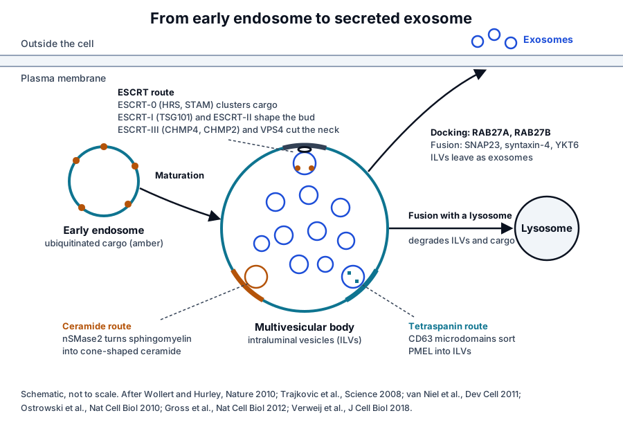
During maturation the limiting membrane buds inward to form intraluminal vesicles, or ILVs (Fig. 1). Ubiquitin marks the membrane proteins that enter these invaginations, and the ESCRT machinery sorts them and releases each vesicle from the perimeter membrane.[4]
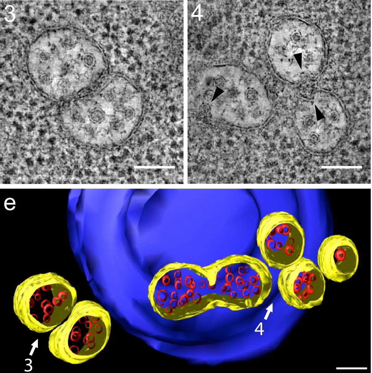
Electron tomography resolves this geometry directly. Adell and colleagues combined light microscopy with tomography of yeast multivesicular bodies and captured ILVs in the lumen alongside buds still attached to the limiting membrane (Fig. 2).[5]
A multivesicular body can fuse with a lysosome, which degrades its contents, or with the plasma membrane, which releases its ILVs as exosomes. Vesicles shed directly from the plasma membrane form a separate class called microvesicles.[1]
ESCRT-0 and ESCRT-I recognize ubiquitinated cargo
The ESCRT pathway was defined in budding yeast. Katzmann and colleagues showed in 2001 that ubiquitination is a signal for sorting into multivesicular bodies, and they identified ESCRT-I, a complex of Vps23, Vps28 and Vps37 that recognizes ubiquitinated cargo through a UBC-like domain in Vps23.[6]
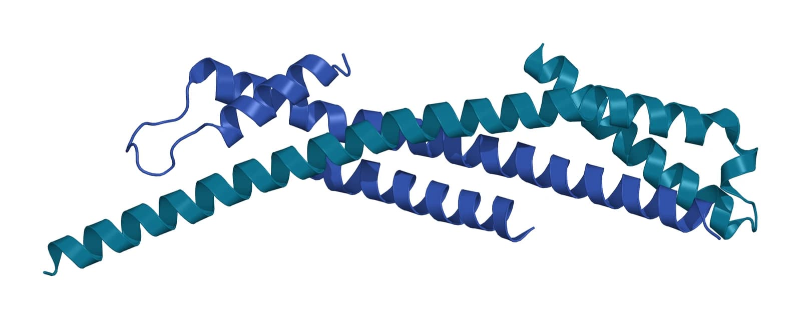
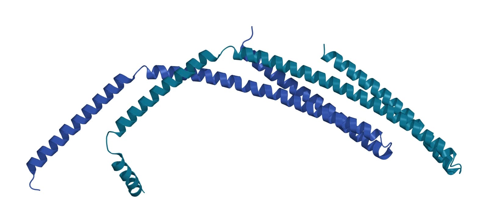
In human cells ESCRT-0 pairs HRS, the product of the HGS gene, with STAM, and HRS sorts monoubiquitinated membrane proteins into the multivesicular body.[7] Ren and colleagues solved the ESCRT-0 core at 2.3 Å, two domain-swapped GAT domains joined by an antiparallel coiled coil, within a flexible 1:1 complex (Fig. 3).[8]
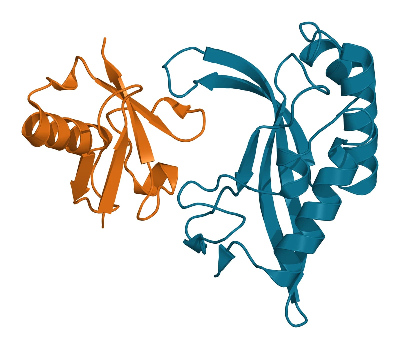
TSG101 is the human counterpart of yeast Vps23.[9] Sundquist and colleagues solved its UEV domain bound to ubiquitin at 2.0 Å: the domain contacts the Ile44 surface of ubiquitin and an adjacent loop through a highly solvated interface, and mutations at that interface inhibited sorting into multivesicular bodies (Fig. 4).[10]
Wollert and Hurley reconstituted the pathway on giant unilamellar vesicles carrying a membrane-tethered ubiquitin cargo. ESCRT-0 gathered the cargo into domains, and ESCRT-I and ESCRT-II together bent the membrane into buds that confined it, with both complexes positioned at the bud neck.[11]
- ESCRT-0 (HRS and STAM) binds ubiquitinated cargo and clusters it on the endosomal membrane.[11]
- ESCRT-I, which contains TSG101, and ESCRT-II shape the bud and sit at its neck.[11]
- ESCRT-III subunits assemble at the neck and cut it to release the ILV.[11]
- VPS4 binds ESCRT-III, drives the final constriction of the neck and recycles the subunits.[12]
ESCRT-III and VPS4 cut the neck of the intraluminal vesicle
In the reconstituted system, ESCRT-III subunits localized to the bud neck and cleaved it. The released ILVs carried the cargo and were free of ESCRT proteins, so the machinery works from the cytoplasmic side of the bud without being consumed.[11]
Wollert and colleagues had shown a year earlier that three yeast ESCRT-III subunits, Vps20, Snf7 and Vps24, suffice to detach ILVs. Vps2 recruits the ATPase Vps4, whose activity recycles ESCRT-III for further rounds of budding, and this minimal set is conserved from archaea to animals.[13]
Human ESCRT-III is built from the charged multivesicular body proteins, and the CHMP4 subfamily assembles into membrane-attached filaments.[14] Azad and colleagues solved membrane-coated CHMP2A and CHMP3 filaments by cryo-EM at 3.3 and 3.6 Å, showing heterodimers in the open conformation that present a positively charged surface to the membrane (Fig. 5).[15]
In those filaments the C-terminal sequence that VPS4 recognizes points toward the tube interior. VPS4 constricted and cleaved CHMP2A and CHMP3 membrane tubes, and the authors concluded that the three proteins form a minimal fission machinery.[15]
Humans carry two paralogs of yeast Vps4, VPS4A and VPS4B, and both associate with endosomal compartments.[16] Adell and colleagues found by electron tomography in yeast that ESCRT-III assembly induces or stabilizes the necks of growing ILVs, and that Vps4 binding, coordinated through Vps2 and Snf7, is coupled to neck constriction.[12]
Lattice light-sheet microscopy then measured the assemblies on yeast endosomes: each lasted 3 to 45 s and gathered 75 to 200 Snf7 and 15 to 50 Vps24 molecules. Productive budding required at least four Vps4 hexamers and continuous exchange of subunits.[5]
Pfitzner and colleagues traced a sequence of subunit exchanges powered by Vps4. Replacing Vps24 with Did2 tilted the polymer and converted flat spirals into helical filaments, and a highly constricted Did2 and Ist1 copolymer promoted fission from inside membrane necks.[17]
The same machinery severs membrane necks from their inner face in cytokinesis, nuclear envelope reformation and autophagy.[18][19] McCullough and colleagues review the structures of ESCRT-III filaments and of Vps4 engaged with an ESCRT-III substrate that underlie this shared mechanism.[20]
ESCRT components shape exosome secretion in mammalian cells
Colombo and colleagues screened 23 ESCRT components and associated proteins by RNA interference in HeLa-CIITA cells. Silencing HRS, STAM1 or TSG101 reduced secretion of vesicle-associated CD63 and MHC class II, and each changed the size or protein composition of the secreted vesicles in a different way.[21]
Depletion of VPS4B increased secretion and left vesicle features unchanged. Silencing ALIX raised MHC class II in vesicles secreted by HeLa-CIITA cells. In primary dendritic cells it raised MHC class II in the cells and left the CD63-positive vesicles unchanged, a difference the authors attributed to vesicle heterogeneity.[21]
Stuffers and colleagues depleted key subunits of all four ESCRT complexes in mammalian cells. EGF-induced multivesicular bodies were highly sensitive to the depletion, yet EGF-independent multivesicular bodies still formed, with enlarged ILVs that excluded the EGF receptor.[22]
The authors concluded that mammalian cells use both ESCRT-dependent and ESCRT-independent mechanisms of multivesicular body biogenesis.[22] Henne and colleagues describe the ESCRT pathway itself as a cargo recognition and membrane sculpting machine.[9]
Ceramide drives ESCRT-independent inward budding
Trajkovic and colleagues found in 2008 that cargo segregates into distinct subdomains on the endosomal membrane. Transfer of the exosome-associated domains into the endosome lumen proceeded independently of ESCRT function and required the sphingolipid ceramide.[23]
Purified exosomes were enriched in ceramide, and inhibiting neutral sphingomyelinases reduced exosome release.[23] The authors proposed that the cone shape of ceramide, with its small head group, and its tendency to coalesce into domains favor the negative curvature of an inward bud.[23]
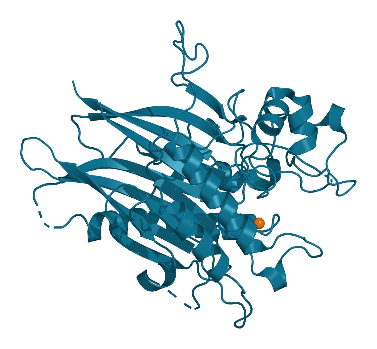
Neutral sphingomyelinase 2 is encoded by SMPD3 and carries sphingomyelin phosphodiesterase activity.[24] Airola and colleagues solved its catalytic domain at 1.85 Å, a DNase-I-type fold whose hydrophobic track to the active site is gated by a conserved element they named the DK switch (Fig. 6).[25]
A membrane-embedded N-terminal domain anchors the enzyme and binds phosphatidylserine. The juxtamembrane region linking it to the catalytic domain was necessary and sufficient for allosteric activation, which suggests that ceramide production follows the lipid composition of the membrane the enzyme sits in.[25]
Tetraspanins and ALIX sort cargo into intraluminal vesicles
Tetraspanin-enriched microdomains offer a third sorting route. In melanocytes, van Niel and colleagues showed that CD63 directly participates in ESCRT-independent sorting of the PMEL luminal domain into ILVs, where it is processed into the amyloid fibrils of melanosomes.[26]
Inactivating CD63 in cultured cells or in mice impaired fibril formation and melanosome morphogenesis. The remaining PMEL transmembrane fragment required functional ESCRTs, and in the absence of CD63 the luminal domain followed it to ESCRT-dependent degradation, one cargo handled by two ILV sorting processes.[26]
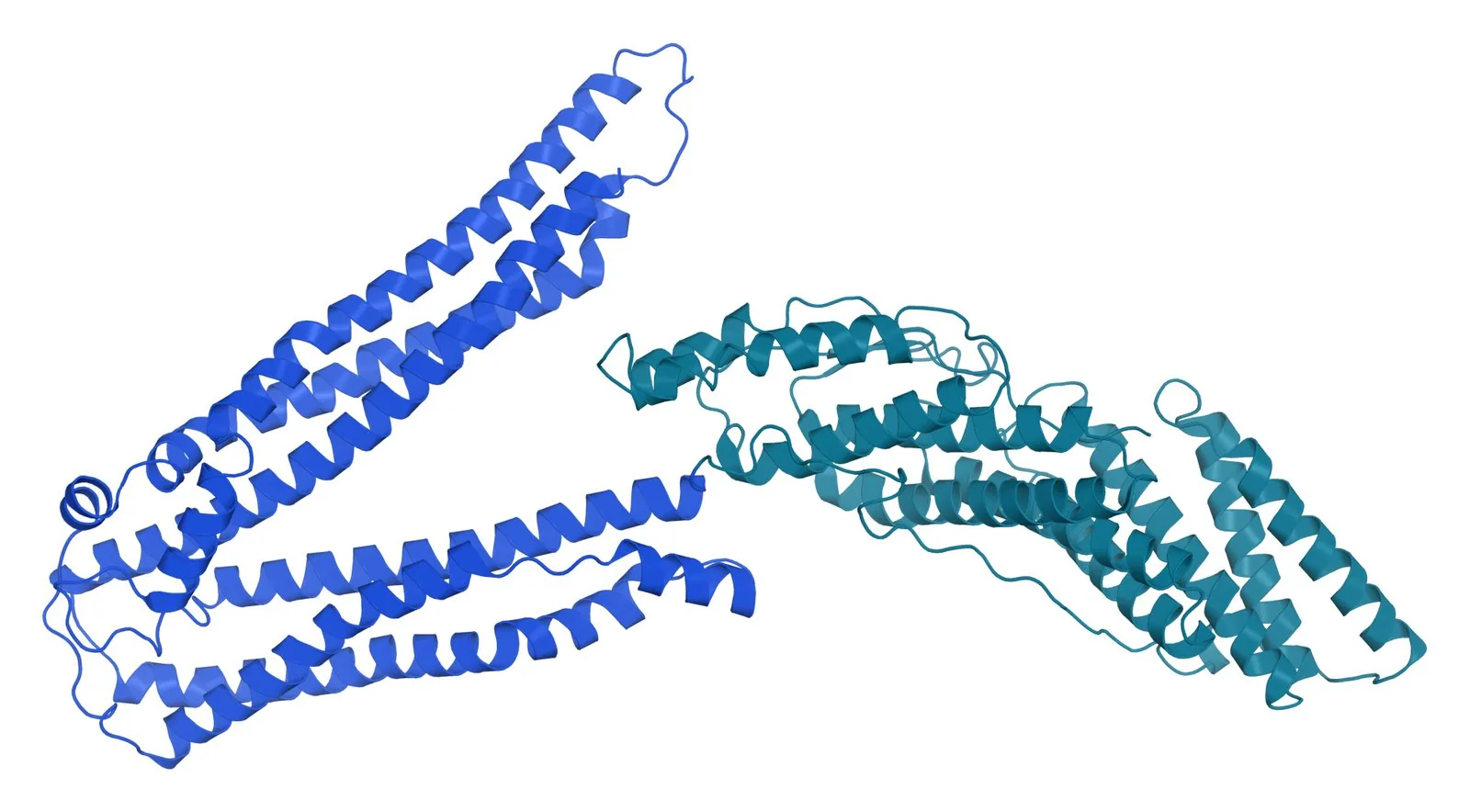
ALIX is one of the early-acting factors that nucleate ESCRT-III assembly (Fig. 7).[20] Baietti and colleagues found that the adaptor syntenin binds ALIX through LYPXnL motifs and links syndecan heparan sulfate proteoglycans to intraluminal budding.[27]
Larios and colleagues showed that an active form of ALIX recruits ESCRT-III to endosomes independently of the other ESCRTs, in a step that requires lysobisphosphatidic acid (LBPA). This ALIX and ESCRT-III pathway promoted the sorting of tetraspanins to exosomes.[28]
Matsuo and colleagues had earlier shown that LBPA can drive the formation of multivesicular liposomes. The process depended on a pH gradient like the one across endosome membranes and was controlled by ALIX.[29]
RAB27 and SNARE proteins deliver multivesicular bodies to the plasma membrane
Ostrowski and colleagues screened Rab GTPases by RNA interference in HeLa cells and identified five that promote exosome secretion. Two of them, Rab27a and Rab27b, act in docking of multivesicular bodies at the plasma membrane.[30]
The two isoforms act at different steps. Rab27a silencing strongly enlarged multivesicular bodies, Rab27b silencing redistributed them toward the perinuclear region, and silencing the effectors Slp4 and Slac2b reproduced the two phenotypes in turn.[30]
Verweij and colleagues imaged fusion directly with tetraspanin-based pH-sensitive reporters and total internal reflection microscopy. Depleting the target membrane SNAREs SNAP23 and syntaxin-4 reduced fusion frequency, and histamine H1 receptor signaling raised it through phosphorylation of SNAP23 at Ser110.[31]
Gross and colleagues found that Wnt proteins travel with their cargo receptor Evi through endosomal compartments onto exosomes in fruit flies and human cells. The exosomes carried Wnts on their surface and induced Wnt signaling in target cells, and the route requires the R-SNARE YKT6.[32]
Multivesicular bodies balance secretion and lysosomal degradation
ILVs are either sorted for degradation in lysosomes or secreted as exosomes. Trajkovic and colleagues found that cargo segregates into distinct subdomains of the endosomal membrane, which places one point of that choice at the moment of budding.[23]
Villarroya-Beltri and colleagues identified ISGylation, a ubiquitin-like modification, as a regulator of this balance. Conjugation of ISG15 decreased the number of multivesicular bodies, moved them toward lysosomes and impaired exosome secretion in cells and in mice.[33]
ISGylation of TSG101 by itself triggered the aggregation and degradation of the protein and was sufficient to impair secretion. Blocking lysosomal function or autophagy restored exosome release, consistent with degradation and secretion drawing on a shared pool of multivesicular bodies.[33]
The fate of a multivesicular body therefore reflects how its ILVs were formed and the docking and degradation signals it receives. These choices help explain why vesicle preparations from different cells differ in size and composition.[1][21]
Where to go from here
The cargo sorting reference follows the signals that load proteins and RNA into ILVs, including the syndecan, syntenin and ALIX route. The lipidome reference describes the ceramide and cholesterol content of the exosome membrane, and the CD63 reference covers the tetraspanin at the center of ESCRT-independent sorting.
For measurement, the quality references on MISEV 2023 and on tetraspanin marker panels explain how vesicle preparations are characterized. Verified practices can request the certificate of analysis for lot-specific data, and our team can explain how each result was measured.
Annotated References
Numbered in the order they are cited in the text. Each source is annotated with what it supports on this page; peer-reviewed articles are listed with journal, volume, issue and pages, and primary sources are regulations, agency documents and reference databases.
- 1.Peer-reviewedvan Niel G, D’Angelo G, Raposo G. Shedding light on the cell biology of extracellular vesicles. Nature Reviews Molecular Cell Biology. 2018;19(4):213-228. https://pubmed.ncbi.nlm.nih.gov/29339798/Annotation: Review of extracellular vesicle cell biology. Cited for the endosomal origin of exosomes, the plasma membrane origin of microvesicles and the two fates of the multivesicular body.
- 2.Peer-reviewedMathieu M, Martin-Jaular L, Lavieu G, et al. Specificities of secretion and uptake of exosomes and other extracellular vesicles for cell-to-cell communication. Nature Cell Biology. 2019;21(1):9-17. https://pubmed.ncbi.nlm.nih.gov/30602770/Annotation: Review of how exosome biogenesis compares with that of other vesicles. Cited for the several machineries that shape budding membranes.
- 3.Peer-reviewedHuotari J, Helenius A. Endosome maturation. The EMBO Journal. 2011;30(17):3481-3500. https://pubmed.ncbi.nlm.nih.gov/21878991/Annotation: Review of the endosome maturation program. Cited for the conversion of early endosomes into late endosomes, or multivesicular bodies.
- 4.Peer-reviewedRaiborg C, Stenmark H. The ESCRT machinery in endosomal sorting of ubiquitylated membrane proteins. Nature. 2009;458(7237):445-452. https://pubmed.ncbi.nlm.nih.gov/19325624/Annotation: Review of ESCRT-mediated sorting. Cited for ubiquitin as the sorting label and for ESCRT release of ILVs from the perimeter membrane.
- 5.Peer-reviewedAdell MAY, Migliano SM, Upadhyayula S, et al. Recruitment dynamics of ESCRT-III and Vps4 to endosomes and implications for reverse membrane budding. eLife. 2017;6:e31652. https://pubmed.ncbi.nlm.nih.gov/29019322/Annotation: Lattice light-sheet microscopy and electron tomography of yeast endosomes. Cited for assembly lifetimes, Snf7 and Vps24 counts, the Vps4 hexamer requirement and tomograms of ILV buds.
- 6.Peer-reviewedKatzmann DJ, Babst M, Emr SD. Ubiquitin-dependent sorting into the multivesicular body pathway requires the function of a conserved endosomal protein sorting complex, ESCRT-I. Cell. 2001;106(2):145-155. https://pubmed.ncbi.nlm.nih.gov/11511343/Annotation: Yeast genetics and biochemistry that defined ESCRT-I. Cited for ubiquitination as the sorting signal and for cargo recognition through the UBC-like domain of Vps23.
- 7.DatabaseNCBI Gene 9146, HGS hepatocyte growth factor-regulated tyrosine kinase substrate (Homo sapiens). Accessed 20 October 2026. https://www.ncbi.nlm.nih.gov/gene/9146
- 8.Peer-reviewedRen X, Kloer DP, Kim YC, et al. Hybrid structural model of the complete human ESCRT-0 complex. Structure. 2009;17(3):406-416. https://pubmed.ncbi.nlm.nih.gov/19278655/Annotation: Crystal structure of the Hrs and STAM core with a solution model of ESCRT-0. Cited for the domain-swapped GAT domains, the coiled coil and the flexible 1:1 complex.
- 9.Peer-reviewedHenne WM, Buchkovich NJ, Emr SD. The ESCRT pathway. Developmental Cell. 2011;21(1):77-91. https://pubmed.ncbi.nlm.nih.gov/21763610/Annotation: Review of ESCRT proteins, their cargo and the membranes they deform. Cited for the correspondence of yeast and human subunits and for the pathway as a cargo recognition and membrane sculpting machine.
- 10.Peer-reviewedSundquist WI, Schubert HL, Kelly BN, et al. Ubiquitin recognition by the human TSG101 protein. Molecular Cell. 2004;13(6):783-789. https://pubmed.ncbi.nlm.nih.gov/15053872/Annotation: Crystal structure of the TSG101 UEV domain with ubiquitin. Cited for the Ile44 contact surface, the solvated interface and the effect of interface mutations on multivesicular body sorting.
- 11.Peer-reviewedWollert T, Hurley JH. Molecular mechanism of multivesicular body biogenesis by ESCRT complexes. Nature. 2010;464(7290):864-869. https://pubmed.ncbi.nlm.nih.gov/20305637/Annotation: Reconstitution of multivesicular body biogenesis on giant unilamellar vesicles. Cited for cargo clustering by ESCRT-0, budding by ESCRT-I and ESCRT-II, and neck cleavage by ESCRT-III.
- 12.Peer-reviewedAdell MA, Vogel GF, Pakdel M, et al. Coordinated binding of Vps4 to ESCRT-III drives membrane neck constriction during MVB vesicle formation. Journal of Cell Biology. 2014;205(1):33-49. https://pubmed.ncbi.nlm.nih.gov/24711499/Annotation: Yeast genetics, biochemistry and electron tomography. Cited for ESCRT-III stabilizing ILV necks and Vps4 binding coupled to neck constriction.
- 13.Peer-reviewedWollert T, Wunder C, Lippincott-Schwartz J, et al. Membrane scission by the ESCRT-III complex. Nature. 2009;458(7235):172-177. https://pubmed.ncbi.nlm.nih.gov/19234443/Annotation: Reconstitution of ESCRT-III scission on giant unilamellar vesicles. Cited for Vps20, Snf7 and Vps24 sufficing to detach ILVs and for Vps4 recycling ESCRT-III.
- 14.DatabaseNCBI Gene 128866, CHMP4B charged multivesicular body protein 4B (Homo sapiens). Accessed 20 October 2026. https://www.ncbi.nlm.nih.gov/gene/128866
- 15.Peer-reviewedAzad K, Guilligay D, Boscheron C, et al. Structural basis of CHMP2A-CHMP3 ESCRT-III polymer assembly and membrane cleavage. Nature Structural and Molecular Biology. 2023;30(1):81-90. https://pubmed.ncbi.nlm.nih.gov/36604498/Annotation: Cryo-EM of membrane-coated human CHMP2A and CHMP3 filaments. Cited for the open heterodimer, the membrane-facing surface and VPS4-driven constriction and cleavage of membrane tubes.
- 16.DatabaseNCBI Gene 27183, VPS4A vacuolar protein sorting 4 homolog A (Homo sapiens). Accessed 20 October 2026. https://www.ncbi.nlm.nih.gov/gene/27183
- 17.Peer-reviewedPfitzner AK, Mercier V, Jiang X, et al. An ESCRT-III Polymerization Sequence Drives Membrane Deformation and Fission. Cell. 2020;182(5):1140-1155.e18. https://pubmed.ncbi.nlm.nih.gov/32814015/Annotation: Reconstitution of sequential ESCRT-III polymerization. Cited for Vps4-powered subunit exchange, the spiral-to-helix transition and Did2 and Ist1 fission from inside necks.
- 18.Peer-reviewedHurley JH. ESCRTs are everywhere. The EMBO Journal. 2015;34(19):2398-2407. https://pubmed.ncbi.nlm.nih.gov/26311197/Annotation: Review of ESCRT functions across the cell. Cited for neck scission from the inner face in cytokinesis, nuclear envelope reformation and autophagy.
- 19.Peer-reviewedVietri M, Radulovic M, Stenmark H. The many functions of ESCRTs. Nature Reviews Molecular Cell Biology. 2020;21(1):25-42. https://pubmed.ncbi.nlm.nih.gov/31705132/Annotation: Review of inverse membrane remodeling by ESCRT and VPS4. Cited for the shared mechanism across cellular processes.
- 20.Peer-reviewedMcCullough J, Frost A, Sundquist WI. Structures, Functions, and Dynamics of ESCRT-III/Vps4 Membrane Remodeling and Fission Complexes. Annual Review of Cell and Developmental Biology. 2018;34:85-109. https://pubmed.ncbi.nlm.nih.gov/30095293/Annotation: Review of ESCRT-III and Vps4 structures. Cited for ALIX among the early-acting factors that nucleate ESCRT-III and for filament and Vps4 substrate structures.
- 21.Peer-reviewedColombo M, Moita C, van Niel G, et al. Analysis of ESCRT functions in exosome biogenesis, composition and secretion highlights the heterogeneity of extracellular vesicles. Journal of Cell Science. 2013;126(Pt 24):5553-5565. https://pubmed.ncbi.nlm.nih.gov/24105262/Annotation: RNA interference screen of 23 ESCRT components and associated proteins. Cited for the effects of HRS, STAM1, TSG101, VPS4B and ALIX silencing on vesicle secretion and composition.
- 22.Peer-reviewedStuffers S, Sem Wegner C, Stenmark H, et al. Multivesicular endosome biogenesis in the absence of ESCRTs. Traffic. 2009;10(7):925-937. https://pubmed.ncbi.nlm.nih.gov/19490536/Annotation: Electron and confocal microscopy after depletion of all four ESCRT complexes. Cited for EGF-independent multivesicular bodies forming with enlarged ILVs.
- 23.Peer-reviewedTrajkovic K, Hsu C, Chiantia S, et al. Ceramide triggers budding of exosome vesicles into multivesicular endosomes. Science. 2008;319(5867):1244-1247. https://pubmed.ncbi.nlm.nih.gov/18309083/Annotation: Cell biology and lipid analysis of exosome budding. Cited for cargo subdomains, ESCRT-independent and ceramide-dependent budding, ceramide enrichment of exosomes and the effect of sphingomyelinase inhibition.
- 24.DatabaseNCBI Gene 55512, SMPD3 sphingomyelin phosphodiesterase 3 (Homo sapiens). Accessed 20 October 2026. https://www.ncbi.nlm.nih.gov/gene/55512
- 25.Peer-reviewedAirola MV, Shanbhogue P, Shamseddine AA, et al. Structure of human nSMase2 reveals an interdomain allosteric activation mechanism for ceramide generation. Proceedings of the National Academy of Sciences of the United States of America. 2017;114(28):E5549-E5558. https://pubmed.ncbi.nlm.nih.gov/28652336/Annotation: Crystal structure and biochemistry of human nSMase2. Cited for the catalytic fold, the DK switch, phosphatidylserine binding and juxtamembrane activation.
- 26.Peer-reviewedvan Niel G, Charrin S, Simoes S, et al. The tetraspanin CD63 regulates ESCRT-independent and -dependent endosomal sorting during melanogenesis. Developmental Cell. 2011;21(4):708-721. https://pubmed.ncbi.nlm.nih.gov/21962903/Annotation: Cell culture and knockout mouse studies of PMEL sorting. Cited for CD63-dependent, ESCRT-independent sorting of the PMEL luminal domain into ILVs.
- 27.Peer-reviewedBaietti MF, Zhang Z, Mortier E, et al. Syndecan-syntenin-ALIX regulates the biogenesis of exosomes. Nature Cell Biology. 2012;14(7):677-685. https://pubmed.ncbi.nlm.nih.gov/22660413/Annotation: Cell biology of syndecan, syntenin and ALIX. Cited for syntenin binding ALIX through LYPXnL motifs and supporting intraluminal budding.
- 28.Peer-reviewedLarios J, Mercier V, Roux A, et al. ALIX- and ESCRT-III-dependent sorting of tetraspanins to exosomes. Journal of Cell Biology. 2020;219(3). https://pubmed.ncbi.nlm.nih.gov/32049272/Annotation: Cell biology and supported bilayer reconstitution. Cited for LBPA-dependent recruitment of ESCRT-III by active ALIX and the sorting of tetraspanins to exosomes.
- 29.Peer-reviewedMatsuo H, Chevallier J, Mayran N, et al. Role of LBPA and Alix in multivesicular liposome formation and endosome organization. Science. 2004;303(5657):531-534. https://pubmed.ncbi.nlm.nih.gov/14739459/Annotation: Liposome and cell studies of LBPA. Cited for pH-dependent formation of multivesicular liposomes controlled by ALIX.
- 30.Peer-reviewedOstrowski M, Carmo NB, Krumeich S, et al. Rab27a and Rab27b control different steps of the exosome secretion pathway. Nature Cell Biology. 2010;12(1):19-30; sup pp 1-13. https://pubmed.ncbi.nlm.nih.gov/19966785/Annotation: RNA interference screen of Rab GTPases in exosome secretion. Cited for Rab27a and Rab27b in docking, their distinct phenotypes and their effectors Slp4 and Slac2b.
- 31.Peer-reviewedVerweij FJ, Bebelman MP, Jimenez CR, et al. Quantifying exosome secretion from single cells reveals a modulatory role for GPCR signaling. Journal of Cell Biology. 2018;217(3):1129-1142. https://pubmed.ncbi.nlm.nih.gov/29339438/Annotation: Live imaging of multivesicular body fusion with pH-sensitive tetraspanin reporters. Cited for the roles of SNAP23 and syntaxin-4 and for histamine receptor signaling through SNAP23 phosphorylation.
- 32.Peer-reviewedGross JC, Chaudhary V, Bartscherer K, et al. Active Wnt proteins are secreted on exosomes. Nature Cell Biology. 2012;14(10):1036-1045. https://pubmed.ncbi.nlm.nih.gov/22983114/Annotation: Drosophila and human cell studies of Wnt secretion. Cited for Wnt transport with Evi onto exosomes through a route requiring the R-SNARE YKT6.
- 33.Peer-reviewedVillarroya-Beltri C, Baixauli F, Mittelbrunn M, et al. ISGylation controls exosome secretion by promoting lysosomal degradation of MVB proteins. Nature Communications. 2016;7:13588. https://pubmed.ncbi.nlm.nih.gov/27882925/Annotation: Cell and mouse studies of ISG15 conjugation. Cited for ISGylation moving multivesicular bodies toward lysosomes and for TSG101 ISGylation reducing exosome secretion.
Cite this page
ExaVeyra Sciences Editorial Team. Exosome Biogenesis: ESCRT, Ceramide and the Multivesicular Body. ExaVeyra Sciences; 20 October 2026. Available from: https://exaveyra.com/science/exosome-biochemistry/exosome-biogenesis-escrt-ceramide. Accessed [date accessed].
Figure sources
- 34.PDB 3F1I, Human ESCRT-0 core complex. ExaVeyra Sciences, rendered in PyMOL from PDB 3F1I. Structure data: RCSB Protein Data Bank (rcsb.org), CC0. Structure publication: Ren X, Kloer DP, Kim YC, et al. Hybrid structural model of the complete human ESCRT-0 complex. Structure. 2009;17(3):406-416.
- 35.PDB 1S1Q, TSG101 UEV domain bound to ubiquitin. ExaVeyra Sciences, rendered in PyMOL from PDB 1S1Q. Structure data: RCSB Protein Data Bank (rcsb.org), CC0. Structure publication: Sundquist WI, Schubert HL, Kelly BN, et al. Ubiquitin recognition by the human TSG101 protein. Mol Cell. 2004;13(6):783-789.
- 36.PDB 7ZCG, ESCRT-III CHMP2A and CHMP3 heterodimer. ExaVeyra Sciences, rendered in PyMOL from PDB 7ZCG. Structure data: RCSB Protein Data Bank (rcsb.org), CC0. Structure publication: Azad K, Guilligay D, Boscheron C, et al. Structural basis of CHMP2A-CHMP3 ESCRT-III polymer assembly and membrane cleavage. Nat Struct Mol Biol. 2023;30(1):81-90.
- 37.PDB 5UVG, Human nSMase2 catalytic domain. ExaVeyra Sciences, rendered in PyMOL from PDB 5UVG. Structure data: RCSB Protein Data Bank (rcsb.org), CC0. Structure publication: Airola MV, Shanbhogue P, Shamseddine AA, et al. Structure of human nSMase2 reveals an interdomain allosteric activation mechanism for ceramide generation. Proc Natl Acad Sci U S A. 2017;114(28):E5549-E5558.
- 38.PDB 2OEV, ALIX (Bro1 and V domains). ExaVeyra Sciences, rendered in PyMOL from PDB 2OEV. Structure data: RCSB Protein Data Bank (rcsb.org), CC0. Structure publication: Fisher RD, Chung HY, Zhai Q, et al. Structural and biochemical studies of ALIX/AIP1 and its role in retrovirus budding. Cell. 2007;128(5):841-852.