EV Storage Stability: Freezing, Lyophilization and Trehalose
By Benn Bluestein-Veyra, M.Sc. Organic Chemistry · Updated 20 October 2026
ExaVeyra Sciences is a Miami-based B2B supplier of exosomes for topical aesthetic use to licensed clinics, and it refers licensed prescribers to a 503A compounding pharmacy that prepares formulations against individual prescriptions.
| Question | Answer |
|---|---|
| What this page covers | What studies measure about extracellular vesicle stability in storage: temperature, freeze-thaw cycles, cryoprotectants such as trehalose, and lyophilization. |
| Who can order | Licensed clinics and practitioners whose active professional license and NPI ExaVeyra has verified. Exosome orders also require a signed professional-use acknowledgment. |
| What ships, and how | Exosome material for topical aesthetic use, characterized by lot, ships overnight in a cold chain. Delivery can take 1 to 3 business days. A certificate of analysis is issued for every lot and provided on request. |
Extracellular vesicle storage is a measured experimental variable. Temperature, freeze-thaw history, buffer composition and drying each change the particle count, size, cargo and activity that a later assay reports.[1][2] Jeyaram and Jay concluded in an early review that −80 °C was the most promising condition and that storage effects varied with the vesicle source.[3]
This page summarizes what storage studies measure, how temperature, freeze-thaw cycles and buffers compare, how trehalose protects membranes, what lyophilization studies report, and how MISEV2023 asks authors to document storage.
Extracellular vesicle storage studies measure particles, cargo and activity
A storage study divides one vesicle preparation into aliquots, holds each under a defined condition, and compares it with fresh material. Gelibter and colleagues, for example, measured concentration, size and zeta potential by tunable resistive pulse sensing, together with contaminating protein.[4]
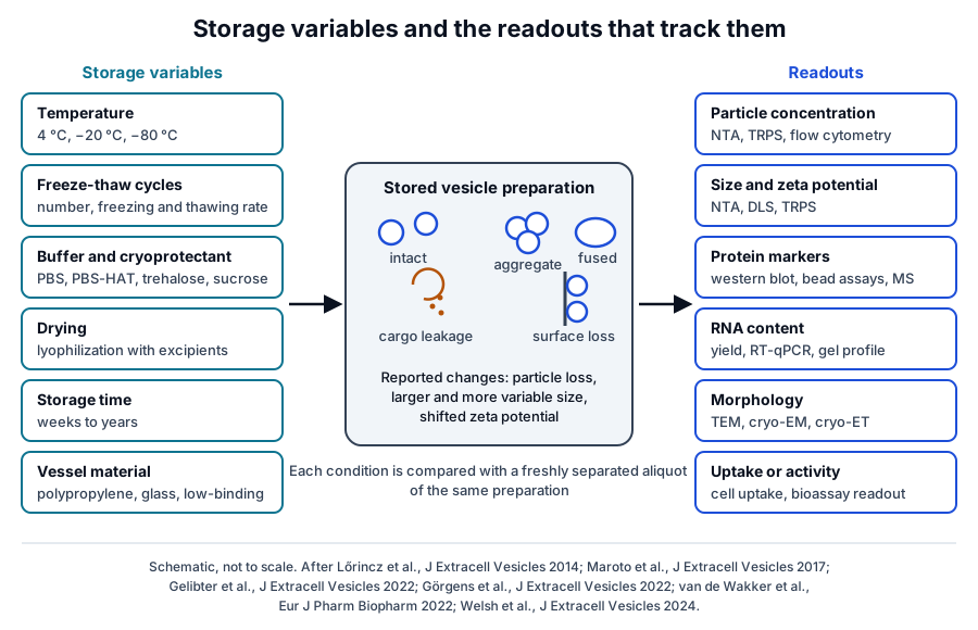
The readouts fall into a few groups, each sensitive to a different kind of change (Fig. 1). Most studies combine at least three of them, because particle number can stay constant while size, cargo or activity shifts.[5] Electron microscopy then shows whether the remaining particles are single, aggregated or multilamellar.[6]
- Particle concentration, by nanoparticle tracking analysis, resistive pulse sensing or calibrated flow cytometry.[7][8]
- Size distribution and zeta potential, which reveal aggregation, fusion and surface changes.[4][6]
- Protein markers such as ALIX, HSP70 and TSG101 by western blot, and whole proteomes by mass spectrometry.[6][9]
- RNA content and morphology by transmission or cryo-electron microscopy.[10][11]
- Cellular uptake, biodistribution in mice, or a defined biological readout in cell culture.[9][12]
Storage temperature shapes vesicle recovery and structure
Temperature comparisons usually test 4 °C, −20 °C and −80 °C against fresh material. A systematic review of 50 studies by Ahmadian and colleagues found that rapid freezing and a constant temperature, optimally −80 °C, best preserved vesicle quantity and cargo.[2]
Short holds and refrigeration
Cheng and colleagues held HEK293T exosomes for 24 h at five temperatures. Material kept at 4 °C retained the highest particle concentration and the highest ALIX, HSP70 and TSG101 signal.[9]
Mean size stayed similar across conditions, so the authors attributed the losses to degradation of whole vesicles. Over about a month, the same markers declined at every temperature, most slowly at −80 °C.[9] Sokolova and colleagues reported that exosome size from three human cell types decreased during storage at 4 °C and 37 °C.[13]
Cheng and colleagues also varied pH. Concentration and marker levels fell most at pH 4, less at pH 10 and least at pH 7, which the authors took to mean that acidic conditions are the most disruptive for stored exosomes.[9]
Freezing at −20 °C and −80 °C
Lőrincz and colleagues stored vesicles from human neutrophils at four temperatures. At −20 °C the vesicle count held for 4 weeks while size shifted and antibacterial activity was almost entirely lost, and at −80 °C count and size were stable with partial retention of activity.[5]
Wu and colleagues compared 4 °C, −20 °C and −80 °C over 4 weeks. Each condition changed size distribution, quantity, cargo, cellular uptake and biodistribution relative to fresh vesicles, and the authors favored −80 °C for long-term storage.[12]
Maroto and colleagues found that mouse airway exosomes grew about 10 percent in diameter at 4 °C and about 25 percent at −80 °C, with multilamellar structures after freezing. Distinct protein groups appeared in the supernatant at each temperature, consistent with leakage or loss of loosely bound proteins.[6]
Gelibter and colleagues reported that −80 °C storage of plasma and microglial vesicles reduced concentration and purity over time and increased size and its variability. These effects appeared under all eight strategies they tested, and the authors advised processing fresh samples in most cases.[4]
Freeze-thaw cycles reduce particle counts and enlarge vesicles
Repeated freezing and thawing is the most consistently reported source of change. In the Gelibter study, concentration fell after the first cycle and particle size rose with each further cycle.[4] Cheng and colleagues saw concentration and marker protein levels decline across five cycles, while uptake per remaining vesicle changed little in their cell assay.[9]
Gelibter and colleagues also mixed GFP-labeled and mCherry-labeled vesicles before storage. After storage a significant population carried both labels, which the authors interpreted as evidence of vesicle fusion during storage. Two-color designs of this kind separate fusion from aggregation, which bulk size measurements report together.[4]
Responses vary by source and buffer. Sokolova and colleagues found exosome size unchanged through repeated freezing to −20 °C,[13] while Bosch and colleagues saw concentration and size spread increase after freeze-thaw in PBS and remain steady in PBS with trehalose.[10]
Storage buffers and excipients govern vesicle recovery
Görgens and colleagues compared storage buffers at several temperatures for up to 2 years, using vesicles from stromal and HEK293T cells. Plain PBS led to sharply reduced recovery over time, and PBS used as a diluent reduced recovery within minutes.[7]
Their lead formulation, PBS-HAT, adds 25 mM HEPES, 0.2 percent human albumin and 25 mM trehalose to PBS. It improved short-term and long-term recovery at −80 °C, preserved vesicles through several freeze-thaw cycles, and improved recovery when used as a diluent.[7]
Van de Wakker and colleagues found higher recovery with HEPES-buffered saline than PBS, and with polypropylene tubes than glass. Bovine albumin and Tween 20 improved preservation without changing activity in an endothelial migration assay, and both 4 °C and −80 °C were suitable for short-term storage.[14]
Additive effects depend on the vesicle type. Lőrincz and colleagues reported that snap-freezing gave no added benefit for neutrophil vesicles, and that the widely used cryoprotectants they tested caused vesicle lysis.[5]
Surface adsorption explains part of the buffer and vessel effects. MISEV2023 asks authors to report storage vessels and their materials, because vesicles can be lost by attaching to surfaces,[1] and the higher recovery from polypropylene over glass fits that picture.[14]
Trehalose protects membranes by hydrogen bonding and glass formation
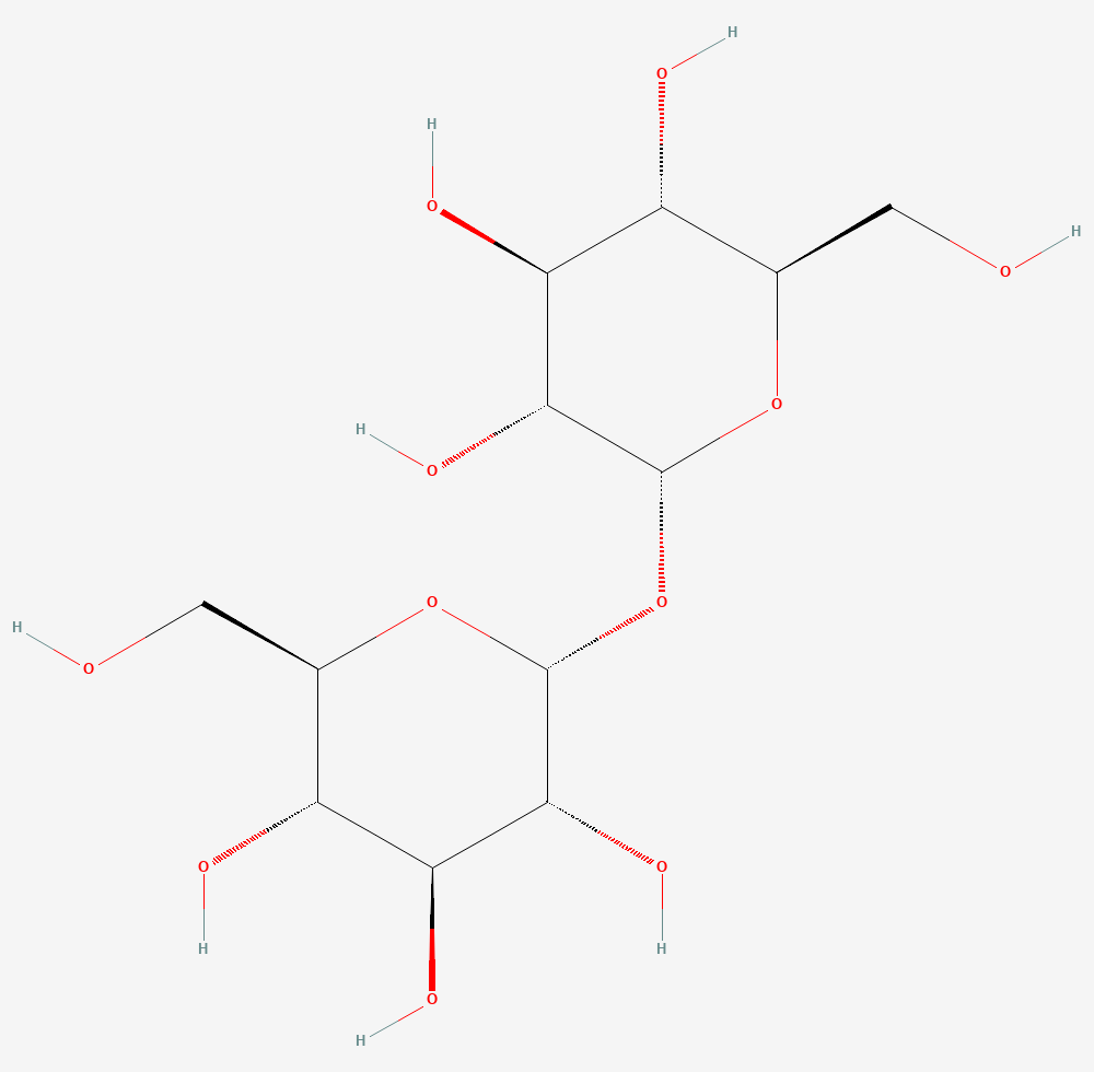
Trehalose is a nonreducing disaccharide of two glucose units joined through their anomeric carbons, C12H22O11 (Fig. 2).[15] It accumulates at high concentrations in organisms that survive near-complete drying, and Crowe and colleagues studied it as a stabilizer of dry membranes in such organisms.[16]
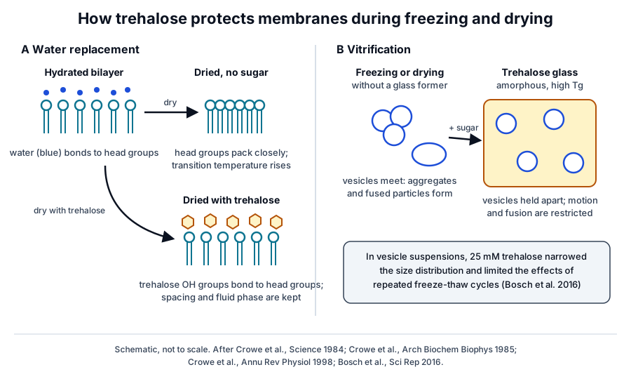
Crowe and colleagues showed in 1984 that dry dipalmitoylphosphatidylcholine with trehalose kept a phase transition temperature close to that of the hydrated lipid, about 30 K lower than lipid dried alone. Infrared spectra indicated hydrogen bonds between trehalose hydroxyl groups and the phospholipid head groups.[16]
In freeze-dried liposomes, trehalose inhibited fusion between vesicles and allowed almost all trapped solute to be retained after rehydration. Retention needed more than ten times the trehalose required to block fusion, which tied it to keeping the lipids in their fluid phase.[17]
A later review by Crowe, Carpenter and Crowe concluded that both direct hydrogen bonding and vitrification, the formation of an amorphous glass, are required (Fig. 3). Trehalose has a high glass transition temperature and forms a dihydrate crystal that shields the remaining glass from absorbed water.[18]
Bosch and colleagues brought trehalose to vesicle work in 2016. Adding 25 mM trehalose to the isolation and storage buffer for pancreatic beta-cell vesicles narrowed the size distribution and raised the particle count per microgram of protein, and cryo-electron tomography showed intact vesicles.[10]
Lyophilization holds vesicles in a dry sugar matrix
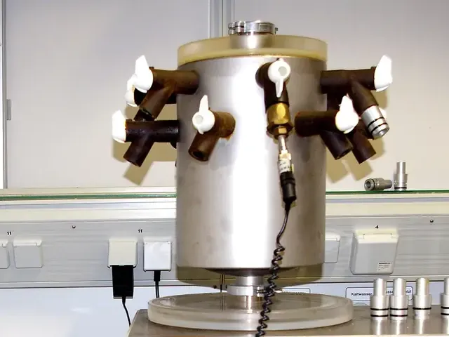
Lyophilization, or freeze-drying, removes water from a frozen sample by sublimation under vacuum (Fig. 4). Reviews of vesicle preservation describe it as an alternative to −80 °C storage and as a reliable method for preserving thermolabile materials.[3][19]
Charoenviriyakul and colleagues found that exosomes lyophilized without a cryoprotectant aggregated, and that trehalose prevented the aggregation. Protein and RNA profiles were retained, and luciferase-labeled exosomes kept their reporter activity for about 4 weeks at 25 °C.[11]
Trenkenschuh and colleagues screened formulations by freeze-thawing first. Sucrose or potassium phosphate buffer and neutral pH limited aggregation and fusion, a low level of poloxamer 188 helped further, and polyvinylpyrrolidone gave little protection on freeze-drying.[20]
Their lyophilizates containing 10 mM phosphate buffer, 0.02 percent poloxamer 188 and 5 percent sucrose retained particle size and concentration for 6 months at 40 °C. The activity of a vesicle-associated enzyme, beta-glucuronidase, held for 1 month and declined by 6 months.[20]
Bari and colleagues described a pilot process for freeze-dried stromal cell secretome under good manufacturing practice conditions. Nanoparticle tracking found a non-aggregated vesicle population in the powder, infrared spectra showed both protein and lipid, and calorimetry confirmed the drying process.[21]
Biofluid handling before isolation sets the starting population
Many vesicle studies begin with stored plasma, urine or saliva, so pre-isolation storage is a variable of its own. Witwer and colleagues called for standardized specimen handling and well-controlled reporting in an early ISEV workshop paper.[22]
Yuana and colleagues measured vesicles in plasma, urine and saliva before and after a single freeze-thaw cycle and after storage for up to 1 year. These steps had no significant effect on measured concentration or diameter, and they recommended freezing and storing all samples identically.[8]
Kim and colleagues found that freezing plasma with residual platelets generated new vesicle subpopulations and cell-free mRNA ex vivo, changes that later processing could not remove.[23] Kalra and colleagues reported a stable TSG101 signal for plasma exosomes over about three months of storage.[24]
Ahmadian and colleagues noted across the literature that vesicles kept in their native biofluid were more stable than purified vesicles in buffer.[2] MISEV2023 advises pre-processing source material before storage to remove cells and other interfering material.[1]
MISEV2023 asks studies to report every storage parameter
The MISEV2023 guidelines from Welsh and colleagues give storage its own section. They ask authors to report all storage parameters before and after separation, including preservatives or cryoprotectants, temperature, time, freezing procedure and storage vessel.[1]
The guidelines list the freezing method, suspension buffer, temperature, duration, thawing method and number of freeze-thaw cycles as conditions that may need optimizing for each vesicle source. They also ask that the effects of storage and formulation on activity be assessed in functional studies.[1]
- Freezing method, such as snap-freezing in liquid nitrogen or gradual freezing.[1]
- Suspension buffer, cryoprotectants and other additives.[1][7]
- Temperature, storage duration and vessel material.[1][14]
- Thawing method, and the number of freeze-thaw cycles each aliquot has seen.[1]
Reporting these details lets readers compare studies whose storage designs differ. The cold-chain validation reference covers the separate question of how temperature is documented during shipping.
Where to go from here
The isolation methods reference explains how the vesicle preparations described here are separated, and the nomenclature and protein marker references define what is being counted. The quality references on particle counting and MISEV2023 compliance show how these measurements are documented.
The cold-chain validation reference covers storage and shipping documentation. Verified practices can request lot documentation, and our team is glad to discuss the methods described on this page.
Annotated References
Numbered in the order they are cited in the text. Each source is annotated with what it supports on this page; peer-reviewed articles are listed with journal, volume, issue and pages, and primary sources are regulations, agency documents and reference databases.
- 1.Peer-reviewedWelsh JA, Goberdhan DCI, O'Driscoll L, et al. Minimal information for studies of extracellular vesicles (MISEV2023): From basic to advanced approaches. Journal of Extracellular Vesicles. 2024;13(2):e12404. https://pubmed.ncbi.nlm.nih.gov/38326288/Annotation: ISEV position paper on vesicle research methods. Cited for its section on pre-separation and post-separation storage, the storage parameters to report, freeze-thaw comparability and surface losses to storage vessels.
- 2.Peer-reviewedAhmadian S, Jafari N, Tamadon A, et al. Different storage and freezing protocols for extracellular vesicles: a systematic review. Stem Cell Research & Therapy. 2024;15(1):453. https://pubmed.ncbi.nlm.nih.gov/39593194/Annotation: Systematic review of 50 storage studies. Cited for rapid freezing and constant −80 °C preserving quantity and cargo, and for greater stability of vesicles kept in native biofluids.
- 3.Peer-reviewedJeyaram A, Jay SM. Preservation and Storage Stability of Extracellular Vesicles for Therapeutic Applications. The AAPS Journal. 2017;20(1):1. https://pubmed.ncbi.nlm.nih.gov/29181730/Annotation: Mini-review of vesicle preservation. Cited for −80 °C as the most promising condition, for storage effects varying with the vesicle source, and for lyophilization as an alternative.
- 4.Peer-reviewedGelibter S, Marostica G, Mandelli A, et al. The impact of storage on extracellular vesicles: A systematic study. Journal of Extracellular Vesicles. 2022;11(2):e12162. https://pubmed.ncbi.nlm.nih.gov/35102719/Annotation: Systematic comparison of eight storage strategies for plasma and microglial vesicles. Cited for time-dependent loss of concentration and purity at −80 °C, freeze-thaw effects and the two-color evidence of fusion.
- 5.Peer-reviewedLőrincz ÁM, Timár CI, Marosvári KA, et al. Effect of storage on physical and functional properties of extracellular vesicles derived from neutrophilic granulocytes. Journal of Extracellular Vesicles. 2014;3:25465. https://pubmed.ncbi.nlm.nih.gov/25536933/Annotation: Storage study of human neutrophil vesicles. Cited for stable counts with shifted size and lost activity at −20 °C, and for stable count and size with partial activity at −80 °C.
- 6.Peer-reviewedMaroto R, Zhao Y, Jamaluddin M, et al. Effects of storage temperature on airway exosome integrity for diagnostic and functional analyses. Journal of Extracellular Vesicles. 2017;6(1):1359478. https://pubmed.ncbi.nlm.nih.gov/28819550/Annotation: Light scattering, electron microscopy and proteomics of mouse airway exosomes. Cited for diameter increases at 4 °C and −80 °C, multilamellar structures and temperature-specific protein leakage.
- 7.Peer-reviewedGörgens A, Corso G, Hagey DW, et al. Identification of storage conditions stabilizing extracellular vesicles preparations. Journal of Extracellular Vesicles. 2022;11(6):e12238. https://pubmed.ncbi.nlm.nih.gov/35716060/Annotation: Comparison of storage buffers and temperatures for up to 2 years. Cited for reduced recovery in plain PBS and for the composition and performance of PBS-HAT.
- 8.Peer-reviewedYuana Y, Böing AN, Grootemaat AE, et al. Handling and storage of human body fluids for analysis of extracellular vesicles. Journal of Extracellular Vesicles. 2015;4:29260. https://pubmed.ncbi.nlm.nih.gov/26563735/Annotation: Comparison of handling and storage of plasma, urine and saliva. Cited for the small effect of one freeze-thaw cycle and storage up to 1 year, and for identical freezing of all samples.
- 9.Peer-reviewedCheng Y, Zeng Q, Han Q, et al. Effect of pH, temperature and freezing-thawing on quantity changes and cellular uptake of exosomes. Protein & Cell. 2019;10(4):295-299. https://pubmed.ncbi.nlm.nih.gov/29616487/Annotation: Comparison of temperature, freeze-thaw and pH for HEK293T exosomes. Cited for the 24 h temperature results, the slower decline of ALIX, HSP70 and TSG101 at −80 °C, and losses across five freeze-thaw cycles.
- 10.Peer-reviewedBosch S, de Beaurepaire L, Allard M, et al. Trehalose prevents aggregation of exosomes and cryodamage. Scientific Reports. 2016;6:36162. https://pubmed.ncbi.nlm.nih.gov/27824088/Annotation: Study of 25 mM trehalose in the buffer for beta-cell vesicles. Cited for a narrower size distribution, more particles per microgram of protein, steadier freeze-thaw behavior and intact vesicles by cryo-electron tomography.
- 11.Peer-reviewedCharoenviriyakul C, Takahashi Y, Nishikawa M, et al. Preservation of exosomes at room temperature using lyophilization. International Journal of Pharmaceutics. 2018;553(1-2):1-7. https://pubmed.ncbi.nlm.nih.gov/30316791/Annotation: Lyophilization of mouse cell line exosomes with and without trehalose. Cited for aggregation without a cryoprotectant, retained protein and RNA with trehalose, and reporter activity after storage at 25 °C.
- 12.Peer-reviewedWu JY, Li YJ, Hu XB, et al. Preservation of small extracellular vesicles for functional analysis and therapeutic applications: a comparative evaluation of storage conditions. Drug Delivery. 2021;28(1):162-170. https://pubmed.ncbi.nlm.nih.gov/33427518/Annotation: Comparison of 4 °C, −20 °C and −80 °C storage with uptake and mouse biodistribution readouts. Cited for storage changing size, quantity, cargo, uptake and biodistribution.
- 13.Peer-reviewedSokolova V, Ludwig AK, Hornung S, et al. Characterisation of exosomes derived from human cells by nanoparticle tracking analysis and scanning electron microscopy. Colloids and Surfaces B: Biointerfaces. 2011;87(1):146-150. https://pubmed.ncbi.nlm.nih.gov/21640565/Annotation: Nanoparticle tracking of exosomes from three human cell types. Cited for size decreasing at 4 °C and 37 °C and holding through repeated freezing to −20 °C.
- 14.Peer-reviewedvan de Wakker SI, van Oudheusden J, Mol EA, et al. Influence of short term storage conditions, concentration methodsand excipients on extracellular vesicle recovery and function. European Journal of Pharmaceutics and Biopharmaceutics. 2022;170:59-69. https://pubmed.ncbi.nlm.nih.gov/34864197/Annotation: Comparison of buffers, tubes and excipients for vesicle recovery. Cited for HEPES-buffered saline over PBS, polypropylene over glass, albumin and Tween 20 as excipients, and 4 °C and −80 °C for short holds.
- 15.DatabasePubChem CID 7427, Trehalose (National Center for Biotechnology Information). Accessed 20 October 2026. https://pubchem.ncbi.nlm.nih.gov/compound/7427
- 16.Peer-reviewedCrowe JH, Crowe LM, Chapman D. Preservation of membranes in anhydrobiotic organisms: the role of trehalose. Science. 1984;223(4637):701-703. https://pubmed.ncbi.nlm.nih.gov/17841031/Annotation: Calorimetry and infrared spectroscopy of dry phospholipid with trehalose. Cited for the preserved transition temperature and hydrogen bonding to head groups.
- 17.Peer-reviewedCrowe LM, Crowe JH, Rudolph A, et al. Preservation of freeze-dried liposomes by trehalose. Archives of Biochemistry and Biophysics. 1985;242(1):240-247. https://pubmed.ncbi.nlm.nih.gov/4051504/Annotation: Freeze-drying of liposomes with trehalose. Cited for inhibition of fusion, near-complete solute retention and the higher trehalose level needed to prevent leakage.
- 18.Peer-reviewedCrowe JH, Carpenter JF, Crowe LM. The role of vitrification in anhydrobiosis. Annual Review of Physiology. 1998;60:73-103. https://pubmed.ncbi.nlm.nih.gov/9558455/Annotation: Review of sugar glasses in dry biological systems. Cited for the requirement for both direct interaction and vitrification, and for the high glass transition temperature and dihydrate of trehalose.
- 19.Peer-reviewedKusuma GD, Barabadi M, Tan JL, et al. To Protect and to Preserve: Novel Preservation Strategies for Extracellular Vesicles. Frontiers in Pharmacology. 2018;9:1199. https://pubmed.ncbi.nlm.nih.gov/30420804/Annotation: Review of vesicle preservation, formulation and cold chain strategies. Cited for freeze-drying as a reliable method for preserving thermolabile materials, including vesicles.
- 20.Peer-reviewedTrenkenschuh E, Richter M, Heinrich E, et al. Enhancing the Stabilization Potential of Lyophilization for Extracellular Vesicles. Advanced Healthcare Materials. 2022;11(5):e2100538. https://pubmed.ncbi.nlm.nih.gov/34310074/Annotation: Formulation screen and 6-month stability study of lyophilized vesicles. Cited for the effects of sucrose, buffer, pH, poloxamer 188 and polyvinylpyrrolidone, and for size, concentration and enzyme activity over time.
- 21.Peer-reviewedBari E, Perteghella S, Di Silvestre D, et al. Pilot Production of Mesenchymal Stem/Stromal Freeze-Dried Secretome for Cell-Free Regenerative Nanomedicine: A Validated GMP-Compliant Process. Cells. 2018;7(11):190. https://pubmed.ncbi.nlm.nih.gov/30380806/Annotation: Pilot freeze-drying of stromal cell secretome under GMP conditions. Cited for a non-aggregated vesicle population in the powder and physical characterization of the lyophilizate.
- 22.Peer-reviewedWitwer KW, Buzás EI, Bemis LT, et al. Standardization of sample collection, isolation and analysis methods in extracellular vesicle research. Journal of Extracellular Vesicles. 2013;2:20360. https://pubmed.ncbi.nlm.nih.gov/24009894/Annotation: ISEV workshop paper on vesicle methods. Cited for the call to standardize specimen handling and report well-controlled experiments.
- 23.Peer-reviewedKim HJ, Rames MJ, Tassi Yunga S, et al. Irreversible alteration of extracellular vesicle and cell-free messenger RNA profiles in human plasma associated with blood processing and storage. Scientific Reports. 2022;12(1):2099. https://pubmed.ncbi.nlm.nih.gov/35136102/Annotation: Calibrated flow cytometry and RT-qPCR of processed plasma. Cited for vesicle subpopulations and cell-free mRNA generated when plasma with residual platelets is frozen.
- 24.Peer-reviewedKalra H, Adda CG, Liem M, et al. Comparative proteomics evaluation of plasma exosome isolation techniques and assessment of the stability of exosomes in normal human blood plasma. Proteomics. 2013;13(22):3354-3364. https://pubmed.ncbi.nlm.nih.gov/24115447/Annotation: Proteomic comparison of plasma exosome isolation methods with a storage arm. Cited for a stable TSG101 signal in stored plasma over about three months.
Cite this page
ExaVeyra Sciences Editorial Team. EV Storage Stability: Freezing, Lyophilization and Trehalose. ExaVeyra Sciences; 20 October 2026. Available from: https://exaveyra.com/science/exosome-biochemistry/extracellular-vesicle-storage-stability. Accessed [date accessed].
Figure sources
- 25.PubChem CID 7427, Trehalose. National Center for Biotechnology Information, PubChem. Public domain.