MSC Immunobiology: HLA Expression, IDO, PGE2 and TSG-6
By Benn Bluestein-Veyra, M.Sc. Organic Chemistry · Updated 20 October 2026
ExaVeyra Sciences is a Miami-based B2B supplier of exosomes for topical aesthetic use to licensed clinics, and it refers licensed prescribers to a 503A compounding pharmacy that prepares formulations against individual prescriptions.
| Question | Answer |
|---|---|
| What this page covers | How MSCs interact with immune cells: low HLA class II expression, tryptophan metabolism by IDO, the PGE2 pathway and TSG-6 binding to hyaluronan. |
| Who can order | Licensed clinics and practitioners whose active professional license and NPI ExaVeyra has verified. Exosome orders also require a signed professional-use acknowledgment. |
| What ships, and how | Exosome material for topical aesthetic use, characterized by lot, ships overnight in a cold chain. Delivery can take 1 to 3 business days. A certificate of analysis is issued for every lot and provided on request. |
MSC immunobiology describes how mesenchymal stromal cells sense and answer immune signaling in culture. Resting cells carry HLA class I with little HLA class II and low costimulatory molecules.[1][2] IFN-gamma and TNF-alpha then license them to express IDO1, COX-2 and TSG-6.[3][4][5]
This reference covers each molecule in turn, the protein structures behind them and the co-culture assays that measure them. The International Society for Cellular Therapy (ISCT) has proposed standardized immune assays for exactly this work.[6][7]
Mesenchymal stromal cells suppress T cell proliferation in co-culture
Di Nicola and colleagues showed in 2002 that bone marrow stromal cells, autologous or allogeneic, reduced T cell proliferation driven by dendritic cells, allogeneic lymphocytes or polyclonal activators. The suppressed T cells remained viable and proliferated again on restimulation.[8]
Inhibition persisted when a Transwell membrane kept the two cell types apart, which pointed to soluble factors; in that system, neutralizing antibodies identified TGF-beta1 and hepatocyte growth factor as mediators.[8] Krampera and colleagues found that MSCs suppressed the proliferation of CD4+ and CD8+ T cells and of NK cells, and left resting B cell proliferation unchanged.[3]
Klyushnenkova and colleagues found that MSCs added to a mixed lymphocyte reaction suppressed proliferation in a dose-dependent, genetically unrestricted way.[2] Aggarwal and Pittenger extended the picture to dendritic cells, T helper subsets and NK cells, whose cytokine output shifted toward an anti-inflammatory profile in co-culture.[9]
Understanding HLA class I and class II expression on mesenchymal stromal cells
Le Blanc and colleagues used flow cytometry to show that undifferentiated MSCs express HLA class I with no detectable surface HLA class II, while Western blot found class II protein inside the cells.[1] HLA-DR, the main class II isotype, pairs an alpha chain encoded by the HLA-DRA gene on chromosome 6p21.32 with a beta chain.[10]
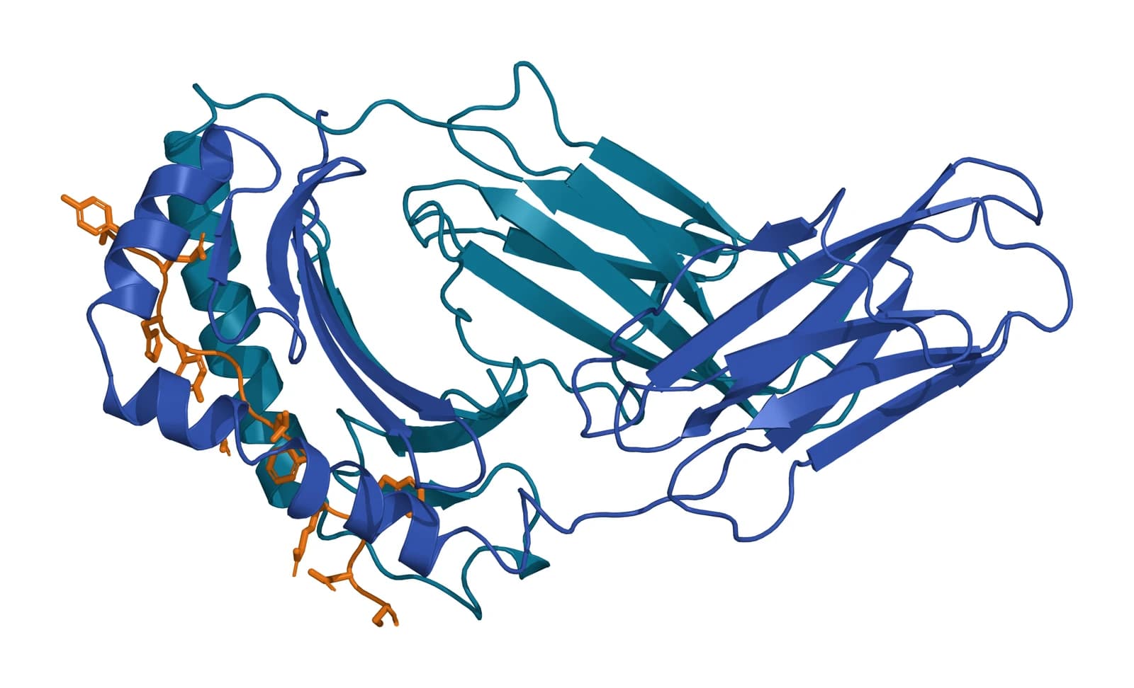
Class II proteins bind peptides and present them to T cells. Murthy and Stern solved HLA-DR1 bound to an endogenous peptide at 2.45 Å resolution, with the peptide in a polyproline II-like conformation and its side chains seated in pockets of the binding groove (Fig. 1).[11]
Interferon-gamma induces HLA-DR in culture
Adding IFN-gamma to the culture brought HLA class II to the surface of more than 90% of MSCs, and these cells still elicited no lymphocyte proliferation as stimulators.[1] Chan and colleagues described a narrow window in which low, autocrine IFN-gamma supported class II expression and antigen presentation, with class II falling as IFN-gamma levels rose.[12]
HLA-DR level and suppressive function vary independently. Across eleven MSC products, Bloom and colleagues found that suppression of T cell proliferation did not correlate with HLA-DR expression.[13]
Costimulatory molecules CD80, CD86 and CD40 remain low
Klyushnenkova and colleagues measured MHC class I on MSCs together with minimal class II and minimal CD80 (B7-1), CD86 (B7-2) and CD40. T cells purified from peripheral blood did not proliferate in response to allogeneic MSCs.[2]
T cells cultured with MSCs produced IFN-gamma and showed secondary kinetics on restimulation, a sign of priming. The authors concluded that MSCs can initiate T cell activation while active suppressive mechanisms hold proliferation down.[2]
IFN-gamma and TNF-alpha license the immunomodulatory program
Krampera and colleagues showed in 2006 that MSC suppression required IFN-gamma produced by activated T and NK cells, which acted by inducing IDO activity.[3] Ren and colleagues found that IFN-gamma combined with TNF-alpha, IL-1alpha or IL-1beta induced chemokines and inducible nitric oxide synthase in mouse MSCs, and the chemokines drew T cells close to the stromal cells.[14]
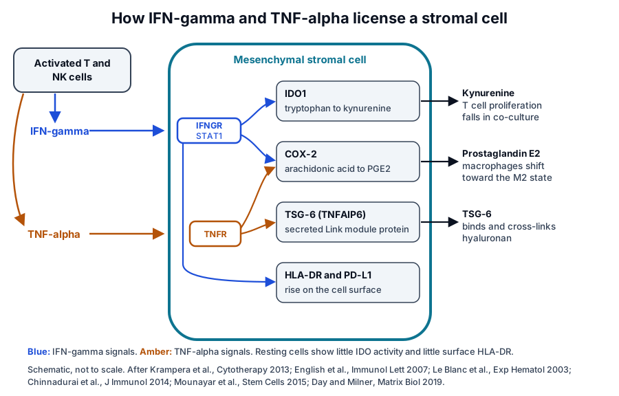
The literature calls this cytokine priming licensing (Fig. 2).[15][16] Ryan and colleagues found that IFN-gamma kept MSC suppression of mixed lymphocyte reactions intact, raised HGF and TGF-beta1 and induced IDO.[17]
The two cytokines act on different targets. In mouse MSCs, English and colleagues found COX-2 and PGE2 raised by both IFN-gamma and TNF-alpha, and functional IDO and PD-L1 induced by IFN-gamma alone.[4]
STAT1 and PI3K-alpha set the strength of the IDO response
Mounayar and colleagues traced IFN-gamma-induced IDO production in human MSCs to an interplay of STAT1 and PI3K-alpha. PI3K-alpha raised STAT1 protein and its phosphorylation at Ser727, which set the level of IFN-gamma-induced IDO.[18]
Licensing also adds surface ligands. Chinnadurai and colleagues found that IFN-gamma-licensed MSCs inhibited T cell effector cytokines through B7-H1 (PD-L1) and B7-DC, the ligands for PD-1, independently of IDO catalytic activity.[15]
IDO1 converts tryptophan to kynurenine in licensed human stromal cells
Meisel and colleagues reported in 2004 that human bone marrow stromal cells express IDO protein with functional activity after IFN-gamma stimulation. In mixed lymphocyte reactions, tryptophan fell and kynurenine appeared in the co-culture supernatant, and added tryptophan restored allogeneic T cell proliferation.[19]
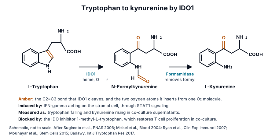
IDO1 cleaves the pyrrole ring of L-tryptophan and incorporates both atoms of one oxygen molecule, the first step of the kynurenine pathway (Fig. 3).[20][21] Badawy notes that IDO rate-limits this pathway outside the liver and responds to IFN-gamma, other cytokines and nitric oxide.[21] The human gene, IDO1, maps to chromosome 8p11.21.[22]
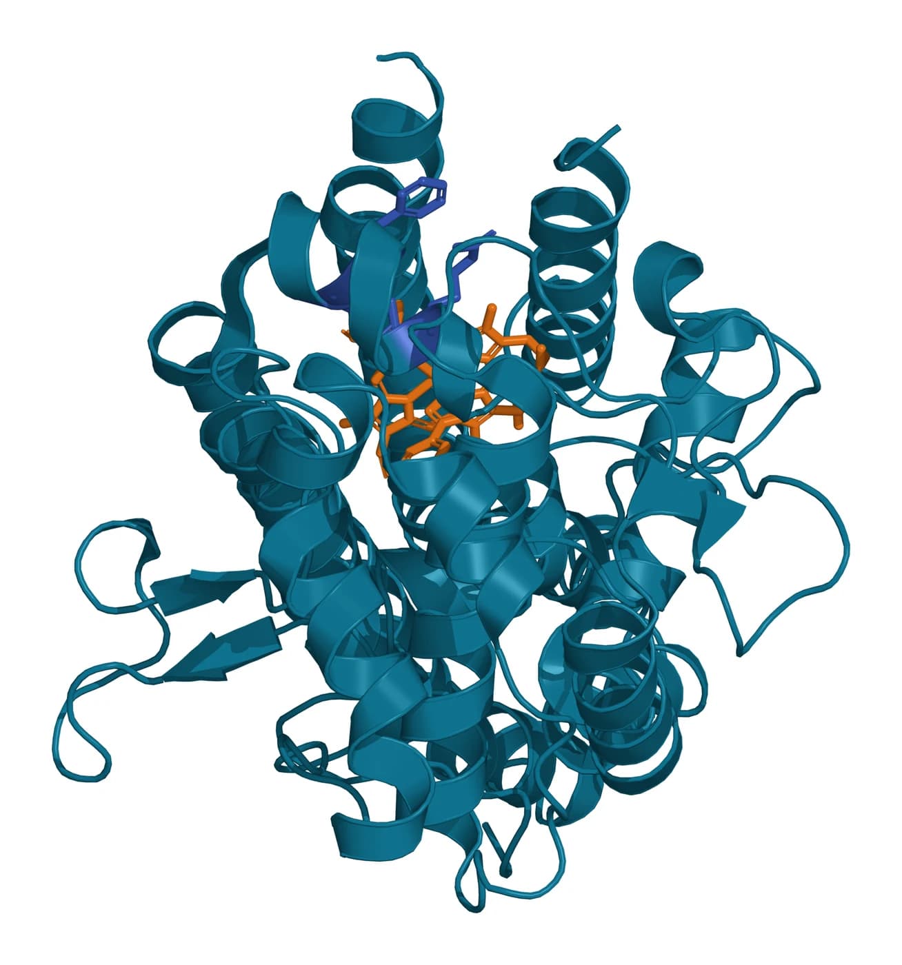
Sugimoto and colleagues solved human IDO bound to the ligand 4-phenylimidazole, showing two alpha-helical domains with the heme between them (Fig. 4).[20] Mutants at F226, F227 and R231 kept substrate binding with no activity, which the authors linked to a precise fit between the indole ring and the pocket.[20]
Munn and Mellor describe IDO activity as metabolic control: by catabolizing an essential amino acid, IDO-expressing cells shape local innate and adaptive immune responses.[23] Ryan and colleagues found that kynurenine added to mixed lymphocyte reactions blocked proliferation, and that the IDO antagonist 1-methyl-L-tryptophan restored it.[17]
Human and mouse stromal cells use different effector enzymes
Ren and colleagues compared species under the same culture conditions. Human and monkey MSCs suppressed T cells through IDO, and mouse MSCs through nitric oxide.[24]
After cytokine stimulation, human MSCs expressed very high IDO and very low iNOS, and mouse MSCs expressed abundant iNOS with very little IDO.[24] Mouse studies of the iNOS route and human studies of the IDO route therefore describe two species-specific forms of one cytokine-induced program.[14][24]
IDO1 induction varies between donors
François and colleagues found that MSCs from different adult donors, licensed with TNF-alpha and IFN-gamma, suppressed T cell proliferation to different degrees in step with their IDO induction.[16] MSC IDO activity also drove monocytes to differentiate into IL-10-secreting M2 macrophages in the same cultures.[16]
COX-2 produces prostaglandin E2, which acts on macrophages, dendritic cells and NK cells
Prostaglandins are lipid mediators made from arachidonic acid by the cyclooxygenase isoenzymes, COX-1 and COX-2.[25] COX-2 is encoded by PTGS2, prostaglandin-endoperoxide synthase 2, on chromosome 1q31.1.[26]
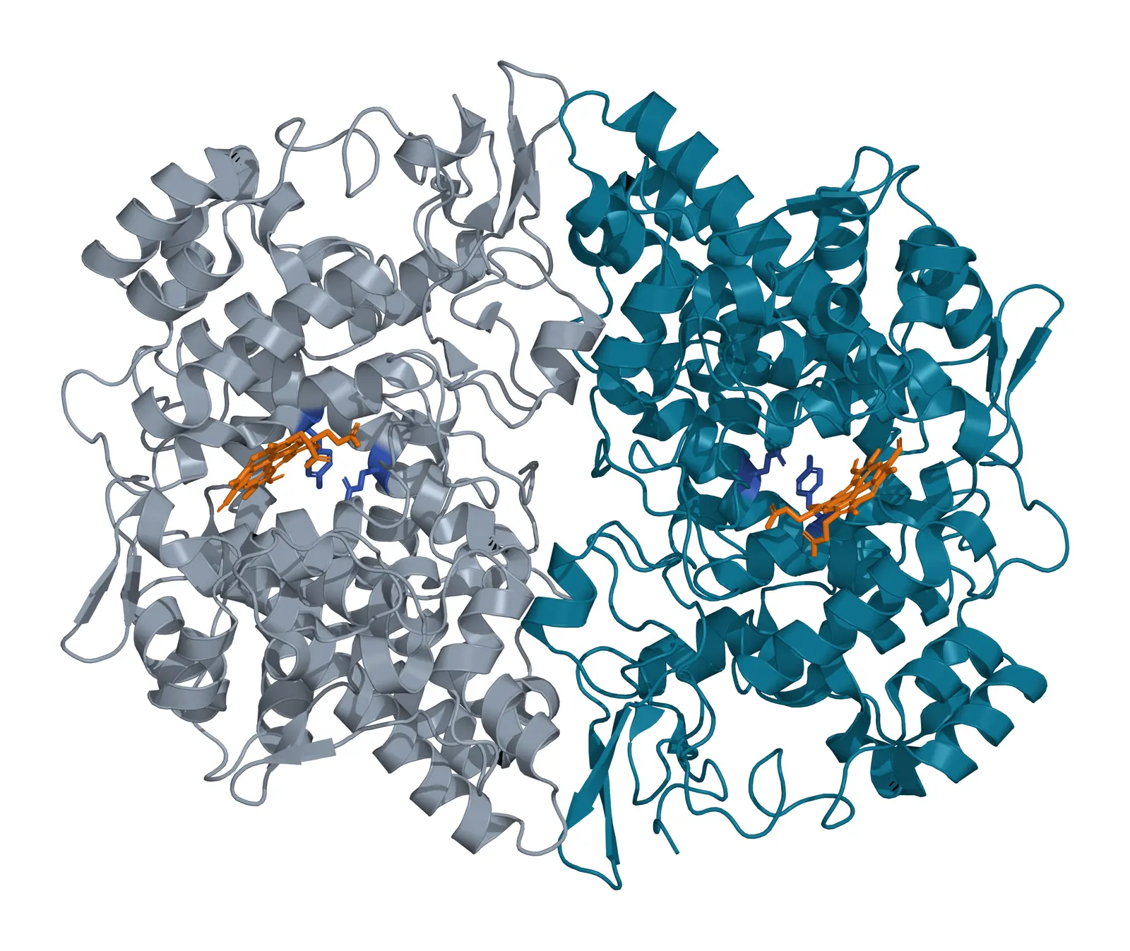
Lucido and colleagues solved human COX-2 at 2.0 Å resolution with Ser530 of the cyclooxygenase active site acetylated by aspirin (Fig. 5).[27] Kalinski describes the PGE2 signal as a balance between COX-2-regulated synthesis and degradation by 15-hydroxyprostaglandin dehydrogenase, read through the PGE2 receptors each cell expresses.[28]
PGE2 supports dendritic cell activation, promotes Th2, Th17 and regulatory T cell responses and dampens type 1 immunity.[28] Ryan and colleagues found that human MSCs express COX-1 and COX-2 and produce PGE2 constitutively, and indomethacin showed that prostaglandins contribute to suppression of mixed lymphocyte reactions.[17]
Aggarwal and Pittenger measured elevated PGE2 in MSC co-cultures, in which mature type 2 dendritic cells raised IL-10 secretion; inhibitors of PGE2 production lessened these cytokine shifts.[9] Spaggiari and colleagues identified IDO and PGE2 as key mediators of MSC inhibition of NK cell proliferation, cytotoxicity and cytokine production.[29]
Vasandan and colleagues followed macrophages in co-culture. In the absence of MSC COX-2 activity, the effect on M1 macrophages and their metabolic programs was lost, placing MSC-secreted PGE2 at the center of macrophage plasticity in that system.[30]
TSG-6 binds hyaluronan through its Link module
TSG-6, encoded by the TNFAIP6 gene on chromosome 2q23.3, is a secreted protein induced by TNF.[5][31] Day and Milner review it as a multifunctional protein that mediates many immunomodulatory activities of MSCs, with functions that depend on binding glycosaminoglycans and other ligands.[5]
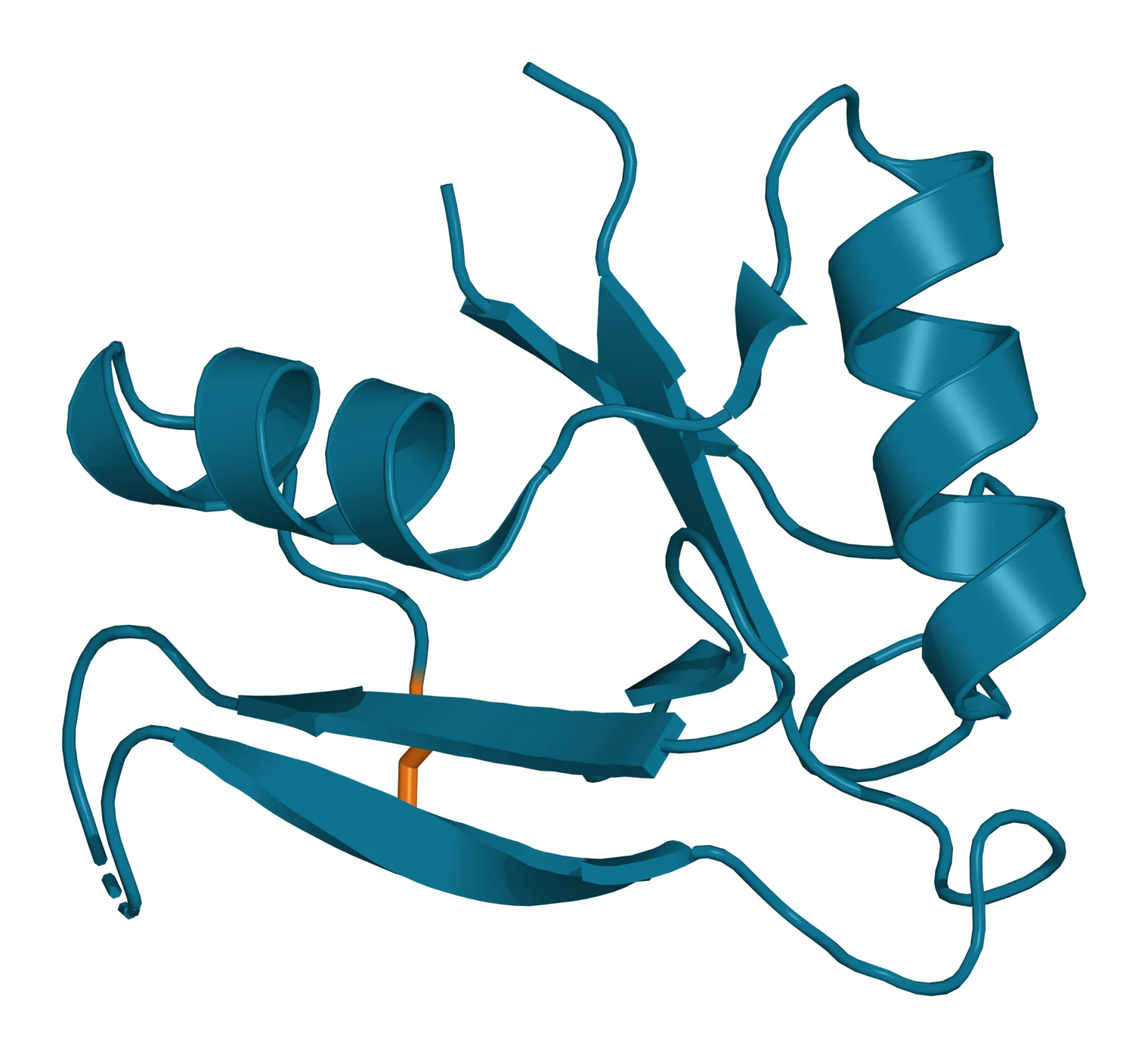
Kohda and colleagues solved the solution structure of the TSG-6 Link module in 1996: two alpha helices and two antiparallel beta sheets around a large hydrophobic core. This fold defines a superfamily that includes CD44 and aggrecan, and it resembles the C-type lectin domain.[32]
Higman and colleagues solved crystal structures of the free Link module and measured its dynamics by NMR, finding high mobility in the beta4/beta5 loop and at the Cys47-Cys68 disulfide, both in the hyaluronan-binding region (Fig. 6).[33] Their refined model showed hyaluronan bending around two faces of the Link module, much as it binds CD44, with a glucuronic acid ring stacking on a histidine.[34]
Baranova and colleagues found that full-length TSG-6 binds hyaluronan films with pronounced positive cooperativity, forms hyaluronan-induced oligomers and cross-links the chains. Both full-length TSG-6 and the Link module alone condensed and stiffened the films, the Link module with weaker binding.[35]
Mixed lymphocyte reactions and kynurenine assays measure MSC immune function
Assays of MSC immunobiology pair a co-culture with a readout. The mixed lymphocyte reaction cultures responder lymphocytes with allogeneic stimulator cells, and MSCs are added to measure how far the resulting proliferation falls.[2][17]
Bloom and colleagues built an immunopotency assay that stimulates T cells with anti-CD3 and anti-CD28 antibodies and scores suppression of CD4+ T cell proliferation against a reference MSC product. Across eleven products from three centers, suppression ranged from 27% to 88%.[13]
Kynurenine in the co-culture supernatant reports IDO activity directly.[19] Menard and colleagues compared MSCs expanded in fetal calf serum or platelet lysate with fully standardized assays and found significant differences in immunosuppressive molecules and in T cell inhibition.[36]
| Assay | Readout | What it reports |
|---|---|---|
| Mixed lymphocyte reaction | Responder proliferation with and without MSCs | Suppression of allogeneic T cell responses |
| Anti-CD3 and anti-CD28 T cell assay | CD4+ T cell proliferation against a reference product | A reproducible T cell suppression value |
| Kynurenine and tryptophan in supernatant | Kynurenine released, tryptophan consumed | IDO activity after IFN-gamma licensing |
| Flow cytometry of licensed cells | HLA-DR, PD-L1 and other surface markers | The surface response to IFN-gamma |
| Quantitative RNA analysis | Transcripts of selected licensing genes | The licensing response at the gene level |
| Secretome protein assays | PGE2, TSG-6 and cytokines in medium | Secreted immune mediators |
Galipeau and colleagues, reporting an ISCT workshop, proposed measuring potency by licensing MSCs in vitro and reading selected markers with a matrix of assays. Their three preferred methods were quantitative RNA analysis, flow cytometry of functionally relevant surface markers and protein assays of the secretome.[7]
The earlier ISCT working proposal, led by Krampera, set out a standardized approach to immune assays so that laboratories could produce shared, reproducible and consistent data.[6]
Tissue source, culture supplements and differentiation shape the immune profile
Prasanna and colleagues primed bone marrow and Wharton’s jelly MSCs with IFN-gamma or TNF-alpha, and both sources kept their suppressive activity, with Wharton’s jelly MSCs suppressing more strongly.[37] The two sources differed in IFN-gamma-inducible IDO activity, HGF and PGE2 levels, which the authors proposed could reflect distinct mechanisms.[37]
In the Menard comparison, adipose-derived MSCs expanded in platelet lysate were the most potent inhibitors of T cell growth, a property ascribed to IFN-gamma-dependent IDO activity. Resting MSCs inhibited T and NK cell proliferation and left B cell proliferation unchanged.[36]
Differentiation changes the profile as well. Le Blanc and colleagues found that osteogenic, chondrogenic or adipogenic culture raised HLA class I and reduced the IFN-gamma induction of class II to about 10% of cells, while the cells continued to inhibit mixed lymphocyte cultures.[1]
Where to go from here
The ISCT minimal criteria reference defines the cells these assays start from, and the stem versus stromal reference explains the two names behind the MSC abbreviation. The source tissues reference compares bone marrow, adipose tissue, umbilical cord and placenta, and the secretome reference follows the soluble factors and vesicles that MSCs release into culture medium.
The perinatal tissue donor eligibility reference explains the federal framework for placental and umbilical cord material, and the surface receptor chemistry reference covers the membrane proteins that flow cytometry reads. Verified practices can request lot documentation, and our team is glad to discuss the methods described on this page.
Annotated References
Numbered in the order they are cited in the text. Each source is annotated with what it supports on this page; peer-reviewed articles are listed with journal, volume, issue and pages, and primary sources are regulations, agency documents and reference databases.
- 1.Peer-reviewedLe Blanc K, Tammik C, Rosendahl K, et al. HLA expression and immunologic properties of differentiated and undifferentiated mesenchymal stem cells. Experimental Hematology. 2003;31(10):890-896. https://pubmed.ncbi.nlm.nih.gov/14550804/Annotation: Flow cytometry and co-culture study of MSC HLA expression. Cited for class I without surface class II, IFN-gamma induction of class II, and the effect of trilineage differentiation on both.
- 2.Peer-reviewedKlyushnenkova E, Mosca JD, Zernetkina V, et al. T cell responses to allogeneic human mesenchymal stem cells: immunogenicity, tolerance, and suppression. Journal of Biomedical Science. 2005;12(1):47-57. https://pubmed.ncbi.nlm.nih.gov/15864738/Annotation: Phenotyping and mixed lymphocyte reaction study. Cited for minimal CD80, CD86 and CD40, T cell priming without proliferation, and dose-dependent suppression of mixed lymphocyte reactions.
- 3.Peer-reviewedKrampera M, Cosmi L, Angeli R, et al. Role for interferon-gamma in the immunomodulatory activity of human bone marrow mesenchymal stem cells. Stem Cells. 2006;24(2):386-398. https://pubmed.ncbi.nlm.nih.gov/16123384/Annotation: Co-culture study of lymphocyte subsets. Cited for suppression of CD4+, CD8+ and NK cell proliferation and its dependence on IFN-gamma from activated cells acting through IDO.
- 4.Peer-reviewedEnglish K, Barry FP, Field-Corbett CP, et al. IFN-gamma and TNF-alpha differentially regulate immunomodulation by murine mesenchymal stem cells. Immunology Letters. 2007;110(2):91-100. https://pubmed.ncbi.nlm.nih.gov/17507101/Annotation: Cytokine stimulation study of mouse MSCs. Cited for COX-2 and PGE2 raised by both IFN-gamma and TNF-alpha, and functional IDO and PD-L1 induced by IFN-gamma alone.
- 5.Peer-reviewedDay AJ, Milner CM. TSG-6: A multifunctional protein with anti-inflammatory and tissue-protective properties. Matrix Biology. 2019;78-79:60-83. https://pubmed.ncbi.nlm.nih.gov/29362135/Annotation: Review of TSG-6 expression, structure and ligand binding. Cited for TSG-6 as a TNF-induced secreted protein that mediates many immunomodulatory activities of MSCs.
- 6.Peer-reviewedKrampera M, Galipeau J, Shi Y, et al. Immunological characterization of multipotent mesenchymal stromal cells--The International Society for Cellular Therapy (ISCT) working proposal. Cytotherapy. 2013;15(9):1054-1061. https://pubmed.ncbi.nlm.nih.gov/23602578/Annotation: ISCT MSC Committee working proposal for standardized immune assays. Cited for the call for shared, reproducible and consistent immunological characterization data.
- 7.Peer-reviewedGalipeau J, Krampera M, Barrett J, et al. International Society for Cellular Therapy perspective on immune functional assays for mesenchymal stromal cells as potency release criterion for advanced phase clinical trials. Cytotherapy. 2016;18(2):151-159. https://pubmed.ncbi.nlm.nih.gov/26724220/Annotation: ISCT workshop perspective on MSC potency assays. Cited for in vitro licensing read with a matrix of RNA, flow cytometry and secretome protein assays, and for open publication of potency assays.
- 8.Peer-reviewedDi Nicola M, Carlo-Stella C, Magni M, et al. Human bone marrow stromal cells suppress T-lymphocyte proliferation induced by cellular or nonspecific mitogenic stimuli. Blood. 2002;99(10):3838-3843. https://pubmed.ncbi.nlm.nih.gov/11986244/Annotation: Co-culture and Transwell study of T cell proliferation. Cited for suppression by autologous or allogeneic stromal cells, recovery on restimulation, and TGF-beta1 and HGF as soluble mediators.
- 9.Peer-reviewedAggarwal S, Pittenger MF. Human mesenchymal stem cells modulate allogeneic immune cell responses. Blood. 2005;105(4):1815-1822. https://pubmed.ncbi.nlm.nih.gov/15494428/Annotation: Co-culture study with purified dendritic cell, T cell and NK cell subsets. Cited for cytokine shifts including IL-10 from type 2 dendritic cells, elevated PGE2 and the effect of PGE2 synthesis inhibitors.
- 10.DatabaseNCBI Gene 3122, HLA-DRA major histocompatibility complex, class II, DR alpha (Homo sapiens). Accessed 20 October 2026. https://www.ncbi.nlm.nih.gov/gene/3122
- 11.Peer-reviewedMurthy VL, Stern LJ. The class II MHC protein HLA-DR1 in complex with an endogenous peptide: implications for the structural basis of the specificity of peptide binding. Structure. 1997;5(10):1385-1396. https://pubmed.ncbi.nlm.nih.gov/9351812/Annotation: Crystal structure of HLA-DR1 with an endogenous peptide (PDB 1AQD). Cited for peptide presentation by class II proteins and the polyproline II-like peptide conformation.
- 12.Peer-reviewedChan JL, Tang KC, Patel AP, et al. Antigen-presenting property of mesenchymal stem cells occurs during a narrow window at low levels of interferon-gamma. Blood. 2006;107(12):4817-4824. https://pubmed.ncbi.nlm.nih.gov/16493000/Annotation: Antigen presentation and flow cytometry study of MSCs. Cited for MHC class II expression supported by low autocrine IFN-gamma and its decline at higher IFN-gamma levels.
- 13.Peer-reviewedBloom DD, Centanni JM, Bhatia N, et al. A reproducible immunopotency assay to measure mesenchymal stromal cell-mediated T-cell suppression. Cytotherapy. 2015;17(2):140-151. https://pubmed.ncbi.nlm.nih.gov/25455739/Annotation: Development of an anti-CD3 and anti-CD28 T cell suppression assay across eleven MSC products. Cited for the 27% to 88% range and the absence of correlation with HLA-DR expression.
- 14.Peer-reviewedRen G, Zhang L, Zhao X, et al. Mesenchymal stem cell-mediated immunosuppression occurs via concerted action of chemokines and nitric oxide. Cell Stem Cell. 2008;2(2):141-150. https://pubmed.ncbi.nlm.nih.gov/18371435/Annotation: Mouse MSC study with knockout cells. Cited for the requirement for IFN-gamma plus TNF-alpha, IL-1alpha or IL-1beta, and the induction of chemokines and iNOS in mouse MSCs.
- 15.Peer-reviewedChinnadurai R, Copland IB, Patel SR, et al. IDO-independent suppression of T cell effector function by IFN-γ-licensed human mesenchymal stromal cells. Journal of Immunology. 2014;192(4):1491-1501. https://pubmed.ncbi.nlm.nih.gov/24403533/Annotation: Co-culture study of IFN-gamma-licensed human MSCs and T cells. Cited for the licensing terminology and for B7-H1 and B7-DC inhibiting T cell effector cytokines independently of IDO.
- 16.Peer-reviewedFrançois M, Romieu-Mourez R, Li M, et al. Human MSC suppression correlates with cytokine induction of indoleamine 2,3-dioxygenase and bystander M2 macrophage differentiation. Molecular Therapy. 2012;20(1):187-195. https://pubmed.ncbi.nlm.nih.gov/21934657/Annotation: Multi-donor study of TNF-alpha and IFN-gamma licensed human MSCs. Cited for donor variation in T cell suppression linked to IDO induction, and IDO-driven IL-10-secreting M2 macrophages.
- 17.Peer-reviewedRyan JM, Barry F, Murphy JM, et al. Interferon-gamma does not break, but promotes the immunosuppressive capacity of adult human mesenchymal stem cells. Clinical and Experimental Immunology. 2007;149(2):353-363. https://pubmed.ncbi.nlm.nih.gov/17521318/Annotation: Mixed lymphocyte reaction study of human MSCs. Cited for constitutive COX-1, COX-2 and PGE2, IFN-gamma induction of IDO, HGF and TGF-beta1, and the effects of kynurenine and 1-methyl-L-tryptophan.
- 18.Peer-reviewedMounayar M, Kefaloyianni E, Smith B, et al. PI3kα and STAT1 Interplay Regulates Human Mesenchymal Stem Cell Immune Polarization. Stem Cells. 2015;33(6):1892-1901. https://pubmed.ncbi.nlm.nih.gov/25753288/Annotation: Signaling study of IFN-gamma-induced IDO in human MSCs. Cited for PI3K-alpha raising STAT1 protein and Ser727 phosphorylation, which set the level of IFN-gamma-induced IDO.
- 19.Peer-reviewedMeisel R, Zibert A, Laryea M, et al. Human bone marrow stromal cells inhibit allogeneic T-cell responses by indoleamine 2,3-dioxygenase-mediated tryptophan degradation. Blood. 2004;103(12):4619-4621. https://pubmed.ncbi.nlm.nih.gov/15001472/Annotation: Mixed lymphocyte reaction study of human stromal cells. Cited for IFN-gamma-induced IDO activity, tryptophan depletion and kynurenine in supernatants, and restoration of proliferation by tryptophan.
- 20.Peer-reviewedSugimoto H, Oda S, Otsuki T, et al. Crystal structure of human indoleamine 2,3-dioxygenase: catalytic mechanism of O2 incorporation by a heme-containing dioxygenase. Proceedings of the National Academy of Sciences of the United States of America. 2006;103(8):2611-2616. https://pubmed.ncbi.nlm.nih.gov/16477023/Annotation: Crystal structure of human IDO with 4-phenylimidazole (PDB 2D0T). Cited for pyrrole ring cleavage with both oxygen atoms incorporated, the two helical domains around the heme, and the F226, F227 and R231 mutants.
- 21.Peer-reviewedBadawy AA. Kynurenine Pathway of Tryptophan Metabolism: Regulatory and Functional Aspects. International Journal of Tryptophan Research. 2017;10:1178646917691938. https://pubmed.ncbi.nlm.nih.gov/28469468/Annotation: Review of the kynurenine pathway. Cited for IDO as the rate-limiting enzyme outside the liver and its regulation by IFN-gamma, other cytokines and nitric oxide.
- 22.DatabaseNCBI Gene 3620, IDO1 indoleamine 2,3-dioxygenase 1 (Homo sapiens). Accessed 20 October 2026. https://www.ncbi.nlm.nih.gov/gene/3620
- 23.Peer-reviewedMunn DH, Mellor AL. Indoleamine 2,3 dioxygenase and metabolic control of immune responses. Trends in Immunology. 2013;34(3):137-143. https://pubmed.ncbi.nlm.nih.gov/23103127/Annotation: Review of IDO biology. Cited for tryptophan catabolism as a form of metabolic control over local innate and adaptive immune responses.
- 24.Peer-reviewedRen G, Su J, Zhang L, et al. Species variation in the mechanisms of mesenchymal stem cell-mediated immunosuppression. Stem Cells. 2009;27(8):1954-1962. https://pubmed.ncbi.nlm.nih.gov/19544427/Annotation: Cross-species comparison of human, monkey and mouse MSCs under the same culture conditions. Cited for IDO in human and monkey cells and nitric oxide in mouse cells.
- 25.Peer-reviewedRicciotti E, FitzGerald GA. Prostaglandins and inflammation. Arteriosclerosis, Thrombosis, and Vascular Biology. 2011;31(5):986-1000. https://pubmed.ncbi.nlm.nih.gov/21508345/Annotation: Review of prostaglandin biosynthesis and receptors. Cited for prostaglandins as lipid mediators made from arachidonic acid by the cyclooxygenase isoenzymes.
- 26.DatabaseNCBI Gene 5743, PTGS2 prostaglandin-endoperoxide synthase 2 (Homo sapiens). Accessed 20 October 2026. https://www.ncbi.nlm.nih.gov/gene/5743
- 27.Peer-reviewedLucido MJ, Orlando BJ, Vecchio AJ, et al. Crystal Structure of Aspirin-Acetylated Human Cyclooxygenase-2: Insight into the Formation of Products with Reversed Stereochemistry. Biochemistry. 2016;55(8):1226-1238. https://pubmed.ncbi.nlm.nih.gov/26859324/Annotation: Crystal structures of COX-2 (human entry PDB 5F19). Cited for the 2.0 Å structure of human COX-2 with Ser530 acetylated in the cyclooxygenase active site.
- 28.Peer-reviewedKalinski P. Regulation of immune responses by prostaglandin E2. Journal of Immunology. 2012;188(1):21-28. https://pubmed.ncbi.nlm.nih.gov/22187483/Annotation: Review of PGE2 in immunity. Cited for the balance of COX-2 synthesis and 15-PGDH degradation, receptor patterns, and PGE2 effects on dendritic cells and T helper responses.
- 29.Peer-reviewedSpaggiari GM, Capobianco A, Abdelrazik H, et al. Mesenchymal stem cells inhibit natural killer-cell proliferation, cytotoxicity, and cytokine production: role of indoleamine 2,3-dioxygenase and prostaglandin E2. Blood. 2008;111(3):1327-1333. https://pubmed.ncbi.nlm.nih.gov/17951526/Annotation: Co-culture study of MSCs and NK cells. Cited for IDO and PGE2 as key mediators of the effect on NK cell proliferation, cytotoxicity and cytokine production.
- 30.Peer-reviewedVasandan AB, Jahnavi S, Shashank C, et al. Human Mesenchymal stem cells program macrophage plasticity by altering their metabolic status via a PGE(2)-dependent mechanism. Scientific Reports. 2016;6:38308. https://pubmed.ncbi.nlm.nih.gov/27910911/Annotation: Co-culture study of MSCs with human macrophages in defined states. Cited for the loss of the effect on M1 macrophages in the absence of MSC COX-2 activity and PGE2 as the mediator of macrophage plasticity.
- 31.DatabaseNCBI Gene 7130, TNFAIP6 TNF alpha induced protein 6 (Homo sapiens). Accessed 20 October 2026. https://www.ncbi.nlm.nih.gov/gene/7130
- 32.Peer-reviewedKohda D, Morton CJ, Parkar AA, et al. Solution structure of the link module: a hyaluronan-binding domain involved in extracellular matrix stability and cell migration. Cell. 1996;86(5):767-775. https://pubmed.ncbi.nlm.nih.gov/8797823/Annotation: NMR structure of the TSG-6 Link module. Cited for the two helices and two antiparallel sheets around a hydrophobic core, the superfamily fold and the similarity to C-type lectins.
- 33.Peer-reviewedHigman VA, Blundell CD, Mahoney DJ, et al. Plasticity of the TSG-6 HA-binding loop and mobility in the TSG-6-HA complex revealed by NMR and X-ray crystallography. Journal of Molecular Biology. 2007;371(3):669-684. https://pubmed.ncbi.nlm.nih.gov/17585936/Annotation: Crystal structures (PDB 2PF5) and NMR dynamics of the TSG-6 Link module. Cited for mobility of the beta4/beta5 loop and the Cys47-Cys68 disulfide in the hyaluronan-binding region.
- 34.Peer-reviewedHigman VA, Briggs DC, Mahoney DJ, et al. A refined model for the TSG-6 link module in complex with hyaluronan: use of defined oligosaccharides to probe structure and function. Journal of Biological Chemistry. 2014;289(9):5619-5634. https://pubmed.ncbi.nlm.nih.gov/24403066/Annotation: NMR-restrained model of the Link module with hyaluronan oligosaccharides. Cited for hyaluronan bending around two faces of the domain, as with CD44, and glucuronic acid stacking on a histidine.
- 35.Peer-reviewedBaranova NS, Nilebäck E, Haller FM, et al. The inflammation-associated protein TSG-6 cross-links hyaluronan via hyaluronan-induced TSG-6 oligomers. Journal of Biological Chemistry. 2011;286(29):25675-25686. https://pubmed.ncbi.nlm.nih.gov/21596748/Annotation: Biophysical study of TSG-6 on hyaluronan films. Cited for cooperative binding, hyaluronan-induced oligomers, cross-linking, and condensation by full-length protein and the Link module.
- 36.Peer-reviewedMenard C, Pacelli L, Bassi G, et al. Clinical-grade mesenchymal stromal cells produced under various good manufacturing practice processes differ in their immunomodulatory properties: standardization of immune quality controls. Stem Cells and Development. 2013;22(12):1789-1801. https://pubmed.ncbi.nlm.nih.gov/23339531/Annotation: Standardized immune assay comparison of MSCs expanded in fetal calf serum or platelet lysate. Cited for process differences and IFN-gamma-dependent IDO activity in adipose-derived MSCs.
- 37.Peer-reviewedPrasanna SJ, Gopalakrishnan D, Shankar SR, et al. Pro-inflammatory cytokines, IFNgamma and TNFalpha, influence immune properties of human bone marrow and Wharton jelly mesenchymal stem cells differentially. PLoS One. 2010;5(2):e9016. https://pubmed.ncbi.nlm.nih.gov/20126406/Annotation: Comparison of cytokine-primed bone marrow and Wharton’s jelly MSCs. Cited for retained suppression after priming and source differences in IDO activity, HGF and PGE2.
Cite this page
ExaVeyra Sciences Editorial Team. MSC Immunobiology: HLA Expression, IDO, PGE2 and TSG-6. ExaVeyra Sciences; 20 October 2026. Available from: https://exaveyra.com/science/stem-cells/msc-immunobiology-hla-ido-pge2. Accessed [date accessed].
Figure sources
- 38.PDB 1AQD, Human HLA-DR1 bound to an endogenous peptide. ExaVeyra Sciences, rendered in PyMOL from PDB 1AQD. Structure data: RCSB Protein Data Bank (rcsb.org), CC0. Structure publication: Murthy VL, Stern LJ. The class II MHC protein HLA-DR1 in complex with an endogenous peptide: implications for the structural basis of the specificity of peptide binding. Structure. 1997;5(10):1385-1396.
- 39.PDB 2D0T, Human indoleamine 2,3-dioxygenase 1 (IDO1). ExaVeyra Sciences, rendered in PyMOL from PDB 2D0T. Structure data: RCSB Protein Data Bank (rcsb.org), CC0. Structure publication: Sugimoto H, Oda S, Otsuki T, et al. Crystal structure of human indoleamine 2,3-dioxygenase: catalytic mechanism of O2 incorporation by a heme-containing dioxygenase. Proc Natl Acad Sci U S A. 2006;103(8):2611-2616.
- 40.PDB 5F19, Human cyclooxygenase-2 (COX-2). ExaVeyra Sciences, rendered in PyMOL from PDB 5F19. Structure data: RCSB Protein Data Bank (rcsb.org), CC0. Structure publication: Lucido MJ, Orlando BJ, Vecchio AJ, Malkowski MG. Crystal Structure of Aspirin-Acetylated Human Cyclooxygenase-2: Insight into the Formation of Products with Reversed Stereochemistry. Biochemistry. 2016;55(8):1226-1238.
- 41.PDB 2PF5, Human TSG-6 Link module. ExaVeyra Sciences, rendered in PyMOL from PDB 2PF5. Structure data: RCSB Protein Data Bank (rcsb.org), CC0. Structure publication: Higman VA, Blundell CD, Mahoney DJ, et al. Plasticity of the TSG-6 HA-binding loop and mobility in the TSG-6-HA complex revealed by NMR and X-ray crystallography. J Mol Biol. 2007;371(3):669-684.