Mitochondrial Transfer by Mesenchymal Stromal Cells
By Benn Bluestein-Veyra, M.Sc. Organic Chemistry · Updated 20 October 2026
ExaVeyra Sciences is a Miami-based B2B supplier of exosomes for topical aesthetic use to licensed clinics, and it refers licensed prescribers to a 503A compounding pharmacy that prepares formulations against individual prescriptions.
| Question | Answer |
|---|---|
| What this page covers | How stromal cells pass mitochondria to other cells through tunneling nanotubes and extracellular vesicles, and the 2025 nomenclature for describing it. |
| Who can order | Licensed clinics and practitioners whose active professional license and NPI ExaVeyra has verified. Exosome orders also require a signed professional-use acknowledgment. |
| What ships, and how | Exosome material for topical aesthetic use, characterized by lot, ships overnight in a cold chain. Delivery can take 1 to 3 business days. A certificate of analysis is issued for every lot and provided on request. |
Mitochondrial transfer from MSCs is the movement of whole mitochondria or mitochondrial DNA from mesenchymal stromal cells into neighboring cells. Spees and colleagues reported in 2006 that human MSCs and skin fibroblasts restored aerobic respiration in cells lacking functional mitochondrial DNA.[1] Tunneling nanotubes, extracellular vesicles, gap junctions and cell fusion have since been described as routes.[2]
This reference covers MSC mitochondrial transfer as normal cell biology: the 2025 consensus vocabulary, the landmark co-culture experiments, each route and its molecules, and the assays that measure transfer. The findings come from co-culture, structural and physiology studies and from the reviews that collect them.[3][4]
Defining mitochondrial transfer with the 2025 consensus nomenclature
Brestoff, Singh and a group of more than thirty investigators published a consensus statement on mitochondria transfer nomenclature in Nature Metabolism in 2025. It defines intercellular mitochondria transfer as an evolutionarily conserved process in which one cell delivers some of its mitochondria to another cell in the absence of cell division.[3]
The statement notes that many transfer mechanisms had been described under a variety of names, which generated confusion. It defines the relevant terms and proposes a nomenclature framework for transfer and for mitochondria transplantation as a foundation for the field.[3]
Donor and acceptor cells frame every description
The consensus vocabulary names a donor cell, which delivers mitochondria, and an acceptor cell, which receives them. Transfer was first shown to give metabolic support to acceptor cells, and later work added roles in donor mitochondrial quality and in tissue homeostasis and remodeling.[3]
Berridge and colleagues describe horizontal transfer of mitochondria and mitochondrial DNA in several mammalian cell culture systems, and they propose that it may be a fundamental physiological process in development and tissue homeostasis.[4] Torralba and colleagues add that mitochondria carry their own circular genome, so horizontal transfer moves genetic material between cells alongside the organelle.[2]
MSCs restore aerobic respiration in cells lacking mitochondrial DNA
Spees and colleagues used rho0 cells, a human cell line whose mitochondrial DNA had been depleted with ethidium bromide, so they grew only in medium supplemented with uridine and pyruvate. After co-culture with human bone marrow MSCs or skin fibroblasts, colonies grew in restrictive medium without those supplements.[1]
Using MSCs from six donors, the authors isolated more than a hundred such colonies. Restriction fragment length polymorphism and SNP-PCR assays found donor MSC mitochondrial DNA in the rescued clones, with 76 of 79 clones positive by SNP-PCR, and the clones raised ATP at least threefold over the rho0 cells.[1]
Nuclear genotyping with a collagen gene polymorphism and nine microsatellite markers showed acceptor nuclei in every clone, which excluded cell fusion as the main mechanism. MSCs expressing a mitochondria-targeted DsRed2 sent cytoplasmic extensions toward the rho0 cells, and labeled mitochondria streamed through them.[1]
Early co-culture studies saw mitochondria inside intercellular tubes
Plotnikov and colleagues co-cultured human MSCs with rat cardiomyocytes and observed intercellular contacts by light, fluorescence and electron microscopy. Mitochondria moved in one direction only, toward the cardiomyocytes, and transmission electron microscopy found mitochondria-like structures inside nanotubes of variable diameter.[5]
Koyanagi and colleagues saw similar connections between human endothelial progenitor cells and neonatal rat cardiomyocytes. The tubes measured 50 to 800 nm across and 5 to 120 µm long, formed transiently, and carried MitoTracker-labeled structures between the cells.[6]
Mitochondria move between cells through four described routes
Reviews of the field group the mechanisms into four routes: tunneling nanotubes, extracellular vesicles, cell fusion and gap junctions (Fig. 1).[2] Each route has its own structure and molecular machinery, and one co-culture can use several at once.[7]
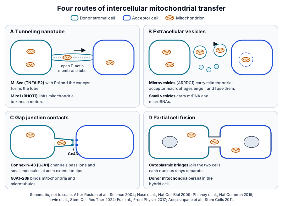
| Route | Structure | Molecules named in the literature | Example co-culture |
|---|---|---|---|
| Tunneling nanotube | Open F-actin membrane tube between two cells | M-Sec (TNFAIP2), RalA, the exocyst; Miro1 (RHOT1) | Bone marrow and lung MSCs with bronchial epithelial cells |
| Extracellular vesicles | Microvesicles carrying mitochondria; small vesicles carrying mtDNA | ARRDC1, TSG101 | Bone marrow MSCs with macrophages |
| Gap junction contacts | Connexin-43 channels at the tips of actin extensions | Connexin-43 (GJA1), GJA1-20k | Bone marrow MSCs with chondrocytes |
| Partial cell fusion | Cytoplasmic bridges with F-actin and microtubules | Not yet defined | Adipose-derived stem cells with cardiomyocytes |
Sinclair and colleagues tested several routes in one system and found that MSCs from three sources donated mitochondria through nanotubes and endocytosis, with a gap junction contribution in some co-cultures.[7] The sections below take each route in turn.
Tunneling nanotubes carry mitochondria along open F-actin bridges
Rustom and colleagues described tunneling nanotubes in 2004 as thin structures formed de novo between cells, building complex networks. They carried membrane vesicles and organelles selectively and seemed to impede the flow of small molecules, which the authors proposed as a principle of cell-to-cell interaction based on membrane continuity.[8]
Sartori-Rupp and colleagues used correlative FIB-SEM, light and cryo-electron microscopy to resolve neuronal nanotubes. Each was a bundle of open-ended individual tubes filled with parallel actin bundles, on which membrane-bound compartments and mitochondria appeared to move.[9]
Austefjord and colleagues review nanotubes as diverse in morphology and composition, varying between and within cell systems.[10]
Zurzolo notes that several TNT-like protrusions have been described and that most studies relied on low-resolution imaging. She identifies consistent proof of material transfer between two distinct cell populations as a central requirement for the field.[11]
Stromal cells from several tissues build nanotubes in co-culture
In the Sinclair study, F-actin tubes extended from bone marrow, lung tissue and bronchoalveolar lavage MSCs to BEAS2B bronchial epithelial cells within 45 min of co-culture (Fig. 2). All three MSC populations donated mitochondria with similar efficiency, and the epithelial cells donated none back to the MSCs.[7]
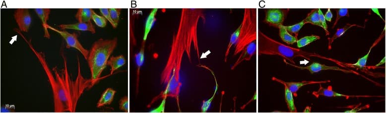
Cytochalasin D, which blocks the actin tubes, and dynasore, which blocks endocytosis, each reduced mitochondrial transfer in all three co-cultures.[7] Transfer in this system therefore drew on nanotubes and on vesicle uptake together.[7]
Immune cells take up stromal mitochondria in co-culture
Jackson and Krasnodembskaya describe a direct co-culture of human bone marrow MSCs with monocyte-derived macrophages, in which MSC mitochondria moved to the macrophages through nanotubes. Transfer raised macrophage oxidative phosphorylation and phagocytosis, and cytochalasin B, which blocks nanotube formation, abolished the phagocytosis effect.[12]
Luz-Crawford and colleagues found that Th17 cells took up mitochondria from bone marrow MSCs within 4 h of co-culture. IL-17 production fell in a contact-dependent way, together with a rise in Th17 oxygen consumption and conversion toward regulatory T cells.[13]
Court and colleagues measured dose-dependent transfer from mitochondria-labeled MSCs mainly to CD4+ T cells, with less to CD8+ T cells and CD19+ B cells. Isolated MSC mitochondria delivered artificially raised FOXP3, IL2RA, CTLA4 and TGFB1 transcripts and expanded a CD25+ FoxP3+ population.[14]
M-Sec and Miro1 build and load the nanotube route
Hase and colleagues identified M-Sec as a protein that induces membrane protrusions de novo, some of which tether to adjacent cells and form TNT-like structures. RNA interference against M-Sec reduced endogenous nanotube formation in a macrophage cell line, and blocking its interaction with Ral and the exocyst complex attenuated tube formation.[15]
M-Sec is a 73 kDa cytosolic protein also known as TNFAIP2 or B94, with sequence similarity to the exocyst subunit Sec6.[16] The human TNFAIP2 gene maps to chromosome 14q32.32.[17]
Kimura and colleagues found that the N-terminal polybasic region of M-Sec binds phosphatidylinositol 4,5-bisphosphate to reach the plasma membrane. Their crystal structure showed helix bundles in a straight rod, like membrane tethering complex subunits, with a positively charged C-terminal surface that recruits active RalA.[16]
Miro1 links mitochondria to microtubule motors
Fransson and colleagues named Miro-1 and Miro-2, for mitochondrial Rho, in 2003: GTPases with tandem GTP-binding domains separated by a linker carrying calcium-binding EF hand motifs, conserved from yeast to mammals.[18] A C-terminal transmembrane domain targets Miro to mitochondria, and Miro binds the kinesin-binding proteins GRIF-1 and OIP106.[19]
MacAskill and colleagues showed that Miro1 links mitochondria to KIF5 motors, which move them along microtubules. Micromolar calcium binding to Miro1 inhibits this link, and Miro1 with mutated EF hands no longer stopped mitochondria in response to glutamate or neuronal activity.[20] The human gene, RHOT1, maps to chromosome 17q11.2.[21]
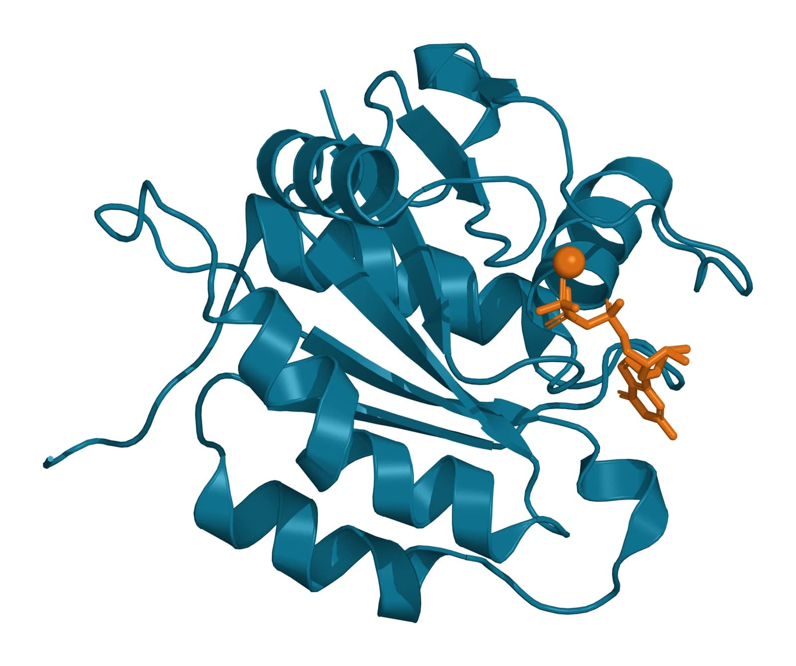
Torralba and colleagues review evidence that Miro1 levels in MSCs set the efficiency of intercellular mitochondrial movement, which ties the motor adaptor to the nanotube route.[2] Smith and colleagues solved the human Miro1 N-terminal GTPase domain at 1.7 Å, bound to GTP in a non-catalytic configuration of the active site (Fig. 3). Small-angle X-ray scattering of the soluble Miro1 and Miro2 proteins allowed modeling of a crescent-shaped assembly.[22]
Extracellular vesicles carry mitochondria and mitochondrial DNA
Phinney and colleagues found that MSCs manage intracellular oxidative stress by targeting depolarized mitochondria to the plasma membrane in microvesicles mediated by arrestin domain-containing protein 1 (ARRDC1). Macrophages engulfed these vesicles and reused the mitochondria through fusion, which raised macrophage bioenergetics.[23]
The microvesicles carried TSG101 and ARRDC1, and two-color MitoTracker labeling followed MSC mitochondria fusing with the macrophage network.[23] The same MSCs shed microRNA-containing exosomes that dampened macrophage Toll-like receptor signaling, and these smaller vesicles carried mitochondrial DNA without whole mitochondria.[23]
Vesicle-borne mitochondria support tissue physiology
Rosina and colleagues found that thermogenically stressed brown adipocytes release extracellular vesicles carrying oxidatively damaged mitochondrial parts. Resident macrophages cleared them, and depleting the macrophages let the vesicles accumulate and blunted the thermogenic response to cold.[24]
Nicolás-Ávila and colleagues found that macrophages in the healthy mouse heart take up mitochondria ejected by cardiomyocytes in membranous particles resembling neural exophers. The cardiomyocyte autophagy machinery drove ejection, and the phagocytic receptor Mertk supported clearance.[25]
Gap junction proteins and partial cell fusion shape contact-dependent transfer
Gap junctions are aggregates of intercellular channels that pass ions and small molecules directly between cells, and in chordates each channel half is a hexamer of connexins.[26] Lee and colleagues solved the human connexin-43 channel by cryo-EM in detergent and lipid nanodiscs, showing two docked hexameric hemichannels (Fig. 4).[27]
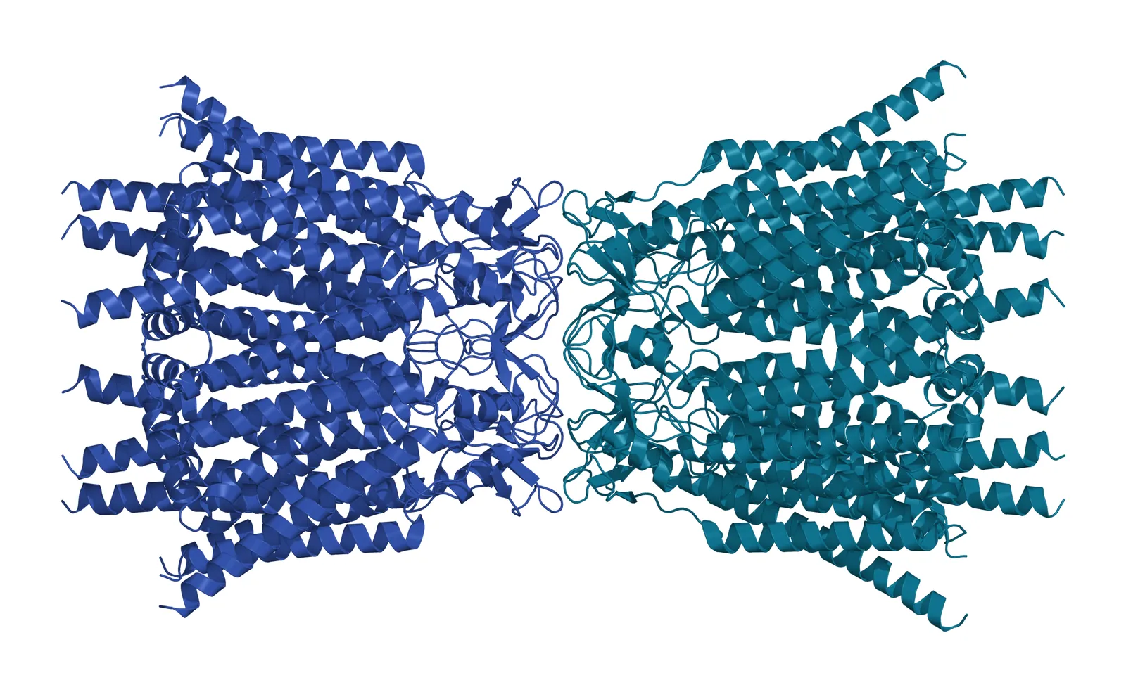
The Lee structures captured three conformations of the connexin-43 N-terminal helix, gate-covering, pore-lining and a flexible intermediate, whose balance shifted with cholesterol analogs, C-terminal truncation and pH.[27] Connexin-43 is encoded by GJA1 on chromosome 6q22.31.[28]
Connexin-43 and its GJA1-20k isoform set the level of transfer
Fu and colleagues found that GJA1 mRNA is alternatively translated into N-terminally truncated isoforms, of which GJA1-20k is the most abundant. GJA1-20k associated strongly with mitochondria, concentrated at the interface between mitochondria and microtubules, and needed its microtubule-binding domain to support mitochondrial transport.[29]
Irwin and colleagues co-cultured bone marrow MSCs carrying fluorescent mitochondria with immortalized human chondrocytes. Transfer was higher in direct contact than across a Transwell insert, and connexin-43 staining concentrated at the tips of MSC actin extensions that contained mitochondria.[30]
Connexin-43 knockdown lowered transfer, and GJA1-20k expression in MSCs correlated with the incidence of transfer.[30] Results vary by system: in the Sinclair co-cultures, the connexin-43 inhibitor GAP26 left mitochondrial transfer unchanged.[7]
Partial cell fusion joins cytoplasm and keeps donor mitochondria
Acquistapace and colleagues co-cultured human multipotent adipose-derived stem cells with mouse cardiomyocytes. Hybrid cells formed mainly through partial cell fusion with intercellular structures of F-actin and microtubules, expressed cardiac progenitor markers and kept a mouse genotype.[31]
Stem cell mitochondria entered the cardiomyocytes, persisted in the hybrids and were required for the shift toward a progenitor-like state. Human bone marrow stromal cells behaved like the adipose cells, and human fibroblasts did not.[31]
Measuring mitochondrial transfer requires labels that follow the organelle
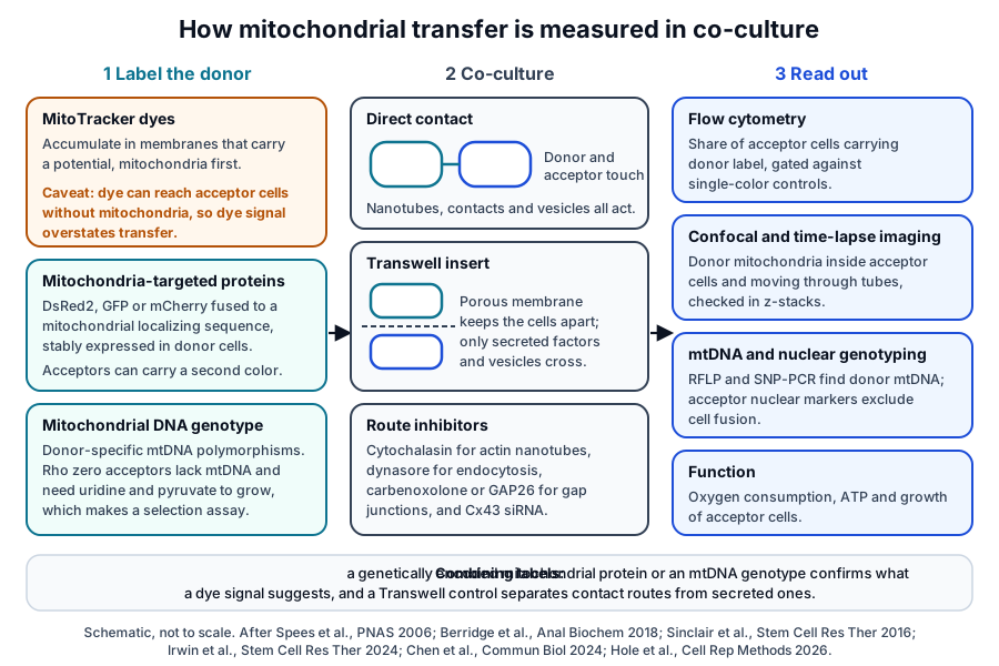
Every transfer assay labels the donor, co-cultures it with an acceptor and reads where the label ends up (Fig. 5). Berridge and colleagues describe three label families: mitochondria-targeting dyes, mitochondrially imported fluorescent proteins and mitochondrial DNA polymorphisms, each with its own limitations.[32]
MitoTracker dyes need confirmation by a second label
Chen and colleagues note that MitoTracker Red accumulates in organelles with a membrane potential and can enrich partially at sites other than mitochondria. Compared with mitochondria-targeted GFP, the dye overstated transfer, and experiments with mitochondria-deficient cells confirmed its non-specificity.[33]
Hole and colleagues labeled astrocyte mitochondria with both GFP and MitoTracker and found that the dye reached neurons independently of mitochondria, including from astrocyte-conditioned medium. They recommend alternative labeling wherever contact-independent transfer is reported.[34]
Genetic labels and genotyping follow the organelle itself
Spees and colleagues imaged a genetically encoded mitochondria-targeted DsRed2, stably expressed in the donor cells, and combined it with RFLP and SNP-PCR assays of donor mitochondrial DNA and nuclear genotyping to exclude fusion.[1] Irwin and colleagues gave donor and acceptor mitochondria different fluorescent proteins and gated transfer events against single-color controls by flow cytometry.[30]
Berridge and colleagues note that mitochondrial DNA polymorphisms suit systems where acceptor mtDNA is depleted, and that they also report the long-term fate of transferred genomes.[32] The rho0 design adds a functional readout, growth without uridine and pyruvate, that only respiring cells pass.[1]
Co-culture design separates contact routes from secreted ones
A Transwell insert keeps donor and acceptor apart while letting medium pass, so comparing it with direct contact separates contact routes from secreted ones.[30] Inhibitors add route detail: Sinclair and colleagues used cytochalasin D for actin tubes, dynasore for endocytosis, carbenoxolone for gap junctions and GAP26 for connexin-43.[7]
Jackson and Krasnodembskaya label MSCs with a mitochondria-specific dye, wash them extensively, seed them with macrophages at a 1:20 ratio and read transfer by confocal microscopy and flow cytometry.[12] Pairing such a protocol with a genetically encoded label answers the dye caveats described above.[33][34]
Where to go from here
The ISCT minimal criteria reference defines the cells these co-cultures start from, and the source tissues reference compares bone marrow, adipose tissue, umbilical cord and placenta. The secretome reference follows the soluble factors and vesicles MSCs release, and the immunobiology reference covers the T cell and macrophage assays met here.
The exosome biochemistry references explain how vesicles are named, isolated and taken up, and the perinatal tissue donor eligibility reference covers the federal framework for perinatal material. Verified practices can request lot documentation, and our team is glad to discuss the methods described on this page.
Annotated References
Numbered in the order they are cited in the text. Each source is annotated with what it supports on this page; peer-reviewed articles are listed with journal, volume, issue and pages, and primary sources are regulations, agency documents and reference databases.
- 1.Peer-reviewedSpees JL, Olson SD, Whitney MJ, et al. Mitochondrial transfer between cells can rescue aerobic respiration. Proceedings of the National Academy of Sciences of the United States of America. 2006;103(5):1283-1288. https://pubmed.ncbi.nlm.nih.gov/16432190/Annotation: Co-culture of human MSCs or skin fibroblasts with mtDNA-depleted rho0 cells. Cited for colony growth in restrictive medium, donor mtDNA by RFLP and SNP-PCR, nuclear genotyping, ATP and DsRed2 imaging.
- 2.Peer-reviewedTorralba D, Baixauli F, Sánchez-Madrid F. Mitochondria Know No Boundaries: Mechanisms and Functions of Intercellular Mitochondrial Transfer. Frontiers in Cell and Developmental Biology. 2016;4:107. https://pubmed.ncbi.nlm.nih.gov/27734015/Annotation: Review of intercellular mitochondrial transfer. Cited for the mitochondrial genome and the four routes: tunneling nanotubes, extracellular vesicles, cell fusion and gap junctions.
- 3.Peer-reviewedBrestoff JR, Singh KK, Aquilano K, et al. Recommendations for mitochondria transfer and transplantation nomenclature and characterization. Nature Metabolism. 2025;7(1):53-67. https://pubmed.ncbi.nlm.nih.gov/39820558/Annotation: Consensus statement on mitochondria transfer terminology. Cited for its definition of intercellular mitochondria transfer, donor and acceptor cells, the functions it lists and its nomenclature framework.
- 4.Peer-reviewedBerridge MV, McConnell MJ, Grasso C, et al. Horizontal transfer of mitochondria between mammalian cells: beyond co-culture approaches. Current Opinion in Genetics & Development. 2016;38:75-82. https://pubmed.ncbi.nlm.nih.gov/27219870/Annotation: Review of horizontal mitochondrial and mtDNA transfer. Cited for transfer in several mammalian culture systems and its proposed place in development and tissue homeostasis.
- 5.Peer-reviewedPlotnikov EY, Khryapenkova TG, Vasileva AK, et al. Cell-to-cell cross-talk between mesenchymal stem cells and cardiomyocytes in co-culture. Journal of Cellular and Molecular Medicine. 2008;12(5A):1622-1631. https://pubmed.ncbi.nlm.nih.gov/18088382/Annotation: Co-culture of human MSCs with rat cardiomyocytes. Cited for intercellular contacts, one-way mitochondrial movement toward cardiomyocytes and mitochondria-like structures in nanotubes.
- 6.Peer-reviewedKoyanagi M, Brandes RP, Haendeler J, et al. Cell-to-cell connection of endothelial progenitor cells with cardiac myocytes by nanotubes: a novel mechanism for cell fate changes? Circulation Research. 2005;96(10):1039-1041. https://pubmed.ncbi.nlm.nih.gov/15879310/Annotation: Co-culture of human endothelial progenitor cells with neonatal rat cardiomyocytes. Cited for nanotube dimensions of 50 to 800 nm by 5 to 120 µm and MitoTracker-labeled cargo.
- 7.Peer-reviewedSinclair KA, Yerkovich ST, Hopkins PM, et al. Characterization of intercellular communication and mitochondrial donation by mesenchymal stromal cells derived from the human lung. Stem Cell Research & Therapy. 2016;7(1):91. https://pubmed.ncbi.nlm.nih.gov/27406134/Annotation: Co-culture of bone marrow and lung MSCs with BEAS2B epithelial cells. Cited for nanotube timing, donation by three MSC sources, one-way transfer and the inhibitor panel; Fig. 1 reproduced under CC BY 4.0.
- 8.Peer-reviewedRustom A, Saffrich R, Markovic I, et al. Nanotubular highways for intercellular organelle transport. Science. 2004;303(5660):1007-1010. https://pubmed.ncbi.nlm.nih.gov/14963329/Annotation: First description of tunneling nanotubes. Cited for de novo nanotube networks that carry vesicles and organelles selectively and the principle of membrane continuity.
- 9.Peer-reviewedSartori-Rupp A, Cordero Cervantes D, Pepe A, et al. Correlative cryo-electron microscopy reveals the structure of TNTs in neuronal cells. Nature Communications. 2019;10(1):342. https://pubmed.ncbi.nlm.nih.gov/30664666/Annotation: Correlative FIB-SEM, light and cryo-electron microscopy of neuronal nanotubes. Cited for bundles of open-ended individual tubes filled with parallel actin, carrying compartments and mitochondria.
- 10.Peer-reviewedAustefjord MW, Gerdes HH, Wang X. Tunneling nanotubes: Diversity in morphology and structure. Communicative & Integrative Biology. 2014;7(1):e27934. https://pubmed.ncbi.nlm.nih.gov/24778759/Annotation: Review of nanotube structure. Cited for diversity of morphology and composition between and within cell systems.
- 11.Peer-reviewedZurzolo C. Tunneling nanotubes: Reshaping connectivity. Current Opinion in Cell Biology. 2021;71:139-147. https://pubmed.ncbi.nlm.nih.gov/33866130/Annotation: Review of nanotube properties. Cited for the range of TNT-like protrusions, reliance on low-resolution imaging and the need to show transfer between distinct cell populations.
- 12.Peer-reviewedJackson MV, Krasnodembskaya AD. Analysis of Mitochondrial Transfer in Direct Co-cultures of Human Monocyte-derived Macrophages (MDM) and Mesenchymal Stem Cells (MSC). Bio-protocol. 2017;7(9). https://pubmed.ncbi.nlm.nih.gov/28534038/Annotation: Protocol for MSC and macrophage co-culture. Cited for dye labeling, washing, the 1:20 ratio, confocal and flow readouts, nanotube transfer and the cytochalasin B result.
- 13.Peer-reviewedLuz-Crawford P, Hernandez J, Djouad F, et al. Mesenchymal stem cell repression of Th17 cells is triggered by mitochondrial transfer. Stem Cell Research & Therapy. 2019;10(1):232. https://pubmed.ncbi.nlm.nih.gov/31370879/Annotation: Co-culture of bone marrow MSCs with Th17 cells. Cited for mitochondrial uptake within 4 h, contact-dependent fall in IL-17, raised oxygen consumption and conversion toward regulatory T cells.
- 14.Peer-reviewedCourt AC, Le-Gatt A, Luz-Crawford P, et al. Mitochondrial transfer from MSCs to T cells induces Treg differentiation and restricts inflammatory response. EMBO Reports. 2020;21(2):e48052. https://pubmed.ncbi.nlm.nih.gov/31984629/Annotation: MSC and lymphocyte co-culture with artificial mitochondrial transfer. Cited only for preferential transfer to CD4+ T cells and the FOXP3, IL2RA, CTLA4 and TGFB1 transcript and CD25+ FoxP3+ results.
- 15.Peer-reviewedHase K, Kimura S, Takatsu H, et al. M-Sec promotes membrane nanotube formation by interacting with Ral and the exocyst complex. Nature Cell Biology. 2009;11(12):1427-1432. https://pubmed.ncbi.nlm.nih.gov/19935652/Annotation: Discovery of M-Sec as a nanotube inducer. Cited for de novo protrusions, RNAi depletion results and the Ral and exocyst interaction.
- 16.Peer-reviewedKimura S, Yamashita M, Yamakami-Kimura M, et al. Distinct Roles for the N- and C-terminal Regions of M-Sec in Plasma Membrane Deformation during Tunneling Nanotube Formation. Scientific Reports. 2016;6:33548. https://pubmed.ncbi.nlm.nih.gov/27629377/Annotation: Structural and cell study of M-Sec. Cited for its TNFAIP2 and B94 names, Sec6 similarity, PI(4,5)P2 binding, rod-like helix bundle structure and RalA recruitment.
- 17.DatabaseNCBI Gene 7127, TNFAIP2 TNF alpha induced protein 2 (Homo sapiens). Accessed 20 October 2026. https://www.ncbi.nlm.nih.gov/gene/7127
- 18.Peer-reviewedFransson A, Ruusala A, Aspenström P. Atypical Rho GTPases have roles in mitochondrial homeostasis and apoptosis. The Journal of Biological Chemistry. 2003;278(8):6495-6502. https://pubmed.ncbi.nlm.nih.gov/12482879/Annotation: Characterization of Miro-1 and Miro-2. Cited for the name, tandem GTPase domains, EF hand linker and conservation from yeast to mammals.
- 19.Peer-reviewedFransson S, Ruusala A, Aspenström P. The atypical Rho GTPases Miro-1 and Miro-2 have essential roles in mitochondrial trafficking. Biochemical and Biophysical Research Communications. 2006;344(2):500-510. https://pubmed.ncbi.nlm.nih.gov/16630562/Annotation: Cell study of Miro trafficking. Cited for the C-terminal transmembrane domain and binding to the kinesin-binding proteins GRIF-1 and OIP106.
- 20.Peer-reviewedMacaskill AF, Rinholm JE, Twelvetrees AE, et al. Miro1 is a calcium sensor for glutamate receptor-dependent localization of mitochondria at synapses. Neuron. 2009;61(4):541-555. https://pubmed.ncbi.nlm.nih.gov/19249275/Annotation: Neuronal study of Miro1. Cited for the Miro1 link to KIF5 motors and its inhibition by micromolar calcium at the EF hands.
- 21.DatabaseNCBI Gene 55288, RHOT1 ras homolog family member T1 (Homo sapiens). Accessed 20 October 2026. https://www.ncbi.nlm.nih.gov/gene/55288
- 22.Peer-reviewedSmith KP, Focia PJ, Chakravarthy S, et al. Insight into human Miro1/2 domain organization based on the structure of its N-terminal GTPase. Journal of Structural Biology. 2020;212(3):107656. https://pubmed.ncbi.nlm.nih.gov/33132189/Annotation: Crystal structure of the human Miro1 N-terminal GTPase, PDB 6D71. Cited for the 1.7 Å GTP-bound structure and the SAXS model of Miro1 and Miro2.
- 23.Peer-reviewedPhinney DG, Di Giuseppe M, Njah J, et al. Mesenchymal stem cells use extracellular vesicles to outsource mitophagy and shuttle microRNAs. Nature Communications. 2015;6:8472. https://pubmed.ncbi.nlm.nih.gov/26442449/Annotation: MSC and macrophage co-culture study. Cited only for ARRDC1 microvesicles carrying depolarized mitochondria, macrophage uptake and fusion, TSG101, and exosomes carrying microRNAs and mtDNA.
- 24.Peer-reviewedRosina M, Ceci V, Turchi R, et al. Ejection of damaged mitochondria and their removal by macrophages ensure efficient thermogenesis in brown adipose tissue. Cell Metabolism. 2022;34(4):533-548.e12. https://pubmed.ncbi.nlm.nih.gov/35305295/Annotation: Mouse brown adipose tissue physiology. Cited for vesicles carrying mitochondrial parts, macrophage clearance and the thermogenic response to cold.
- 25.Peer-reviewedNicolás-Ávila JA, Lechuga-Vieco AV, Esteban-Martínez L, et al. A Network of Macrophages Supports Mitochondrial Homeostasis in the Heart. Cell. 2020;183(1):94-109.e23. https://pubmed.ncbi.nlm.nih.gov/32937105/Annotation: Mouse heart physiology. Cited for macrophage uptake of mitochondria ejected by cardiomyocytes in exopher-like particles, autophagy dependence and Mertk.
- 26.Peer-reviewedGoodenough DA, Paul DL. Gap junctions. Cold Spring Harbor Perspectives in Biology. 2009;1(1):a002576. https://pubmed.ncbi.nlm.nih.gov/20066080/Annotation: Review of gap junction biology. Cited for channels that pass ions and small molecules and for connexin hexamers.
- 27.Peer-reviewedLee HJ, Cha HJ, Jeong H, et al. Conformational changes in the human Cx43/GJA1 gap junction channel visualized using cryo-EM. Nature Communications. 2023;14(1):931. https://pubmed.ncbi.nlm.nih.gov/36805660/Annotation: Cryo-EM structures of the human connexin-43 channel, including PDB 7XQF. Cited for docked hemichannels and the three N-terminal helix conformations.
- 28.DatabaseNCBI Gene 2697, GJA1 gap junction protein alpha 1 (Homo sapiens). Accessed 20 October 2026. https://www.ncbi.nlm.nih.gov/gene/2697
- 29.Peer-reviewedFu Y, Zhang SS, Xiao S, et al. Cx43 Isoform GJA1-20k Promotes Microtubule Dependent Mitochondrial Transport. Frontiers in Physiology. 2017;8:905. https://pubmed.ncbi.nlm.nih.gov/29163229/Annotation: Cell study of the GJA1-20k isoform. Cited for alternative translation, mitochondrial association, the mitochondria-microtubule interface and the microtubule-binding domain.
- 30.Peer-reviewedIrwin RM, Thomas MA, Fahey MJ, et al. Connexin 43 regulates intercellular mitochondrial transfer from human mesenchymal stromal cells to chondrocytes. Stem Cell Research & Therapy. 2024;15(1):359. https://pubmed.ncbi.nlm.nih.gov/39390589/Annotation: Co-culture of bone marrow MSCs with chondrocytes. Cited only for contact versus Transwell transfer, connexin-43 at actin extension tips, knockdown results, GJA1-20k and flow gating.
- 31.Peer-reviewedAcquistapace A, Bru T, Lesault PF, et al. Human mesenchymal stem cells reprogram adult cardiomyocytes toward a progenitor-like state through partial cell fusion and mitochondria transfer. Stem Cells. 2011;29(5):812-824. https://pubmed.ncbi.nlm.nih.gov/21433223/Annotation: Co-culture of human adipose-derived and bone marrow stem cells with mouse cardiomyocytes. Cited for partial fusion, F-actin and microtubule bridges, and persistence of donor mitochondria in hybrids.
- 32.Peer-reviewedBerridge MV, Herst PM, Rowe MR, et al. Mitochondrial transfer between cells: Methodological constraints in cell culture and animal models. Analytical Biochemistry. 2018;552:75-80. https://pubmed.ncbi.nlm.nih.gov/29158129/Annotation: Methods review. Cited for the three label families, dye limitations and the uses of mtDNA polymorphisms.
- 33.Peer-reviewedChen C, Li H, Zhang J, et al. Exploring the limitations of mitochondrial dye as a genuine horizontal mitochondrial transfer surrogate. Communications Biology. 2024;7(1):281. https://pubmed.ncbi.nlm.nih.gov/38448655/Annotation: Methods study comparing MitoTracker Red with mitochondria-targeted GFP. Cited for non-specific dye enrichment and lower transfer efficiency by the genetic label.
- 34.Peer-reviewedHole KL, Norkett R, Russell E, et al. MitoTracker transfers from astrocytes to neurons independently of mitochondria. Cell Reports Methods. 2026;6(3):101338. https://pubmed.ncbi.nlm.nih.gov/41831447/Annotation: Astrocyte and neuron co-culture with dual GFP and MitoTracker labels. Cited for dye transfer independent of mitochondria and the call for alternative labels.
Cite this page
ExaVeyra Sciences Editorial Team. Mitochondrial Transfer by Mesenchymal Stromal Cells. ExaVeyra Sciences; 20 October 2026. Available from: https://exaveyra.com/science/stem-cells/mitochondrial-transfer-stromal-cells. Accessed [date accessed].
Figure sources
- 35.PDB 6D71, Human Miro1 N-terminal GTPase bound to GTP. ExaVeyra Sciences, rendered in PyMOL from PDB 6D71. Structure data: RCSB Protein Data Bank (rcsb.org), CC0. Structure publication: Smith KP, Focia PJ, Chakravarthy S, et al. Insight into human Miro1/2 domain organization based on the structure of its N-terminal GTPase. J Struct Biol. 2020;212(3):107656.
- 36.PDB 7XQF, Human connexin-43 gap junction channel in nanodiscs. ExaVeyra Sciences, rendered in PyMOL from PDB 7XQF. Structure data: RCSB Protein Data Bank (rcsb.org), CC0. Structure publication: Lee HJ, Cha HJ, Jeong H, et al. Conformational changes in the human Cx43/GJA1 gap junction channel visualized using cryo-EM. Nat Commun. 2023;14(1):931.