Bone Marrow Aspirate Concentrate and Adipose SVF: Composition
By Benn Bluestein-Veyra, M.Sc. Organic Chemistry · Updated 21 October 2026
ExaVeyra Sciences is a Miami-based B2B supplier of exosomes for topical aesthetic use to licensed clinics, and it refers licensed prescribers to a 503A compounding pharmacy that prepares formulations against individual prescriptions.
| Question | Answer |
|---|---|
| What this page covers | What bone marrow aspirate concentrate and adipose stromal vascular fraction contain: nucleated cell types, stromal cell frequency and how each is characterized. |
| Who can order | Licensed clinics and practitioners whose active professional license and NPI ExaVeyra has verified. Exosome orders also require a signed professional-use acknowledgment. |
| What ships, and how | Exosome material for topical aesthetic use, characterized by lot, ships overnight in a cold chain. Delivery can take 1 to 3 business days. A certificate of analysis is issued for every lot and provided on request. |
Bone marrow aspirate concentrate (BMAC) is the nucleated cell and platelet fraction of aspirated marrow, collected after density centrifugation. Schäfer and colleagues measured enrichment of total nucleated cells, platelets, CFU-F colonies and several stromal cell subsets after concentration.[1] Adipose stromal vascular fraction (SVF) is the cell pellet released from fat tissue, defined by a joint IFATS and ISCT statement.[2]
This reference describes what each preparation contains, how preparation steps shape that content and which assays count the cells. It covers nucleated cell counts, the fibroblast colony-forming unit (CFU-F) assay and flow cytometry panels.[2][3]
Understanding the cell composition of bone marrow aspirate
Marrow aspirate is a mixed suspension of red cells, platelets, myeloid and lymphoid nucleated cells, CD34+ hematopoietic progenitors and a small population of adherent stromal cells.[1][3] Pittenger and colleagues isolated stromal cells from the marrow aspirates of volunteer donors and showed that single cells expanded into colonies retained adipocytic, chondrocytic and osteocytic potential.[4]
CD34 is a highly glycosylated single-pass membrane protein encoded at chromosome 1q32.2, and it may help stem cells attach to marrow matrix or stromal cells.[5] Stromal progenitors are rare: Muschler and colleagues counted a mean of 36 alkaline phosphatase-positive colony-forming units per million nucleated cells in iliac crest aspirates.[6]
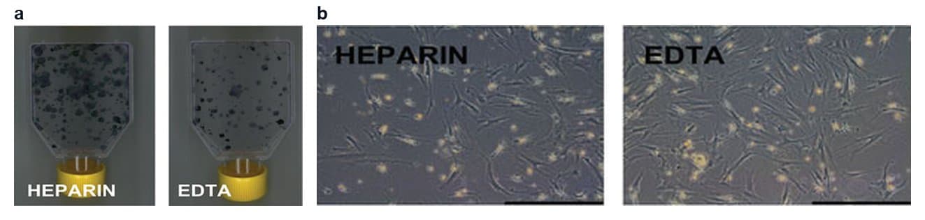
In a later series of 57 donors, the same group harvested a mean of 64 million nucleated cells per aspirate, with a mean prevalence of 55 colonies per million nucleated cells.[7] Colony counts also depend on how marrow is collected and how cells are selected before plating (Fig. 1).[8][9]
Quirici and colleagues reported a median CFU-F frequency of 35 per million low-density mononuclear cells. In the fraction selected with antibodies to the low-affinity nerve growth factor receptor, the median rose to 1,584 per million.[9]
Aspiration volume and site shape marrow aspirate quality
Peripheral blood enters the syringe as aspiration continues, so the first milliliters drawn from a site carry the most marrow. Muschler and colleagues found that raising aspiration volume from one to four milliliters increased total colony-forming units while lowering their concentration by about 50% (Fig. 2).[6]
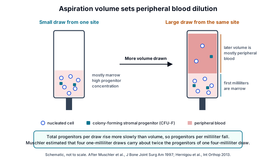
Hernigou and colleagues compared bilateral aspirates from 30 adults and reported progenitor concentrations about 300% higher with a 10-milliliter syringe than with a 50-milliliter syringe.[10] Cuthbert and colleagues likewise reported a several-fold fall in CFU-F and CD271-bright cells per milliliter as aspirated volume grew.[3]
- Site: Pierini and colleagues reported a 1.6-fold greater yield of colony-founding progenitors from the posterior iliac crest than from the anterior crest, with comparable phenotype and differentiation of the cultured cells.[11]
- Insertion technique: Oliver and colleagues compared single-site aspiration with needle redirection against multiple-site aspiration and found no significant difference in cell ratios or stromal cell numbers.[12]
- Anticoagulant: Ferencakova and colleagues found a higher cell yield and more CFU colonies in heparin tubes than in EDTA tubes, with no significant change in the molecular characteristics of the cultured cells.[8]
- Donor: Muschler and colleagues attributed 70% of the variation in progenitor prevalence to differences among donors, and in a later series nucleated cells per aspirate declined with age.[6][7]
Density separation concentrates the buffy coat into BMAC
Centrifugation layers marrow aspirate by density into plasma, a thin buffy coat of nucleated cells and packed red cells. Bone marrow aspirate concentrate is the buffy coat collected with part of the plasma (Fig. 3).[1][13]
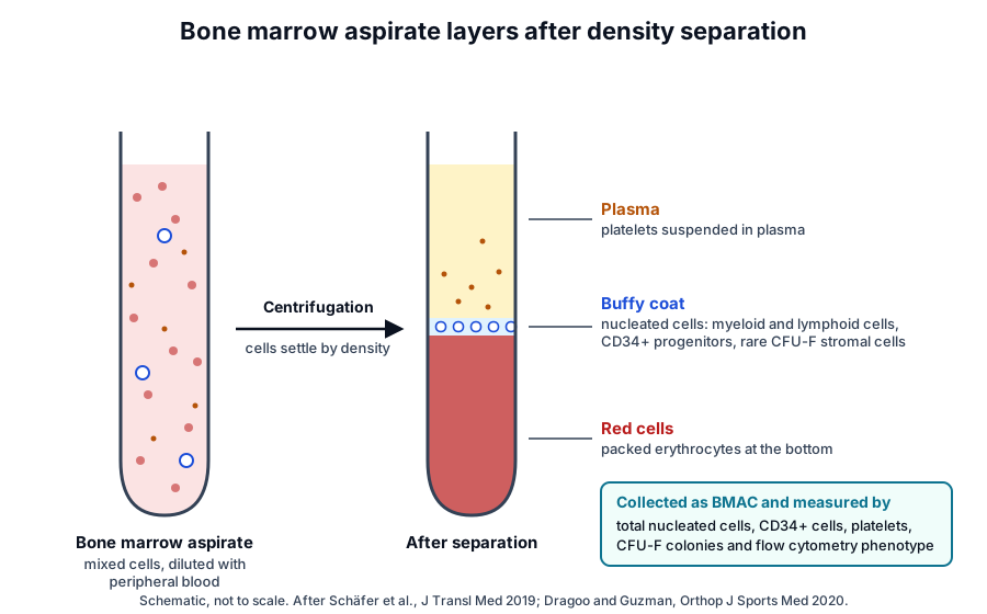
Schäfer and colleagues processed 120-milliliter aspirates from 10 donors on two point-of-care systems. Both systems enriched total nucleated cells, platelets and CD45-CD73+ and CD45-CD73+CD90+ cells, with cell viability near 90% in all groups.[1]
Concentration also raised CFU-F numbers, platelet-derived growth factor, vascular endothelial growth factor, macrophage colony-stimulating factor and VCAM-1. Neither system concentrated red cells, hematopoietic stem cells or bone morphogenetic proteins.[1]
Separation systems yield different products
Dragoo and Guzman compared three systems on combined blood and marrow from 10 donors. The systems differed in recovery of CFU-F, CD34+ cells, leukocytes and platelets, and one system raised platelet concentration about 11 times above baseline.[13]
Hegde and colleagues compared three systems in two paired series of bilateral aspirates from 40 donors and found different numbers and concentrations of connective tissue progenitors.[14] Cassano and colleagues found that two systems both increased colony-forming units over the starting aspirate, and that one concentrate held more interleukin 1 receptor antagonist than platelet-rich plasma prepared with the same system from the same donors.[15]
Characterizing BMAC with counts, colony assays and flow cytometry
An automated hematology analyzer reports total nucleated cells, platelets and red cells in the aspirate and the concentrate, and the ratio gives the fold enrichment.[1][13] Flow cytometry adds CD34+ cell enumeration, and Cuthbert and colleagues noted donor ranges compatible with standard ISHAGE protocols.[3]
The CFU-F assay plates nucleated cells at low density and counts the fibroblast colonies that form. It serves as the functional reference for stromal progenitor frequency.[1][3]
Cuthbert and colleagues enumerated CD45-negative or low, CD271-bright cells with counting beads in a single-platform assay. These cells co-expressed CD73, CD105 and CD90, and their numbers correlated with manual CFU-F counts (R = 0.81).[3] CD271, also called p75NTR, is encoded by the NGFR gene at 17q21.33, and its extracellular domain holds four cysteine-rich repeats that contain the nerve growth factor binding domain.[16]
| Measurement | What it reports | Typical method |
|---|---|---|
| Total nucleated cells | All leukocytes and progenitors per milliliter | Hematology analyzer |
| Platelets and red cells | Platelet enrichment and red cell carryover | Hematology analyzer |
| CD34+ cells | Hematopoietic progenitor content | Flow cytometry with counting beads |
| CD45-/low CD271-bright cells | Native stromal progenitor content | Single-platform flow cytometry |
| CFU-F | Functional frequency of colony-forming stromal cells | Low-density plating and colony count |
Cultured marrow stromal cells are described against the ISCT minimal criteria: plastic adherence, CD105, CD73 and CD90 expression without hematopoietic markers, and trilineage differentiation in vitro.[17] Busser and colleagues enriched native marrow stromal cells by immunomagnetic selection for MSCA-1, SUSD2 or CD271, and also found a distinct CD34+ fresh marrow subset.[18]
Adipose stromal vascular fraction is released by enzymatic or mechanical processing
Zuk and colleagues described processed lipoaspirate cells in 2002, a stromal population from fat tissue that differentiated toward osteogenic, adipogenic, myogenic and chondrogenic lineages in culture.[19] The SVF is the cell pellet left after adipocytes float away, and it holds stromal, perivascular, endothelial and blood-derived cells (Fig. 4).[2][20]
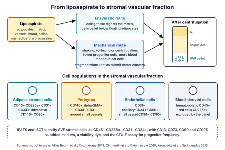
Enzymatic isolation digests the extracellular matrix with collagenase before centrifugation. Aronowitz and colleagues reviewed studies showing that enzymatic digestion separates cells with greater efficiency, while mechanical methods yield more blood mononuclear cells and fewer progenitor cells.[21]
Yoshimura and colleagues characterized cells from both the fatty and fluid portions of liposuction aspirates. Multicolor flow cytometry showed blood-derived CD45+ cells, CD34+ stromal cells and CD146+ pericytes, and the adherent cells from both portions converged in phenotype after plating.[20]
Microfragmented adipose tissue preserves the vascular stroma
Mechanical fragmentation can also reduce adipose tissue to submillimeter clusters without enzyme. Bianchi and colleagues described such clusters with preserved vascular stroma, a higher percentage of pericytes and lower hematopoietic content than enzymatically digested lipoaspirate.[22]
Vezzani and colleagues compared matched samples and found microfragmented tissue enriched in pericytes relative to adventitial cells. In culture, the clusters released more growth factors and cytokines into the medium than isogenic SVF.[23]
Understanding the IFATS and ISCT marker panel for SVF cells
Bourin and colleagues, writing for IFATS and ISCT, identified SVF stromal cells as CD45-negative, CD235a-negative, CD31-negative and CD34-positive. They proposed a viability marker and CD13, CD73, CD90 and CD105 as added value, and the CFU-F assay to measure progenitor frequency.[2]
Cultured adipose stromal cells share CD90, CD73, CD105 and CD44 with other MSCs and stay negative for CD45 and CD31. The statement distinguishes them from marrow-derived MSCs by CD36 positivity and CD106 negativity.[2]
Zimmerlin and colleagues mapped perivascular cells in intact fat. Inner pericytes were CD146+ alpha-SMA+ CD34- CD31-, outer adventitial stromal cells were CD146- CD90+ CD34+ CD31-, and capillary endothelial cells were CD31+ CD34+ CD90+.[25] Corselli and colleagues reviewed how such flow cytometry gates allow prospective purification of perivascular stromal cells.[26]
Mitchell and colleagues measured CFU-F in fresh SVF at a frequency of 1 in 32 cells.[24] Busser and colleagues found that CD34 selection isolated stromal cells from adipose tissue, while SUSD2 and MSCA-1 selection did not.[18]
Comparing marrow and adipose preparations by composition
The two preparations start from different tissues and carry different cell mixtures (Fig. 5). Marrow concentrate holds hematopoietic cells, platelets and rare stromal progenitors, and SVF holds a larger stromal and perivascular fraction alongside blood-derived cells.[1][2][24]
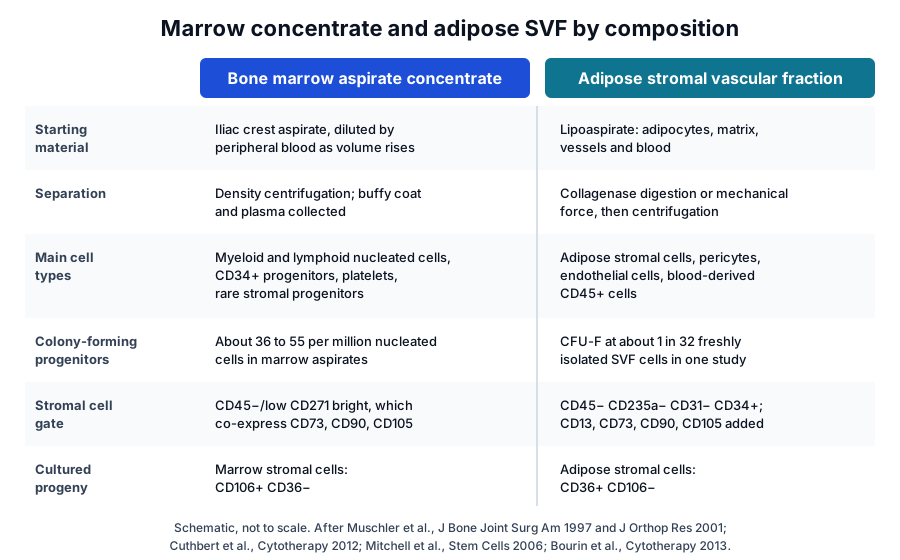
| Feature | Bone marrow aspirate concentrate | Adipose stromal vascular fraction |
|---|---|---|
| Starting material | Iliac crest marrow aspirate | Lipoaspirate |
| Separation | Density centrifugation | Collagenase or mechanical release, then centrifugation |
| Main cells | Nucleated hematopoietic cells, CD34+ progenitors, platelets | Adipose stromal cells, pericytes, endothelial and blood-derived cells |
| Stromal gate | CD45-/low CD271-bright | CD45- CD235a- CD31- CD34+ |
| Cultured progeny | CD106+ CD36- | CD36+ CD106- |
Donor characteristics also shape composition. In marrow aspirates, nucleated cells per aspirate declined with age, and in women colony-forming units per aspirate also declined with age.[7]
Locating these preparations in the federal HCT/P framework
Federal regulation states that “An HCT/P is regulated solely under section 361 of the PHS Act and the regulations in this part if it meets all of the following criteria:” The first criterion reads “(1) The HCT/P is minimally manipulated;”[27] FDA sets out how it reads these terms in its guidance “Regulatory Considerations for Human Cells, Tissues, and Cellular and Tissue-Based Products: Minimal Manipulation and Homologous Use,” dated July 2020.[28]
The 361 HCT/P versus 351 biologic reference on this site quotes all four criteria of 21 CFR 1271.10 in full. It is the place to read the regulatory framework alongside this page.[27]
Where to go from here
The PRP preparation variables reference explains the centrifugation physics behind buffy coat separation, and the platelet granule reference describes the growth factors that platelets in a concentrate carry. The growth factor receptors reference follows those signals to their target cells, and the platelet study reporting reference lists the details a complete methods section includes.
The MSC source tissues and ISCT minimal criteria references describe the cultured stromal cells that marrow and fat give rise to, and the HCT/P reference sets out the federal framework. Our team is glad to discuss the characterization methods described on this page.
Annotated References
Numbered in the order they are cited in the text. Each source is annotated with what it supports on this page; peer-reviewed articles are listed with journal, volume, issue and pages, and primary sources are regulations, agency documents and reference databases.
- 1.Peer-reviewedSchäfer R, DeBaun MR, Fleck E, et al. Quantitation of progenitor cell populations and growth factors after bone marrow aspirate concentration. Journal of Translational Medicine. 2019. https://pubmed.ncbi.nlm.nih.gov/30961655/Annotation: Flow cytometry, CFU-F and analyzer counts on marrow aspirate before and after concentration. Cited for enrichment of nucleated cells, platelets, stromal subsets, CFU-F and growth factors, and for red cells and hematopoietic stem cells left unconcentrated.
- 2.Peer-reviewedBourin P, Bunnell BA, Casteilla L, et al. Stromal cells from the adipose tissue-derived stromal vascular fraction and culture expanded adipose tissue-derived stromal/stem cells: a joint statement of the International Federation for Adipose Therapeutics and Science (IFATS) and the International Society for Cellular Therapy (ISCT). Cytotherapy. 2013;15(6):641-648. https://pubmed.ncbi.nlm.nih.gov/23570660/Annotation: IFATS and ISCT minimal definitions for SVF and cultured adipose stromal cells. Cited for the CD45-CD235a-CD31-CD34+ SVF phenotype, the added markers, the CFU-F assay and the CD36 and CD106 distinction from marrow cells.
- 3.Peer-reviewedCuthbert R, Boxall SA, Tan HB, et al. Single-platform quality control assay to quantify multipotential stromal cells in bone marrow aspirates prior to bulk manufacture or direct therapeutic use. Cytotherapy. 2012. https://pubmed.ncbi.nlm.nih.gov/22268519/Annotation: Bead-based enumeration of CD45-/low CD271-bright cells in marrow aspirates. Cited for their MSC phenotype, correlation with CFU-F, CD34 enumeration and the fall in stromal cell frequency with larger aspirated volume.
- 4.Peer-reviewedPittenger MF, Mackay AM, Beck SC, et al. Multilineage potential of adult human mesenchymal stem cells. Science. 1999;284(5411):143-147. https://pubmed.ncbi.nlm.nih.gov/10102814/Annotation: Isolation of stromal cells from marrow aspirates of volunteer donors. Cited for clonal colonies that retained adipocytic, chondrocytic and osteocytic potential.
- 5.DatabaseNCBI Gene 947, CD34 CD34 molecule (Homo sapiens). Accessed 21 October 2026. https://www.ncbi.nlm.nih.gov/gene/947
- 6.Peer-reviewedMuschler GF, Boehm C, Easley K. Aspiration to obtain osteoblast progenitor cells from human bone marrow: the influence of aspiration volume. The Journal of Bone and Joint Surgery. American Volume. 1997. https://pubmed.ncbi.nlm.nih.gov/9384430/Annotation: Colony-forming unit counts in iliac crest aspirates of one to four milliliters. Cited for a mean of 36 colonies per million nucleated cells, the dilution of larger draws by peripheral blood and the share of variation due to donors.
- 7.Peer-reviewedMuschler GF, Nitto H, Boehm CA, et al. Age- and gender-related changes in the cellularity of human bone marrow and the prevalence of osteoblastic progenitors. Journal of Orthopaedic Research. 2001. https://pubmed.ncbi.nlm.nih.gov/11332607/Annotation: Nucleated cell and colony-forming unit counts across ages and sexes. Cited for 64 million nucleated cells per aspirate, 55 colonies per million cells and the age-related decline in cellularity.
- 8.Peer-reviewedFerencakova M, Benova A, Raska I Jr, et al. Human bone marrow stromal cells: the impact of anticoagulants on stem cell properties. Frontiers in Cell and Developmental Biology. 2023. https://pubmed.ncbi.nlm.nih.gov/37791077/Annotation: Paired marrow aspirates collected into heparin or EDTA and characterized by flow cytometry, RNA-seq and CFU assays. Cited for higher yield and more CFU colonies with heparin and unchanged molecular characteristics, and as the source of Fig. 1.
- 9.Peer-reviewedQuirici N, Soligo D, Bossolasco P, et al. Isolation of bone marrow mesenchymal stem cells by anti-nerve growth factor receptor antibodies. Experimental Hematology. 2002. https://pubmed.ncbi.nlm.nih.gov/12135677/Annotation: Immunomagnetic selection of marrow stromal cells with antibodies to the low-affinity nerve growth factor receptor (CD271). Cited for CFU-F frequency in low-density mononuclear cells and in the selected fraction.
- 10.Peer-reviewedHernigou P, Homma Y, Flouzat Lachaniette CH, et al. Benefits of small volume and small syringe for bone marrow aspirations of mesenchymal stem cells. International Orthopaedics. 2013. https://pubmed.ncbi.nlm.nih.gov/23881064/Annotation: Paired bilateral aspirates drawn with 10-milliliter and 50-milliliter syringes. Cited for higher progenitor concentration with the smaller syringe.
- 11.Peer-reviewedPierini M, Di Bella C, Dozza B, et al. The posterior iliac crest outperforms the anterior iliac crest when obtaining mesenchymal stem cells from bone marrow. The Journal of Bone and Joint Surgery. American Volume. 2013. https://pubmed.ncbi.nlm.nih.gov/23783207/Annotation: Paired anterior and posterior iliac crest aspirates. Cited for the 1.6-fold greater yield of colony-founding progenitors posteriorly and comparable cultured cell properties.
- 12.Peer-reviewedOliver K, Awan T, Bayes M. Single- Versus Multiple-Site Harvesting Techniques for Bone Marrow Concentrate: Evaluation of Aspirate Quality and Pain. Orthopaedic Journal of Sports Medicine. 2017. https://pubmed.ncbi.nlm.nih.gov/28890905/Annotation: Bilateral comparison of single-site and multiple-site aspiration. Cited for no significant difference in cell ratios or stromal cell numbers between techniques.
- 13.Peer-reviewedDragoo JL, Guzman RA. Evaluation of the Consistency and Composition of Commercially Available Bone Marrow Aspirate Concentrate Systems. Orthopaedic Journal of Sports Medicine. 2020. https://pubmed.ncbi.nlm.nih.gov/32010732/Annotation: Three concentration systems compared on the same donor material. Cited for differing recovery of CFU-F, CD34+ cells, leukocytes and platelets, and platelet concentration about 11 times baseline in one system.
- 14.Peer-reviewedHegde V, Shonuga O, Ellis S, et al. A prospective comparison of 3 approved systems for autologous bone marrow concentration demonstrated nonequivalency in progenitor cell number and concentration. Journal of Orthopaedic Trauma. 2014. https://pubmed.ncbi.nlm.nih.gov/24694554/Annotation: Bilateral aspirates processed on paired concentration systems. Cited for different numbers and concentrations of connective tissue progenitors across systems.
- 15.Peer-reviewedCassano JM, Kennedy JG, Ross KA, et al. Bone marrow concentrate and platelet-rich plasma differ in cell distribution and interleukin 1 receptor antagonist protein concentration. Knee Surgery, Sports Traumatology, Arthroscopy. 2018. https://pubmed.ncbi.nlm.nih.gov/26831858/Annotation: Cell counts, colony-forming units, flow cytometry and multiplex ELISA on marrow concentrate and platelet-rich plasma from the same donors. Cited for colony enrichment over aspirate and higher IL-1 receptor antagonist in the concentrate.
- 16.DatabaseNCBI Gene 4804, NGFR nerve growth factor receptor (Homo sapiens). Accessed 21 October 2026. https://www.ncbi.nlm.nih.gov/gene/4804
- 17.Peer-reviewedDominici M, Le Blanc K, Mueller I, et al. Minimal criteria for defining multipotent mesenchymal stromal cells. The International Society for Cellular Therapy position statement. Cytotherapy. 2006;8(4):315-317. https://pubmed.ncbi.nlm.nih.gov/16923606/Annotation: ISCT minimal criteria for cultured MSCs. Cited for plastic adherence, the CD105, CD73 and CD90 panel without hematopoietic markers, and trilineage differentiation.
- 18.Peer-reviewedBusser H, Najar M, Raicevic G, et al. Isolation and Characterization of Human Mesenchymal Stromal Cell Subpopulations: Comparison of Bone Marrow and Adipose Tissue. Stem Cells and Development. 2015. https://pubmed.ncbi.nlm.nih.gov/26086188/Annotation: Single-marker immunomagnetic selection of stromal subpopulations from marrow and adipose tissue. Cited for MSCA-1, SUSD2 and CD271 enrichment in marrow and CD34 selection in adipose tissue.
- 19.Peer-reviewedZuk PA, Zhu M, Ashjian P, et al. Human adipose tissue is a source of multipotent stem cells. Molecular Biology of the Cell. 2002;13(12):4279-4295. https://pubmed.ncbi.nlm.nih.gov/12475952/Annotation: Characterization of processed lipoaspirate cells and clones. Cited for their multilineage differentiation in culture.
- 20.Peer-reviewedYoshimura K, Shigeura T, Matsumoto D, et al. Characterization of freshly isolated and cultured cells derived from the fatty and fluid portions of liposuction aspirates. Journal of Cellular Physiology. 2006. https://pubmed.ncbi.nlm.nih.gov/16557516/Annotation: Multicolor flow cytometry of fresh and cultured cells from the fatty and fluid portions of lipoaspirate. Cited for blood-derived, stromal and pericyte populations in SVF and their convergence after plating.
- 21.Peer-reviewedAronowitz JA, Lockhart RA, Hakakian CS. Mechanical versus enzymatic isolation of stromal vascular fraction cells from adipose tissue. SpringerPlus. 2015. https://pubmed.ncbi.nlm.nih.gov/26636001/Annotation: Review of mechanical and enzymatic SVF isolation. Cited for the greater separation efficiency of enzymatic digestion and the higher blood mononuclear cell and lower progenitor content of mechanical isolates.
- 22.Peer-reviewedBianchi F, Maioli M, Leonardi E, et al. A new nonenzymatic method and device to obtain a fat tissue derivative highly enriched in pericyte-like elements by mild mechanical forces from human lipoaspirates. Cell Transplantation. 2013. https://pubmed.ncbi.nlm.nih.gov/23051701/Annotation: Histology and flow cytometry of mechanically fragmented lipoaspirate. Cited for preserved vascular stroma, a higher percentage of pericytes and lower hematopoietic content than enzymatic digests.
- 23.Peer-reviewedVezzani B, Shaw I, Lesme H, et al. Higher Pericyte Content and Secretory Activity of Microfragmented Human Adipose Tissue Compared to Enzymatically Derived Stromal Vascular Fraction. Stem Cells Translational Medicine. 2018. https://pubmed.ncbi.nlm.nih.gov/30255987/Annotation: Perivascular cell distribution and secretome arrays in microfragmented tissue and isogenic SVF. Cited for pericyte enrichment and greater release of growth factors and cytokines in culture.
- 24.Peer-reviewedMitchell JB, McIntosh K, Zvonic S, et al. Immunophenotype of human adipose-derived cells: temporal changes in stromal-associated and stem cell-associated markers. Stem Cells. 2006. https://pubmed.ncbi.nlm.nih.gov/16322640/Annotation: Flow cytometry of fresh SVF cells and serially passaged adipose stromal cells. Cited for CFU-F at 1 in 32 cells, rising stromal markers with passage and peak CD34 in fresh SVF.
- 25.Peer-reviewedZimmerlin L, Donnenberg VS, Pfeifer ME, et al. Stromal vascular progenitors in adult human adipose tissue. Cytometry Part A. 2010. https://pubmed.ncbi.nlm.nih.gov/19852056/Annotation: Immunohistochemistry and multiparameter flow cytometry of perivascular and endothelial SVF populations. Cited for the pericyte, adventitial stromal and endothelial phenotypes.
- 26.Peer-reviewedCorselli M, Crisan M, Murray IR, et al. Identification of perivascular mesenchymal stromal/stem cells by flow cytometry. Cytometry Part A. 2013. https://pubmed.ncbi.nlm.nih.gov/23818229/Annotation: Review of flow cytometry evidence for a perivascular origin of MSCs. Cited for prospective purification of perivascular stromal cells.
- 27.Regulatory21 CFR 1271.10, criteria for regulation solely under section 361 of the PHS Act. Accessed 21 October 2026. https://www.ecfr.gov/current/title-21/chapter-I/subchapter-L/part-1271/subpart-A/section-1271.10
- 28.RegulatoryFDA guidance, Regulatory Considerations for Human Cells, Tissues, and Cellular and Tissue-Based Products: Minimal Manipulation and Homologous Use (July 2020). Accessed 21 October 2026. https://www.fda.gov/media/109176/download
Cite this page
ExaVeyra Sciences Editorial Team. Bone Marrow Aspirate Concentrate and Adipose SVF: Composition. ExaVeyra Sciences; 21 October 2026. Available from: https://exaveyra.com/science/platelet-biology/bmac-svf-composition. Accessed [date accessed].