Extracellular Vesicle Lipid Composition and Membrane Biophysics
Reviewed by ExaVeyra Sciences editorial team · Updated 20 October 2026
ExaVeyra Sciences is a Miami-based B2B supplier of exosomes for topical aesthetic use to licensed clinics, and it refers licensed prescribers to a 503A compounding pharmacy that prepares formulations against individual prescriptions.
| Question | Answer |
|---|---|
| What this page covers | The lipidome of exosomes and small extracellular vesicles: cholesterol, sphingomyelin, ceramide, phosphatidylserine and BMP, and how they set membrane mechanics. |
| Who can order | Licensed clinics and practitioners whose active professional license and NPI ExaVeyra has verified. Exosome orders also require a signed professional-use acknowledgment. |
| What ships, and how | Exosome material for topical aesthetic use, characterized by lot, ships overnight in a cold chain. Delivery can take 1 to 3 business days. A certificate of analysis is issued for every lot and provided on request. |
Extracellular vesicle lipid composition differs measurably from that of the cells that release the vesicles.1 Small extracellular vesicles, including exosomes, are often enriched in cholesterol, sphingomyelin, glycosphingolipids and phosphatidylserine, lipids that shape both how the vesicles form and how their membranes behave.2
This reference covers the exosome lipidome class by class, how lipidomics measures it, how ceramide and BMP bend the endosomal membrane, and how membrane lipids relate to stability and uptake. Skotland and colleagues describe exosomes as useful models for membrane biology because each carries a single bilayer.3
Small extracellular vesicles concentrate cholesterol, sphingolipids and phosphatidylserine
Skotland and colleagues compared published lipidomes and found exosomes often enriched in cholesterol, sphingomyelin, glycosphingolipids and phosphatidylserine relative to their parent cells.2 Their 2017 review also traced how individual lipid species, defined by their fatty acyl chains, differ between vesicles and cells.1
Cholesterol accumulates in vesicle membranes
Pfrieger and Vitale compiled the evidence that cholesterol (Fig. 1) accumulates in extracellular vesicles, and discussed how it may contribute to their biogenesis, release and uptake.4 The degree of enrichment varies with the source: in mast cell and dendritic cell exosomes, Laulagnier and colleagues measured cholesterol at a proportion similar to the parent cells.5
Sphingomyelin and glycosphingolipids characterize the outer leaflet
Laulagnier and colleagues found sphingomyelin (Fig. 2) enriched in exosomes from both cell types they studied. Phosphatidylcholine was reduced, and disaturated molecular species were enriched.5
Haraszti and colleagues compared exosomes and microvesicles from three cell sources, including bone marrow-derived mesenchymal stem cells. Glycolipids and free fatty acids characterized the exosomes, and ceramides and sphingomyelins characterized the microvesicles.6
The source cell leaves its own mark on the lipidome. In the same study, exosomes from two of the three sources, including the mesenchymal stem cells, were specifically enriched in cardiolipins, and those from the third source in sphingomyelins.6
Phosphatidylserine links the two leaflets
Phosphatidylserine (Fig. 3) is an anionic phospholipid.7 Skotland and colleagues drew attention to one species, PS 18:0/18:1, and proposed that it sits in the inner leaflet, where it interacts with very-long-chain sphingolipids of the outer leaflet.1
Exosome membranes also exchange lipids between leaflets readily. Laulagnier and colleagues measured faster flip-flop than in the plasma membrane and a random distribution of phosphatidylethanolamine across the bilayer.5 Part of the phosphatidylserine is exposed on the vesicle surface, where receptors on other cells can bind it.8
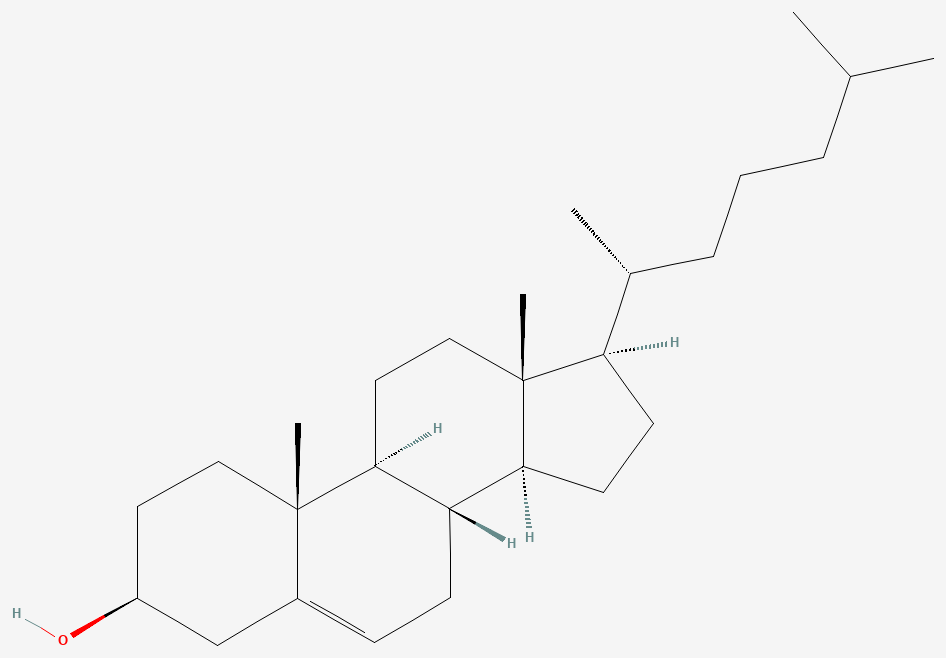
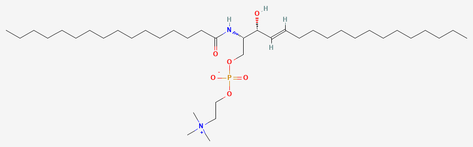
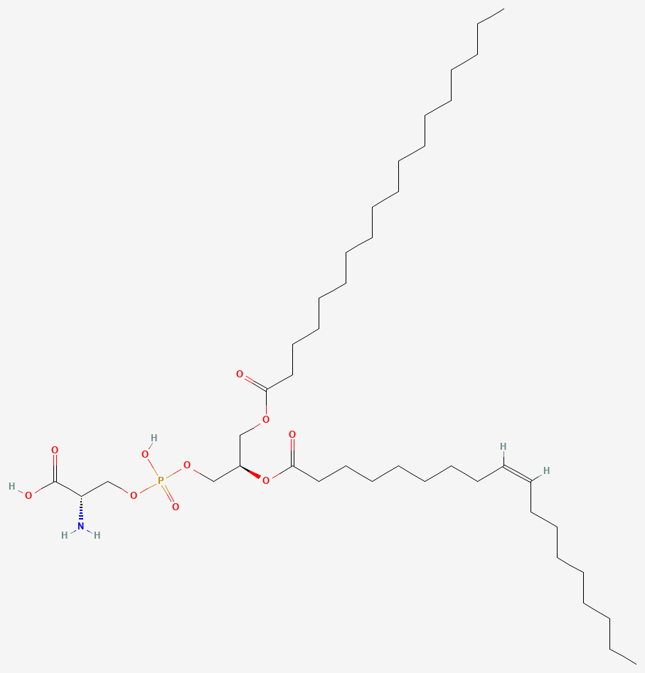
Fig. 1 | Cholesterol, N-Palmitoylsphingomyelin (SM d18:1/16:0) and Phosphatidylserine PS 18:0/18:1.
Bis(monoacylglycero)phosphate marks the late endosomal origin of exosomes
Bis(monoacylglycero)phosphate (BMP), also called lysobisphosphatidic acid (LBPA), is a phospholipid specific to the late endosome and lysosome.9 Matsuo and colleagues showed that LBPA could drive liposomes to form internal vesicles resembling multivesicular endosomes, through a process that depended on a pH gradient across the membrane.10
The same study found that the protein ALIX selectively controlled this LBPA-driven budding and organized LBPA-containing endosomes in cells.10 BMP therefore sits where intraluminal vesicles, the precursors of exosomes, are formed.9
Rabia and colleagues detected BMP on extracellular vesicles from human urine and from HEK293 cell cultures, where it co-localized with the vesicle markers CD63 and ALIX.11 Its level depends on the source: in mast cell and dendritic cell exosomes, BMP was present at a level similar to the parent cells.5
Hullin-Matsuda and colleagues proposed BMP as a lipid signature that can identify the endosomal origin of small extracellular vesicles.9 That makes it a candidate lipid counterpart to the protein markers used to define exosome subpopulations.12
Mass spectrometry and lipid-to-protein ratios measure the exosome lipidome
Quantitative mass spectrometry is the method that determines the lipid content of extracellular vesicles. Skotland and colleagues note that it reports total lipid amount, species composition, sample purity and cellular origin, and they recommend the guidelines of the Lipidomic Standard Initiative.13
Haraszti and colleagues paired high-resolution lipidomics with proteomics on exosomes, microvesicles and source cells, identifying 1,961 lipid species and 3,532 proteins.6 The exosome lipidomes grouped by source differently from the proteomes, so the two profiles describe a vesicle in complementary ways.6
Lipid-to-protein ratios separate vesicle populations
Osteikoetxea and colleagues introduced a plate-based total lipid assay and found protein-to-lipid ratios characteristic of each vesicle subpopulation. The ratios shifted when vesicles were damaged or preparations carried protein contaminants, which makes the measure useful for quality control.14
The same ratio separates vesicles from other particles. Ghosal and colleagues found that non-vesicular extracellular particles have higher protein-to-lipid ratios than vesicle subtypes and are enriched in cholesteryl esters and triacylglycerols.15 Lipid composition therefore reports on the purity of an exosome preparation.2
A complete lipid characterization of a vesicle preparation typically combines the following measurements:
- Total lipid amount, from a lipid assay or quantitative mass spectrometry.13,14
- Lipid class and species composition, from quantitative mass spectrometry.13
- Protein-to-lipid ratio, as an indicator of vesicle subtype and of non-vesicular particles.14,15
- Lipid order, from environment-sensitive membrane probes such as Di-4-ANEPPDHQ.14
Cholesterol and sphingolipids give the exosome membrane raft-like order
Sezgin and colleagues describe membrane rafts as packed, ordered domains that form when cholesterol associates preferentially with saturated lipids, and that selectively recruit certain lipids and proteins.16 Exosome lipids resemble those of detergent-resistant membranes, the classical raft model, with high sphingolipid, cholesterol and phosphatidylserine content.3
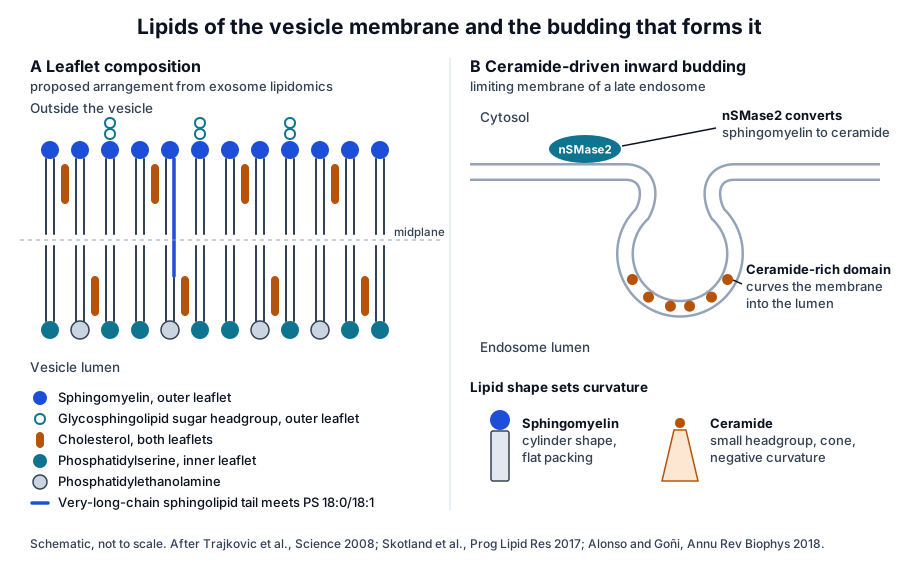
Skotland and colleagues proposed that cholesterol supports the interaction between very-long-chain sphingolipids in the outer leaflet and PS 18:0/18:1 in the inner leaflet, coupling the two halves of the bilayer (Fig. 4).1,3
Osteikoetxea and colleagues measured lipid order with the probe Di-4-ANEPPDHQ and found marked differences between vesicle populations, along with differences in clustered cholesterol and the ganglioside GM1.14 Laulagnier and colleagues found exosome membranes more rigid at pH 7 than at pH 5, consistent with reorganization as vesicles pass from the acidic endosome to the neutral extracellular medium.5
Ceramide drives inward budding of the endosomal membrane
Trajkovic and colleagues showed in 2008 that cargo segregates into distinct domains on the endosomal membrane, and that transfer of exosome-associated domains into the endosome lumen required the sphingolipid ceramide.17 Purified exosomes were enriched in ceramide, and inhibiting neutral sphingomyelinases reduced exosome release.17
The enzyme behind this step, neutral sphingomyelinase 2 (nSMase2), is encoded by the human SMPD3 gene on chromosome 16q22.1. It carries sphingomyelin phosphodiesterase activity, the reaction that converts sphingomyelin to ceramide.18
A cone-shaped lipid curves the membrane
Ceramide (Fig. 5) carries a small hydroxyl headgroup on two long hydrocarbon chains. Alonso and Goñi describe ceramides as strongly hydrophobic lipids that raise the molecular order of neighboring phospholipids, separate into domains and carry a marked intrinsic negative curvature.19
That cone-like geometry offers a physical basis for the budding Trajkovic and colleagues observed: as ceramide-rich domains coalesce, they curve the membrane away from the cytosol and into the endosome lumen (Fig. 4).17,19
The nSMase2 structure explains how phosphatidylserine activates the enzyme
Airola and colleagues solved the crystal structure of the human nSMase2 catalytic domain at 1.85 Å resolution (Fig. 6). It has a DNase-I-type fold, with a hydrophobic track leading to the active site that a conserved motif, the DK switch, can block.7
The catalytic domain binds the membrane-anchored N-terminal domain, which activates it allosterically. Phosphatidylserine binds that N-terminal domain and stimulates the enzyme, which links two of the lipids enriched in exosomes to the same reaction.7

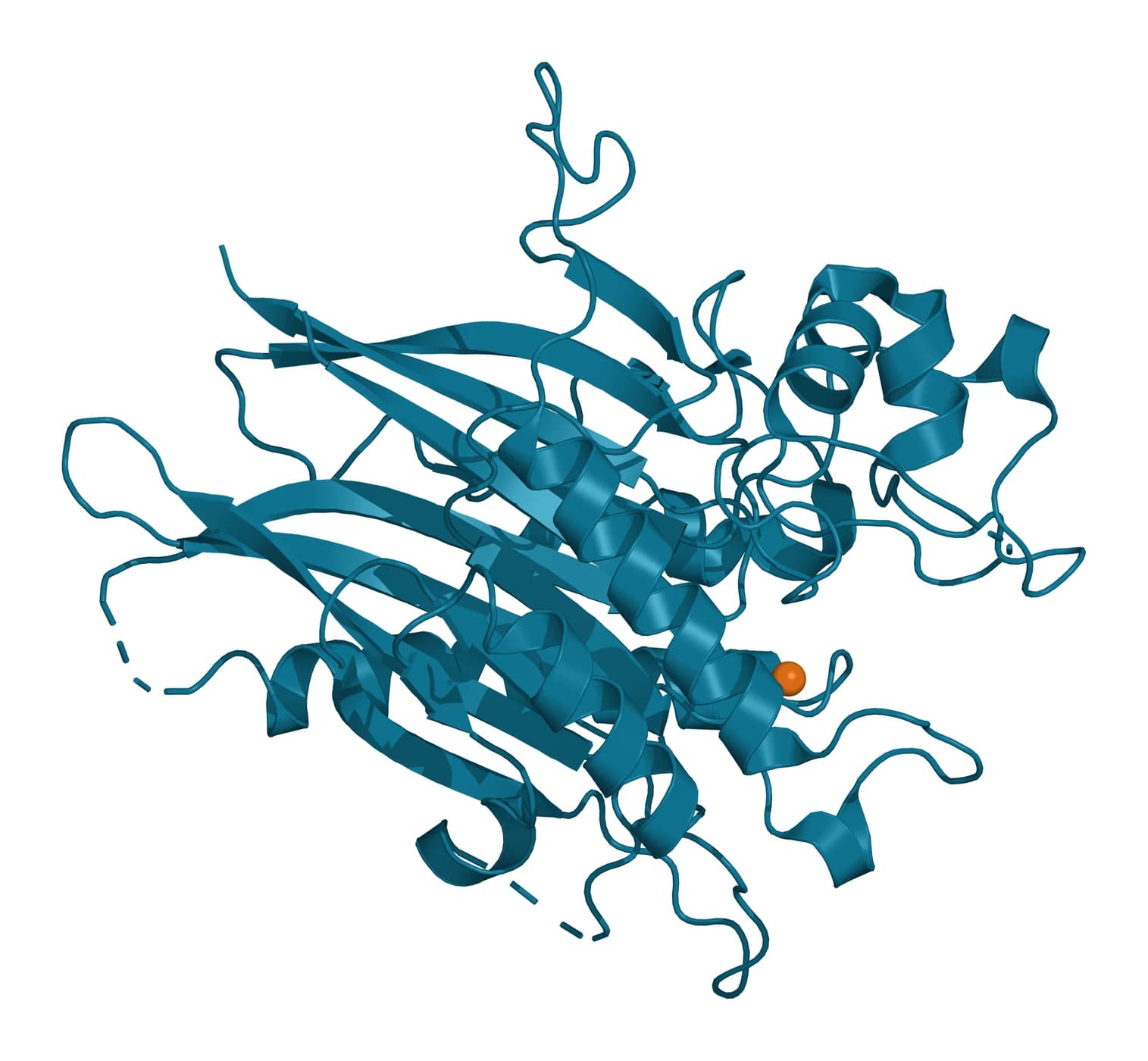
Fig. 3 | C16 ceramide (Cer d18:1/16:0) and Human nSMase2 catalytic domain.
Headgroups and acyl chains set membrane curvature and packing
The shape of a lipid follows from the size of its headgroup relative to its hydrocarbon chains. Ceramide, with its small headgroup, favors inverted hexagonal phases and promotes the transmembrane flip-flop of neighboring lipids.19 The other lipid classes enriched in small vesicles contribute their own structural features:
- Very-long-chain sphingolipids reach across the bilayer midplane toward PS 18:0/18:1 in the opposite leaflet, with cholesterol supporting the contact.3
- Disaturated phospholipid species, enriched in mast cell and dendritic cell exosomes, are consistent with the tight lipid packing measured at neutral pH.5
- Ether-linked phospholipids are abundant in some exosomes and are discussed as part of raft-like structures and membrane trafficking.2,3
- BMP drives internal budding in a manner that depends on the pH gradient across the endosomal membrane.10
- Phosphoinositides help regulate membrane dynamics and take part in the release of exosomes.2
Headgroup chemistry also sets surface charge. The anionic serine headgroup of phosphatidylserine is the feature recognized by nSMase2 inside the cell and by phosphatidylserine receptors on the cell surface.7,8
Tight lipid packing makes small vesicles resistant and stiff
The lipid bilayer of an extracellular vesicle protects its encapsulated proteins, nucleic acids, lipids and metabolites from the extracellular environment.20 Osteikoetxea and colleagues found exosomes more resistant to detergent lysis than microvesicles and apoptotic bodies, and proposed graded detergent lysis as a way to tell subpopulations apart.21
Sorkin and colleagues measured vesicle mechanics by atomic force microscopy force spectroscopy. Natural vesicles of different origins and compositions showed similar mechanical properties, and vesicle stiffness decreased as the protein-to-lipid ratio increased.22
In red blood cell vesicles produced under different conditions, the same group found protein-rich soft vesicles and stiffer vesicles with low membrane protein content, each linked to a different vesiculation route. These measurements tie the mechanics of a vesicle to the lipid fraction of its membrane.22
Membrane lipids and their partner proteins shape vesicle uptake
Recipient cells take up extracellular vesicles by several routes, including clathrin-dependent endocytosis, caveolin-mediated uptake, macropinocytosis, phagocytosis and lipid raft-mediated internalization. Mulcahy and colleagues note that the route may depend on molecules on the surfaces of both the vesicle and the target cell.23
Surface phosphatidylserine is one such molecule. Miyanishi and colleagues identified Tim4 and Tim1 as phosphatidylserine receptors and showed that exosomes bound Tim1- or Tim4-expressing cells through phosphatidylserine.8
Pfrieger and Vitale discuss how cholesterol may contribute to vesicle uptake as well as release, and how vesicles may take part in cellular sterol homeostasis.4 Record and colleagues add that vesicles carry bioactive lipid mediators, including eicosanoids, that can change the phenotype of receiving cells.24
Flotillins and cholesterol-binding tetraspanins
Some vesicle proteins are closely tied to the lipid environment. Kowal and colleagues found flotillin, long used as an exosome marker, at similar levels across all the vesicle populations they separated from human dendritic cells, alongside MHC and HSP70 proteins. The same study proposed immuno-isolation with the tetraspanins CD63, CD81 or CD9 to separate exosomal from other small vesicle subpopulations.12
Tetraspanins connect protein organization to cholesterol. Zimmerman and colleagues solved the structure of full-length CD81 and found a cholesterol molecule bound in an intramembrane pocket where its helices converge at the inner leaflet; cholesterol binding appeared to modulate CD81 activity in cells.25
Where to go from here
The cargo sorting reference explains how proteins and RNA enter the intraluminal vesicles whose membranes this page describes, and the uptake reference follows those vesicles into recipient cells. The tetraspanin family reference covers CD81, CD9 and CD63 in depth.
The MISEV2023 reference explains the reporting framework in which lipid measurements sit. Verified practices can request the certificate of analysis for a lot, and our team can explain how its characterization data were obtained.
References
Numbered in the order they are cited in the text. Each study carries its design and, where people were enrolled, how many, with a line saying what it supports on this page. Primary sources are regulations, agency documents and reference databases.
- 1.Skotland T, Sandvig K, Llorente A. Lipids in exosomes: Current knowledge and the way forward. Progress in Lipid Research. 2017. doi:10.1016/j.plipres.2017.03.001. PMID: 28342835.Narrative reviewReference 1 supports: Review comparing published exosome lipidomes at the class and species level. Cited for lipid enrichment relative to parent cells and for the proposed pairing of very-long-chain sphingolipids with inner leaflet PS 18:0/18:1.
- 2.Skotland T, Hessvik NP, Sandvig K, et al. Exosomal lipid composition and the role of ether lipids and phosphoinositides in exosome biology. Journal of Lipid Research. 2019. doi:10.1194/jlr.R084343. PMID: 30076207.Narrative reviewReference 2 supports: Review of exosome lipid composition. Cited for enrichment in cholesterol, sphingomyelin, glycosphingolipids and phosphatidylserine, for ether lipids and phosphoinositides, and for lipid composition as a purity indicator.
- 3.Skotland T, Llorente A, Sandvig K. Lipids in Extracellular Vesicles: What Can Be Learned about Membrane Structure and Function? Cold Spring Harbor Perspectives in Biology. 2023. doi:10.1101/cshperspect.a041415. PMID: 37277192.Narrative reviewReference 3 supports: Review of vesicles as single-bilayer membrane models. Cited for the similarity to detergent-resistant membranes, interleaflet coupling supported by cholesterol, and ether-linked phospholipids in raft-like structures.
- 4.Pfrieger FW, Vitale N. Cholesterol and the journey of extracellular vesicles. Journal of Lipid Research. 2018. doi:10.1194/jlr.R084210. PMID: 29678958.Narrative reviewReference 4 supports: Review of sterols in vesicle biology. Cited for cholesterol accumulation in vesicles and its possible roles in biogenesis, release, uptake and sterol homeostasis.
- 5.Laulagnier K, Motta C, Hamdi S, et al. Mast cell- and dendritic cell-derived exosomes display a specific lipid composition and an unusual membrane organization. Biochemical Journal. 2004. doi:10.1042/BJ20031594. PMID: 14965343.In vitroLandmarkReference 5 supports: Lipid analysis and membrane biophysics of mast cell and dendritic cell exosomes. Cited for sphingomyelin and disaturated species enrichment, cholesterol and BMP levels, pH-dependent rigidity and rapid flip-flop.
- 6.Haraszti RA, Didiot MC, Sapp E, et al. High-resolution proteomic and lipidomic analysis of exosomes and microvesicles from different cell sources. Journal of Extracellular Vesicles. 2016. doi:10.3402/jev.v5.32570. PMID: 27863537.In vitroReference 6 supports: Paired lipidomic and proteomic analysis of exosomes, microvesicles and source cells. Cited for the 1,961 lipid species identified and the lipid classes that characterize exosomes and microvesicles.
- 7.Airola MV, Shanbhogue P, Shamseddine AA, et al. Structure of human nSMase2 reveals an interdomain allosteric activation mechanism for ceramide generation. Proceedings of the National Academy of Sciences of the United States of America. 2017. doi:10.1073/pnas.1705134114. PMID: 28652336.In vitroReference 7 supports: Crystal structure of the nSMase2 catalytic domain with biochemistry of its activation. Cited for the DNase-I-type fold, the DK switch, allosteric activation by the N-terminal domain and stimulation by phosphatidylserine.
- 8.Miyanishi M, Tada K, Koike M, et al. Identification of Tim4 as a phosphatidylserine receptor. Nature. 2007. doi:10.1038/nature06307. PMID: 17960135.PreclinicalLandmarkReference 8 supports: Identification of Tim4 and Tim1 as phosphatidylserine receptors in mouse cells. Cited for phosphatidylserine on the exosome surface and exosome binding to Tim1- and Tim4-expressing cells.
- 9.Hullin-Matsuda F, Colosetti P, Rabia M, et al. Exosomal lipids from membrane organization to biomarkers: Focus on an endolysosomal-specific lipid. Biochimie. 2022. doi:10.1016/j.biochi.2022.09.016. PMID: 36184001.Narrative reviewReference 9 supports: Review of vesicle lipids with a focus on BMP. Cited for BMP as an endolysosome-specific lipid and a proposed signature of the endosomal origin of small vesicles.
- 10.Matsuo H, Chevallier J, Mayran N, et al. Role of LBPA and Alix in multivesicular liposome formation and endosome organization. Science. 2004. doi:10.1126/science.1092425. PMID: 14739459.In vitroLandmarkReference 10 supports: Liposome and cell studies of LBPA. Cited for LBPA-driven formation of multivesicular liposomes, its dependence on a pH gradient and its control by ALIX.
- 11.Rabia M, Leuzy V, Soulage C, et al. Bis(monoacylglycero)phosphate, a new lipid signature of endosome-derived extracellular vesicles. Biochimie. 2020. doi:10.1016/j.biochi.2020.07.005. PMID: 32659447.Clinical studyReference 11 supports: Human pilot study with cell culture work on urinary and HEK293 vesicles. Cited for BMP on extracellular vesicles co-localizing with CD63 and ALIX.
- 12.Kowal J, Arras G, Colombo M, et al. Proteomic comparison defines novel markers to characterize heterogeneous populations of extracellular vesicle subtypes. Proceedings of the National Academy of Sciences of the United States of America. 2016. doi:10.1073/pnas.1521230113. PMID: 26858453.In vitroReference 12 supports: Quantitative proteomics of separated dendritic cell vesicle populations. Cited for flotillin, MHC and HSP70 present across all vesicle types and for protein markers of exosome subpopulations.
- 13.Skotland T, Ekroos K, Llorente A, et al. Quantitative Lipid Analysis of Extracellular Vesicle Preparations: A Perspective. Journal of Extracellular Vesicles. 2025. doi:10.1002/jev2.70049. PMID: 40091364.Narrative reviewReference 13 supports: Perspective on quantitative lipidomics of vesicle preparations. Cited for what mass spectrometry reports (total lipid, species, purity, origin) and for the Lipidomic Standard Initiative guidelines.
- 14.Osteikoetxea X, Balogh A, Szabó-Taylor K, et al. Improved characterization of EV preparations based on protein to lipid ratio and lipid properties. PLoS One. 2015. doi:10.1371/journal.pone.0121184. PMID: 25798862.In vitroReference 14 supports: Total lipid assay and membrane probe study of vesicle subpopulations. Cited for characteristic protein-to-lipid ratios and for differences in lipid order, clustered cholesterol and GM1.
- 15.Ghosal S, Leporati R, Varga Á, et al. Cholesteryl esters and high protein-to-lipid ratios distinguish non-vesicular extracellular particles from extracellular vesicles. Cell Communication and Signaling. 2026. doi:10.1186/s12964-026-02880-6. PMID: 41992224.In vitroReference 15 supports: Lipidomic and biochemical profiling of non-vesicular particles and vesicle subtypes. Cited for higher protein-to-lipid ratios and cholesteryl ester and triacylglycerol enrichment in non-vesicular particles.
- 16.Sezgin E, Levental I, Mayor S, et al. The mystery of membrane organization: composition, regulation and roles of lipid rafts. Nature Reviews Molecular Cell Biology. 2017. doi:10.1038/nrm.2017.16. PMID: 28356571.Narrative reviewReference 16 supports: Review of membrane raft biophysics. Cited for ordered domains formed by preferential association of cholesterol with saturated lipids.
- 17.Trajkovic K, Hsu C, Chiantia S, et al. Ceramide triggers budding of exosome vesicles into multivesicular endosomes. Science. 2008. doi:10.1126/science.1153124. PMID: 18309083.In vitroLandmarkReference 17 supports: Cell biology and lipidomics of intraluminal vesicle budding. Cited for ceramide-dependent, ESCRT-independent budding, ceramide enrichment in exosomes and reduced release after neutral sphingomyelinase inhibition.
- 18.NCBI Gene 55512, SMPD3 sphingomyelin phosphodiesterase 3 (Homo sapiens). Accessed 20 October 2026.Primary source
- 19.Alonso A, Goñi FM. The Physical Properties of Ceramides in Membranes. Annual Review of Biophysics. 2018. doi:10.1146/annurev-biophys-070317-033309. PMID: 29618220.Narrative reviewReference 19 supports: Review of ceramide membrane biophysics. Cited for increased lipid order, domain formation, intrinsic negative curvature, inverted hexagonal phases and promotion of flip-flop.
- 20.Skotland T, Sagini K, Sandvig K, et al. An emerging focus on lipids in extracellular vesicles. Advanced Drug Delivery Reviews. 2020. doi:10.1016/j.addr.2020.03.002. PMID: 32151658.Narrative reviewReference 20 supports: Review of the state of vesicle lipid research. Cited for the bilayer protecting encapsulated proteins, nucleic acids, lipids and metabolites from the extracellular environment.
- 21.Osteikoetxea X, Sódar B, Németh A, et al. Differential detergent sensitivity of extracellular vesicle subpopulations. Organic & Biomolecular Chemistry. 2015. doi:10.1039/c5ob01451d. PMID: 26264754.In vitroReference 21 supports: Detergent lysis study of size-based vesicle subpopulations. Cited for the greater detergent resistance of exosomes compared with microvesicles and apoptotic bodies.
- 22.Sorkin R, Huisjes R, Bošković F, et al. Nanomechanics of Extracellular Vesicles Reveals Vesiculation Pathways. Small. 2018. doi:10.1002/smll.201801650. PMID: 30160371.In vitroReference 22 supports: Atomic force microscopy force spectroscopy of natural vesicles. Cited for similar mechanics across vesicle origins and for stiffness decreasing as the protein-to-lipid ratio rises.
- 23.Mulcahy LA, Pink RC, Carter DR. Routes and mechanisms of extracellular vesicle uptake. Journal of Extracellular Vesicles. 2014. doi:10.3402/jev.v3.24641. PMID: 25143819.Narrative reviewReference 23 supports: Review of vesicle uptake mechanisms. Cited for the endocytic routes, including lipid raft-mediated internalization, and for surface molecules on both partners shaping the route.
- 24.Record M, Silvente-Poirot S, Poirot M, et al. Extracellular vesicles: lipids as key components of their biogenesis and functions. Journal of Lipid Research. 2018. doi:10.1194/jlr.E086173. PMID: 29764923.Narrative reviewReference 24 supports: Review of lipid pathways in vesicle biogenesis and function. Cited for vesicles carrying bioactive lipid mediators such as eicosanoids to receiving cells.
- 25.Zimmerman B, Kelly B, McMillan BJ, et al. Crystal Structure of a Full-Length Human Tetraspanin Reveals a Cholesterol-Binding Pocket. Cell. 2016. doi:10.1016/j.cell.2016.09.056. PMID: 27881302.In vitroReference 25 supports: Crystal structure of full-length CD81. Cited for the intramembrane pocket holding cholesterol at the inner leaflet and for cholesterol binding modulating CD81 activity.
Cite this page
ExaVeyra Sciences Editorial Team. Extracellular Vesicle Lipid Composition and Membrane Biophysics. ExaVeyra Sciences; 20 October 2026. Available from: https://exaveyra.com/science/exosome-biochemistry/extracellular-vesicle-lipidome. Accessed [date accessed].
Figure sources
- 26.PubChem CID 5997, Cholesterol. National Center for Biotechnology Information, PubChem. Public domain.
- 27.PubChem CID 9939941, N-Palmitoylsphingomyelin. National Center for Biotechnology Information, PubChem. Public domain.
- 28.PubChem CID 9547087, PS(18:0/18:1(9Z)). National Center for Biotechnology Information, PubChem. Public domain.
- 29.PubChem CID 5283564, C16-Ceramide. National Center for Biotechnology Information, PubChem. Public domain.
- 30.PDB 5UVG, Human nSMase2 catalytic domain. ExaVeyra Sciences, rendered in PyMOL from PDB 5UVG. Structure data: RCSB Protein Data Bank (rcsb.org), CC0. Structure publication: Airola MV, Shanbhogue P, Shamseddine AA, et al. Structure of human nSMase2 reveals an interdomain allosteric activation mechanism for ceramide generation. Proc Natl Acad Sci U S A. 2017;114(28):E5549-E5558.